
Create successful ePaper yourself
Turn your PDF publications into a flip-book with our unique Google optimized e-Paper software.
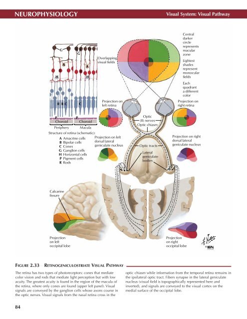
NEUROPHYSIOLOGY<br />
Visual System: Visual Pathway<br />
G<br />
A<br />
B<br />
H<br />
G<br />
B<br />
A<br />
H<br />
Overlapping<br />
visual fields<br />
Central<br />
darker<br />
circle<br />
represents<br />
macular<br />
zone<br />
Lightest<br />
shades<br />
represent<br />
monocular<br />
fields<br />
Each<br />
quadrant<br />
a different<br />
color<br />
R R C<br />
C<br />
Projection on<br />
left retina<br />
Projection on<br />
right retina<br />
P<br />
P<br />
Choroid Choroid<br />
Periphery Macula<br />
Structure of retina (schematic):<br />
A Amacrine cells<br />
B Bipolar cells<br />
C Cones<br />
G Ganglion cells<br />
H Horizontal cells<br />
P Pigment cells<br />
R Rods<br />
Projection on left<br />
dorsal lateral<br />
geniculate nucleus<br />
Optic<br />
(II) nerves<br />
Optic chiasm<br />
Optic tracts<br />
Lateral<br />
geniculate<br />
bodies<br />
Projection on right<br />
dorsal lateral<br />
geniculate nucleus<br />
Calcarine<br />
fissure<br />
Projection<br />
on left<br />
occipital lobe<br />
Projection<br />
on right<br />
occipital lobe<br />
©<br />
FIGURE 2.33<br />
RETINOGENICULOSTRIATE VISUAL PATHWAY •<br />
The retina has two types of photoreceptors: cones that mediate<br />
color vision and rods that mediate light perception but with low<br />
acuity. The greatest acuity is found in the region of the macula of<br />
the retina, where only cones are found (upper left panel). Visual<br />
signals are conveyed by the ganglion cells whose axons course in<br />
the optic nerves. Visual signals from the nasal retina cross in the<br />
optic chiasm while information from the temporal retina remains in<br />
the ipsilateral optic tract. Fibers synapse in the lateral geniculate<br />
nucleus (visual field is topographically represented here and<br />
inverted), and signals are conveyed to the visual cortex on the<br />
medial surface of the occipital lobe.<br />
84