
You also want an ePaper? Increase the reach of your titles
YUMPU automatically turns print PDFs into web optimized ePapers that Google loves.
NEUROPHYSIOLOGY<br />
Sensory Pathways: III<br />
C2<br />
C3<br />
C4<br />
C5<br />
T1<br />
T2<br />
T3<br />
T4<br />
T5<br />
T6<br />
T7<br />
T8<br />
T9<br />
T10<br />
T11<br />
T12<br />
L1<br />
S2, 3<br />
L2<br />
L3<br />
L4<br />
T1<br />
C8<br />
C6<br />
Schematic demarcation of<br />
dermatomes shown as distinct<br />
segments. There is actually<br />
considerable overlap between<br />
any two adjacent dermatomes<br />
C5<br />
C7<br />
C6<br />
C7<br />
C6<br />
C7<br />
C8<br />
S3<br />
S4<br />
S5<br />
C7 C8<br />
C8<br />
L5<br />
L1<br />
L2<br />
L3<br />
C2<br />
C3<br />
C4<br />
C5<br />
C6<br />
T1<br />
T2<br />
T3<br />
T4<br />
T5<br />
T6<br />
T7<br />
T8<br />
T9<br />
T10<br />
T11<br />
T12<br />
L1<br />
L2<br />
L3<br />
L4<br />
L5<br />
S1<br />
S2<br />
S1<br />
S2<br />
L5<br />
S1 S2<br />
L5<br />
S1<br />
L4<br />
L4<br />
S1<br />
L5<br />
L4<br />
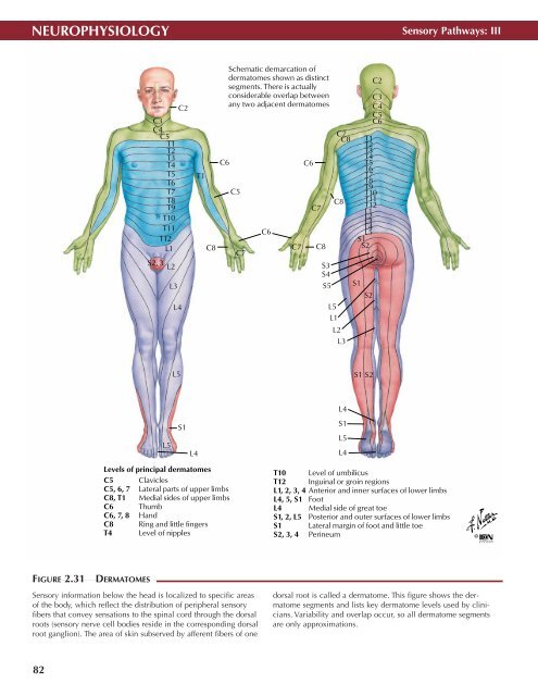
Levels of principal dermatomes<br />
C5 Clavicles<br />
C5, 6, 7 Lateral parts of upper limbs<br />
C8, T1 Medial sides of upper limbs<br />
C6 Thumb<br />
C6, 7, 8 Hand<br />
C8 Ring and little fingers<br />
T4 Level of nipples<br />
T10 Level of umbilicus<br />
T12 Inguinal or groin regions<br />
L1, 2, 3, 4 Anterior and inner surfaces of lower limbs<br />
L4, 5, S1 Foot<br />
L4 Medial side of great toe<br />
S1, 2, L5 Posterior and outer surfaces of lower limbs<br />
S1 Lateral margin of foot and little toe<br />
S2, 3, 4 Perineum<br />
©<br />
FIGURE 2.31<br />
DERMATOMES •<br />
Sensory information below the head is localized to specific areas<br />
of the body, which reflect the distribution of peripheral sensory<br />
fibers that convey sensations to the spinal cord through the dorsal<br />
roots (sensory nerve cell bodies reside in the corresponding dorsal<br />
root ganglion). The area of skin subserved by afferent fibers of one<br />
dorsal root is called a dermatome. This figure shows the dermatome<br />
segments and lists key dermatome levels used by clinicians.<br />
Variability and overlap occur, so all dermatome segments<br />
are only approximations.<br />
82