
Create successful ePaper yourself
Turn your PDF publications into a flip-book with our unique Google optimized e-Paper software.
NEUROPHYSIOLOGY<br />
Autonomic Nervous System: Schema<br />
Oculomotor nerve (III)<br />
Facial nerve (VII)<br />
Glossopharyngeal nerve (IX)<br />
Medulla oblongata<br />
Vagus nerve (X)<br />
Sweat<br />
gland<br />
Peripheral<br />
blood vessel<br />
C1<br />
Arrector (smooth)<br />
muscle of hair follicle<br />
Note: Above three<br />
structures are shown at<br />
only one level but<br />
occur at all levels<br />
C2<br />
C3<br />
C4<br />
C5<br />
C6<br />
C7<br />
C8<br />
T1<br />
T2<br />
T3<br />
T4<br />
T5<br />
T9<br />
T10<br />
T7<br />
T8<br />
T11<br />
T12<br />
T6<br />
L1<br />
L2<br />
L3<br />
L4<br />
L5<br />
Note: Blue-shaded S1<br />
areas indicate zones of<br />
parasympathetic S2<br />
outflow from CNS S3<br />
S4<br />
S5<br />
Coccygeal<br />
Gray rami<br />
communicantes<br />
Gray and white rami communicantes<br />
Gray rami communicantes<br />
Greater<br />
Lesser<br />
Least<br />
Lumbar<br />
splanchnic<br />
nerves<br />
⎧<br />
⎨<br />
⎩<br />
Splanchnic<br />
nerves<br />
Pelvic<br />
splanchnic<br />
nerves<br />
Intracranial vessels<br />
Ciliary ganglion<br />
Eye<br />
Pterygopalatine ganglion<br />
Lacrimal glands<br />
Otic ganglion<br />
Parotid glands<br />
Submandibular ganglion<br />
Sublingual and<br />
submandibular glands<br />
Peripheral cranial<br />
and facial vessels<br />
Larynx<br />
Trachea<br />
Bronchi<br />
Lungs<br />
Heart<br />
Stomach<br />
Liver<br />
Gallbladder<br />
Bile ducts<br />
Pancreas<br />
Suprarenal glands<br />
Kidneys<br />
Intestines<br />
Descending colon<br />
Sigmoid colon<br />
Rectum<br />
Prostate<br />
External genitalia<br />
Urinary bladder<br />
Pulmonary plexus<br />
Cardiac plexus<br />
Celiac ganglion<br />
Aorticorenal ganglion<br />
Superior mesenteric<br />
ganglion<br />
Inferior mesenteric<br />
ganglion<br />
Superior hypogastric<br />
plexus<br />
Inferior<br />
hypogastric<br />
plexus<br />
Sympathetic<br />
fibers<br />
Presynaptic<br />
Postsynaptic<br />
Parasympathetic<br />
fibers<br />
Presynaptic<br />
Postsynaptic<br />
Antidromic<br />
conduction<br />
©<br />
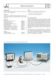
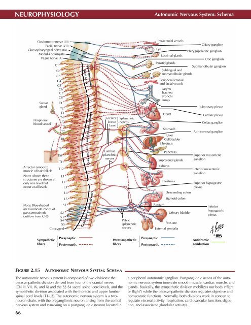
FIGURE 2.15<br />
AUTONOMIC NERVOUS SYSTEM: SCHEMA •<br />
The autonomic nervous system is composed of two divisions: the<br />
parasympathetic division derived from four of the cranial nerves<br />
(CN III, VII, IX, and X) and the S2-S4 sacral spinal cord levels, and the<br />
sympathetic division associated with the thoracic and upper lumbar<br />
spinal cord levels (T1-L2). The autonomic nervous system is a twoneuron<br />
chain, with the preganglionic neuron arising from the central<br />
nervous system and synapsing on a postganglionic neuron located in<br />
66<br />
a peripheral autonomic ganglion. Postganglionic axons of the autonomic<br />
nervous system innervate smooth muscle, cardiac muscle, and<br />
glands. Basically, the sympathetic division mobilizes our body (“fight<br />
or flight”) while the parasympathetic division regulates digestive and<br />
homeostatic functions. Normally, both divisions work in concert to<br />
regulate visceral activity (respiration, cardiovascular function, digestion,<br />
and associated glandular activity).