Jahresbericht 2005 - IPHT Jena
Jahresbericht 2005 - IPHT Jena
Jahresbericht 2005 - IPHT Jena

Create successful ePaper yourself
Turn your PDF publications into a flip-book with our unique Google optimized e-Paper software.
64<br />
a)<br />
b)<br />
Fig. 3.1: a) View of the compact UV-Raman<br />
spectral sensor (circled) with a commercial<br />
Raman spectrometer HR 600 with nearly the<br />
same performance and b) measured spectra of<br />
bacillus pumius, DSM 361, excitation wavelength<br />
257 nm, with both instruments.<br />
MIKROSYSTEME / MICROSYSTEMS<br />
Unconventional micro-imaging<br />
(R. Riesenberg, A. Grjasnow, M. Kanka,<br />
J. Bergmann)<br />
The coherent illumination of a sample by a pinhole<br />
generates an interference image (inlineholography).<br />
Results are given by unconventional<br />
microscopic imaging by illumination with a pinhole<br />
array, see Fig. 3.2. The microscopy by multiplane<br />
interference detection uses three or more<br />
interference pictures to reconstruct the objects.<br />
The actual technique is adapted to microscopic<br />
imaging. No reference is needed and a sub-pixel<br />
algorithm is implemented.<br />
The lensless technique is applied for wide field<br />
imaging. The illumination by a pinhole-array<br />
leads to a homogenous illumination with more<br />
intensity and so with an increased sensitivity. The<br />
field of view can be extended without loosing resolution<br />
(see Fig. 3.3).<br />
Fig. 3.2.: Arrangement of illumination of the<br />
sample by a pinhole-array and interference<br />
detection by a CCD.<br />
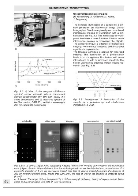
Fig. 3.3: a…d above: Digital inline holography. Objects (diameter of 0.5 µm) at the edge of the illumination<br />
cone (object plane in 10 µm distance from the pinhole-plane) can not be detected and reconstructed. For<br />
a pinhole diameter of 1 µm the aperture is limited. The field of view is limited [hologram at a distance of<br />
220 µm from the pinhole-plane, image area (200 µm) 2 , the field of view in the example is limited to about<br />
(9 µm) 2 .<br />
e…h below: The single pinhole is replaced by a pinhole-array (9 pinholes). Nearly all objects can be illuminated<br />
and reconstructed. The field of view is extended.