Jahresbericht 2005 - IPHT Jena
Jahresbericht 2005 - IPHT Jena
Jahresbericht 2005 - IPHT Jena

You also want an ePaper? Increase the reach of your titles
YUMPU automatically turns print PDFs into web optimized ePapers that Google loves.
68<br />
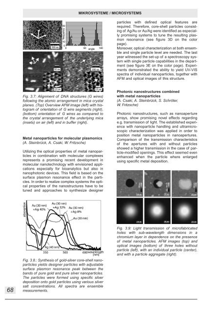
Fig. 3.7: Alignment of DNA structures (G wires)<br />
following the atomic arrangement in mica crystal<br />
planes. (Top) Overview AFM image (left) with histogram<br />
of orientation of G wire segments (right),<br />
(bottom) orientation of G wires as compared to<br />
the crystal arrangement of the underlying mica<br />
(insets) on air (left) and in buffer (right).<br />
Metal nanoparticles for molecular plasmonics<br />
(A. Steinbrück, A. Csaki, W. Fritzsche)<br />
Utilizing the optical properties of metal nanoparticles<br />
in combination with molecular complexes<br />
represents a promising recent development in<br />
molecular nanotechnology with envisioned applications<br />
especially for bioanalytics but also in<br />
nanophotonic devices. This field is based on the<br />
surface plasmon resonance effect in the particles.<br />
In order to realize complex systems the optical<br />
properties of the nanostructures have to be<br />
tuned and approaches to synthesize designer<br />
Fig. 3.8.: Synthesis of gold-silver core-shell nanoparticles<br />
yields designer particles with adjustable<br />
surface plasmon resonance peak between the<br />
bands of pure gold and pure silver nanoparticles.<br />
The particles were formed using specific silver<br />
deposition onto gold particles using various silver<br />
salt concentrations. All spectra are ensemble<br />
measurements.<br />
MIKROSYSTEME / MICROSYSTEMS<br />
particles with defined optical features are<br />
required. Therefore, core-shell particles consisting<br />
of Ag/Au or Au/Ag were identified as especially<br />
promising systems to tune the resulting plasmon<br />
resonance (see figure 3D on the color<br />
page).<br />
Moreover, optical characterization at both ensemble<br />
and single particle level are needed. The last<br />
year witnessed the set-up of a spectroscopy system<br />
with single particle capabilities in the department<br />
(see figure 3E on the color page). Experiments<br />
demonstrated the ability to yield UV-VIS<br />
spectra of individual nanoparticles, together with<br />
AFM and optical images of this structure.<br />
Photonic nanostructures combined<br />
with metal nanoparticles<br />
(A. Csaki, A. Steinbrück, S. Schröter,<br />
W. Fritzsche)<br />
Photonic nanostructures, such as nanoaperture<br />
arrays, show promising novel effects regarding<br />
e.g. transmission of light. The established experience<br />
with nanoparticle handling and ultramicroscopic<br />
characterization was applied in order to<br />
position metal nanoparticles in nanoapertures.<br />
Comparison of the transmission characteristics<br />
of the apertures with and without particles<br />
showed a higher transmission in the case of particle-modified<br />
openings. This effect seemed even<br />
enhanced when the particle where enlarged<br />
using specific metal deposition.<br />
Fig. 3.9: Light transmission of microfabricated<br />
holes with sub-wavelength dimensions in a<br />
chromium layer in dependence on the presence<br />
of metal nanoparticles. AFM images (top) and<br />
optical images (bottom) of three holes without<br />
particle (left), with an individual particle (center),<br />
and with a particle aggregate (right).