From Protein Structure to Function with Bioinformatics.pdf
From Protein Structure to Function with Bioinformatics.pdf
From Protein Structure to Function with Bioinformatics.pdf

Create successful ePaper yourself
Turn your PDF publications into a flip-book with our unique Google optimized e-Paper software.
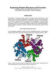
280 J.D. Watson and J.M. Thorn<strong>to</strong>nfold matches were all found <strong>to</strong> be hypothetical proteins <strong>with</strong> no functional annotation,looking further down the list of hits all except one of those remaining were<strong>to</strong> various monooxygenases. No significant matches were found <strong>to</strong> any of theenzyme, DNA or ligand templates but the reverse template scan provided a largenumber of matches. Once again the majority of the <strong>to</strong>p hits were <strong>to</strong> proteins ofunknown function but the first significant hit <strong>with</strong> an assigned function was <strong>to</strong> amonooxygenase from Strep<strong>to</strong>myces coelicolor (PDB entry 1lq9). The combinedresults from the fold comparison and reverse templates methods therefore indicateda monooxygenase function. Subsequent experimental analysis has characterisedthe protein as a haem-degrading enzyme <strong>with</strong> structural similarity <strong>to</strong>monooxygenases (Wu et al. 2005). This is a prime example where the structurecan lend additional supporting evidence <strong>to</strong> one out of many equally confidentsequence-based predictions.More recently a paper by Adams et al. (2007) discussed the structure-basedfunctional annotation of hypothetical proteins. The paper illustrated five case studieswhere a variety of methods used in combination <strong>with</strong> biochemical assays providedfunctional annotations. The first case involved a protein (ChuS) <strong>with</strong> a novelfold where initial operon studies combined <strong>with</strong> gene knockouts suggested that theprotein was involved in haem uptake and utilisation. As the structure solved was theapo-form of the protein and it was a novel fold, the initial structure-based analysesdid not suggest any particular function. However, further biochemical analysis suggesteda haem oxygenase function. A multiple sequence alignment of ChuS <strong>with</strong> itshomologues highlighted four conserved histidine residues which, when mappedon<strong>to</strong> the structure of ChuS, revealed that three of the four conserved histidine residuesare adjacent <strong>to</strong> or pointing in<strong>to</strong> one of two large clefts on opposite sides of thecentral core. This prompted further structural studies <strong>with</strong> haem co-crystallisationand mutagenesis of the conserved histidines. These additional studies indicated thatthe haem coordination is unlike any other haem degradation enzyme and that ChuSis the first haem oxygenase <strong>to</strong> be identified in E. coli.The second example illustrated the case of protein YgiN. The structure of thisprotein was scanned against the SCOP database using the MSDfold (SSM) serverand found <strong>to</strong> have fold similarity <strong>to</strong> ActVA-Orf6, a monooxygenase from S. coelicolor.Members of the ActVA-Orf6 mono-oxygenase family are involved in thesynthesis of large, polyketide compounds in the antibiotic biosynthetic pathwaysof Gram-positive bacteria. The ActVA-Orf6 acts as an enzyme late on in the processtailoring an antifungal compound, dihydrokalafungin, <strong>to</strong> confer its specificactivity (Sciara et al. 2003). As E. coli was not known <strong>to</strong> produce the same compound,the natural substrate for YgiN was expected <strong>to</strong> differ. Initial attempts <strong>to</strong>biochemically characterise the enzyme were fruitless until two prior reports in theliterature were identified as actually referring <strong>to</strong> the YgiN protein. With this additionalexperimental information and structural information on the various substratesused previously, the authors suggested that the YgiN protein may beinvolved in menadione metabolism. Further experimental work resulted in thestructure being co-crystallised <strong>with</strong> menadione, in its apo form and <strong>with</strong> flavinadenine dinucleotide (Adams and Jia 2006).