- Page 3:
Daniel John RigdenEditorFrom Protei
- Page 8 and 9:
ContentsSection I Generating and In
- Page 10 and 11:
Contentsxi4.2.1 Alpha-Helical Bundl
- Page 12 and 13:
Contentsxiii7.4.2 Predicting Bindin
- Page 14 and 15:
Contentsxv12.3 Accuracy and Added V
- Page 16 and 17:
4 J. Lee et al.about 5.3 million pr
- Page 18 and 19:
6 J. Lee et al.Table 1.1 A list of
- Page 20 and 21:
8 J. Lee et al.2000; Sorin and Pand
- Page 22 and 23:
10 J. Lee et al.Although most knowl
- Page 24 and 25:
12 J. Lee et al.Fig. 1.3 Two exampl
- Page 26 and 27:
14 J. Lee et al.rugged containing m
- Page 28 and 29:
16 J. Lee et al.1.3.4 Mathematical
- Page 30 and 31:
18 J. Lee et al.potentials, each re
- Page 32 and 33:
20 J. Lee et al.viewpoint of struct
- Page 34 and 35:
22 J. Lee et al.Hsieh MJ, Luo R (20
- Page 36 and 37:
24 J. Lee et al.Sippl MJ (1990) Cal
- Page 38 and 39:
Chapter 2Fold RecognitionLawrence A
- Page 40 and 41:
2 Fold Recognition 29It has long be
- Page 42 and 43:
2 Fold Recognition 31Fig. 2.2 Graph
- Page 44 and 45:
2 Fold Recognition 33distribute the
- Page 46 and 47:
2 Fold Recognition 35Table 2.1 (a)
- Page 48 and 49:
2 Fold Recognition 37dependent on t
- Page 50 and 51:
2 Fold Recognition 39based on the r
- Page 52 and 53:
2 Fold Recognition 41The idea of co
- Page 54 and 55:
2 Fold Recognition 43query sequence
- Page 56 and 57:
2 Fold Recognition 452.3.4 Consensu
- Page 58 and 59:
2 Fold Recognition 472.4 Alignment
- Page 60 and 61:
2 Fold Recognition 49statistical me
- Page 62 and 63:
2 Fold Recognition 51methods (e.g.
- Page 64 and 65:
2 Fold Recognition 53Berman HM, Wes
- Page 66 and 67:
2 Fold Recognition 55Tress ML, Jone
- Page 68 and 69:
58 A. Fiserwith more than 50% seque
- Page 70 and 71:
60 A. FiserIn contrast to ab initio
- Page 72 and 73:
62 A. FiserTable 3.1 (continued)SWI
- Page 74 and 75:
64 A. Fiserbetween fold recognition
- Page 76 and 77:
66 A. FiserFig. 3.1 Comparing accur
- Page 78 and 79:
68 A. Fiserinto conserved core regi
- Page 80 and 81:
70 A. Fiseralignments are built and
- Page 82 and 83:
72 A. Fiserfold, such as the hyperv
- Page 84 and 85:
74 A. Fiserfunctions delivers impro
- Page 86 and 87:
76 A. Fiserfrom efforts of genome s
- Page 88 and 89:
78 A. FiserA rigorous statistical e
- Page 90 and 91:
80 A. Fiser(Clore et al. 1993). Cha
- Page 92 and 93:
82 A. FiserImproved and new methods
- Page 94 and 95:
84 A. FiserClaessens M, Van Cutsem
- Page 96 and 97:
86 A. FiserHavel TF, Snow ME (1991)
- Page 98 and 99:
88 A. FiserPetrey D, Xiang Z, Tang
- Page 100 and 101:
90 A. Fiservan Vlijmen HW, Karplus
- Page 102 and 103:
92 T. Nugent and D.T. Jones4.2 Stru
- Page 104 and 105:
94 T. Nugent and D.T. JonesFig. 4.2
- Page 106 and 107:
96 T. Nugent and D.T. JonesTable 4.
- Page 108 and 109:
98 T. Nugent and D.T. Jones2000), d
- Page 110 and 111:
100 T. Nugent and D.T. JonesTable 4
- Page 112 and 113:
102 T. Nugent and D.T. JonesFig. 4.
- Page 114 and 115:
104 T. Nugent and D.T. JonesFig. 4.
- Page 116 and 117:
106 T. Nugent and D.T. Jonessubdivi
- Page 118 and 119:
108 T. Nugent and D.T. Jonescomplex
- Page 120 and 121:
110 T. Nugent and D.T. JonesMartell
- Page 122 and 123:
Chapter 5Bioinformatics Approaches
- Page 124 and 125:
5 Structure and Function of Intrins
- Page 126 and 127:
5 Structure and Function of Intrins
- Page 128 and 129:
5 Structure and Function of Intrins
- Page 130 and 131:
5 Structure and Function of Intrins
- Page 132 and 133:
5 Structure and Function of Intrins
- Page 134 and 135:
5 Structure and Function of Intrins
- Page 136 and 137:
5 Structure and Function of Intrins
- Page 138 and 139:
5 Structure and Function of Intrins
- Page 140 and 141:
5 Structure and Function of Intrins
- Page 142 and 143:
5 Structure and Function of Intrins
- Page 144 and 145:
5 Structure and Function of Intrins
- Page 146 and 147:
5 Structure and Function of Intrins
- Page 148 and 149:
5 Structure and Function of Intrins
- Page 150 and 151:
Chapter 6Function Diversity Within
- Page 152 and 153:
6 Function Diversity Within Folds a
- Page 154 and 155:
6 Function Diversity Within Folds a
- Page 156 and 157:
6 Function Diversity Within Folds a
- Page 158 and 159:
6 Function Diversity Within Folds a
- Page 160 and 161:
6 Function Diversity Within Folds a
- Page 162 and 163:
6 Function Diversity Within Folds a
- Page 164 and 165:
6 Function Diversity Within Folds a
- Page 166 and 167:
6 Function Diversity Within Folds a
- Page 168 and 169:
6 Function Diversity Within Folds a
- Page 170 and 171:
6 Function Diversity Within Folds a
- Page 172 and 173:
6 Function Diversity Within Folds a
- Page 174 and 175:
Chapter 7Predicting Protein Functio
- Page 176 and 177:
7 Predicting Protein Function from
- Page 178 and 179:
7 Predicting Protein Function from
- Page 180 and 181:
7 Predicting Protein Function from
- Page 182 and 183:
7 Predicting Protein Function from
- Page 184 and 185:
7 Predicting Protein Function from
- Page 186 and 187:
7 Predicting Protein Function from
- Page 188 and 189:
7 Predicting Protein Function from
- Page 190 and 191:
Table 7.1 Online resources and tool
- Page 192 and 193:
7 Predicting Protein Function from
- Page 194 and 195:
Chapter 83D MotifsElaine C. Meng, B
- Page 196 and 197:
8 3D Motifs 189clustering similar s
- Page 198 and 199:
8 3D Motifs 191To improve the signa
- Page 200 and 201:
8 3D Motifs 193structures together.
- Page 202 and 203:
8 3D Motifs 195Table 8.1 (continued
- Page 204 and 205:
8 3D Motifs 1978.3.1 User-Defined M
- Page 206 and 207:
8 3D Motifs 199Fig. 8.3 The FFF mot
- Page 208 and 209:
8 3D Motifs 2018.3.2 Motif Discover
- Page 210 and 211:
8 3D Motifs 203In addition to SITE
- Page 212 and 213:
8 3D Motifs 205folds; they shared a
- Page 214 and 215:
8 3D Motifs 207from a training set
- Page 216 and 217:
8 3D Motifs 209Table 8.3 Web server
- Page 218 and 219:
8 3D Motifs 211its greater overall
- Page 220 and 221:
8 3D Motifs 213Ideally, a 3D motif
- Page 222 and 223:
8 3D Motifs 215Ivanisenko VA, Pintu
- Page 224 and 225:
Chapter 9Protein Dynamics: From Str
- Page 226 and 227:
9 Protein Dynamics: From Structure
- Page 228 and 229:
9 Protein Dynamics: From Structure
- Page 230 and 231:
9 Protein Dynamics: From Structure
- Page 232 and 233:
9 Protein Dynamics: From Structure
- Page 234 and 235:
9 Protein Dynamics: From Structure
- Page 236 and 237:
9 Protein Dynamics: From Structure
- Page 238 and 239:
9 Protein Dynamics: From Structure
- Page 240 and 241:
9 Protein Dynamics: From Structure
- Page 242 and 243:
9 Protein Dynamics: From Structure
- Page 244 and 245:
9 Protein Dynamics: From Structure
- Page 246 and 247:
9 Protein Dynamics: From Structure
- Page 248 and 249:
9 Protein Dynamics: From Structure
- Page 250 and 251:
9 Protein Dynamics: From Structure
- Page 252 and 253:
9 Protein Dynamics: From Structure
- Page 254 and 255:
9 Protein Dynamics: From Structure
- Page 256 and 257:
9 Protein Dynamics: From Structure
- Page 258 and 259: 252 R.A. LaskowskiConsequently, man
- Page 260 and 261: 254 R.A. Laskowskiucla.edu, and Pro
- Page 262 and 263: 256 R.A. LaskowskiFig. 10.2 Gene On
- Page 264 and 265: 258 R.A. Laskowski10.2.5 Protein In
- Page 266 and 267: 260 R.A. LaskowskiFig. 10.4 Schemat
- Page 268 and 269: 262 R.A. Laskowskiaccessibility and
- Page 270 and 271: 264 R.A. LaskowskiFig. 10.6 A ligan
- Page 272 and 273: 266 R.A. LaskowskiGly97Gly99Tyr98Ty
- Page 274 and 275: 268 R.A. LaskowskiFig. 10.8 Example
- Page 276 and 277: 270 R.A. LaskowskiReferencesAltschu
- Page 278 and 279: 272 R.A. LaskowskiXenarios I, Salwi
- Page 280 and 281: 274 J.D. Watson and J.M. ThorntonTh
- Page 282 and 283: 276 J.D. Watson and J.M. ThorntonTa
- Page 284 and 285: 278 J.D. Watson and J.M. Thorntonva
- Page 286 and 287: 280 J.D. Watson and J.M. Thorntonfo
- Page 288 and 289: 282 J.D. Watson and J.M. Thorntonin
- Page 290 and 291: 284 J.D. Watson and J.M. Thorntonan
- Page 292 and 293: 286 J.D. Watson and J.M. ThorntonFi
- Page 294 and 295: 288 J.D. Watson and J.M. ThorntonPD
- Page 296 and 297: 290 J.D. Watson and J.M. ThorntonKi
- Page 298 and 299: Chapter 12Prediction of Protein Fun
- Page 300 and 301: 12 Prediction of Protein Function f
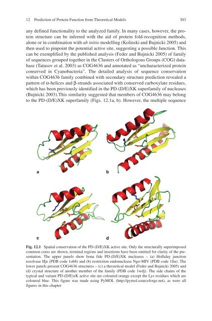
- Page 302 and 303: 12 Prediction of Protein Function f
- Page 304 and 305: 12 Prediction of Protein Function f
- Page 306 and 307: 12 Prediction of Protein Function f
- Page 310 and 311: 12 Prediction of Protein Function f
- Page 312 and 313: 12 Prediction of Protein Function f
- Page 314 and 315: 12 Prediction of Protein Function f
- Page 316 and 317: 12 Prediction of Protein Function f
- Page 318 and 319: 12 Prediction of Protein Function f
- Page 320 and 321: 12 Prediction of Protein Function f
- Page 322 and 323: 12 Prediction of Protein Function f
- Page 324 and 325: 320 IndexConsensus templates, 205Co
- Page 326 and 327: 322 IndexLigand-binding templates,
- Page 328: 324 IndexStructure-function linkage