H e m a t o lo g y E d u c a t io n - European Hematology Association
H e m a t o lo g y E d u c a t io n - European Hematology Association
H e m a t o lo g y E d u c a t io n - European Hematology Association

Create successful ePaper yourself
Turn your PDF publications into a flip-book with our unique Google optimized e-Paper software.
16 th Congress of the <strong>European</strong> Hemato<strong>lo</strong>gy Associat<strong>io</strong>n<br />
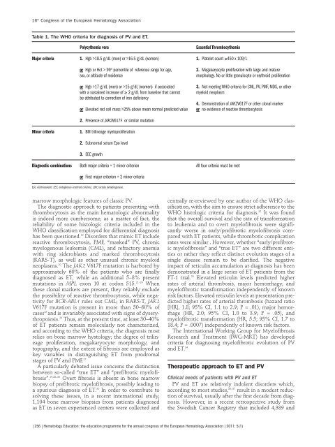
Table 1. The WHO criteria for diagnosis of PV and ET.<br />
Polycythemia vera Essential Thrombocythemia<br />
Major criteria 1. Hgb >18.5 g/dL (men) or >16.5 g/dL (women) 1. Platelet count ≥450 x 109/L<br />
or Hgb or Hct > 99 th percentile of reference range for age, 2. Megakaryocyte proliferat<strong>io</strong>n with large and mature<br />
sex, or altitude of residence morpho<strong>lo</strong>gy. No or little granu<strong>lo</strong>cyte or erythroid proliferat<strong>io</strong>n<br />
or Hgb >17 g/dL (men) or >15 g/dL (women) if associated 3. Not meeting WHO criteria for CML, PV, PMF, MDS, or other<br />
with a sustained increase of ≥ 2 g/dL from baseline that cannot mye<strong>lo</strong>id neoplasm<br />
be attributed to correct<strong>io</strong>n of iron deficiency<br />
4. Demonstrat<strong>io</strong>n of JAK2V617F or other c<strong>lo</strong>nal marker<br />
or Elevated red cell mass >25% above mean normal predicted value or no evidence of reactive thrombocytosis<br />
2. Presence of JAK2V617F or similar mutat<strong>io</strong>n<br />
Minor criteria 1. BM trilineage mye<strong>lo</strong>proliferat<strong>io</strong>n<br />
2. Subnormal serum Epo level<br />
3. EEC growth<br />
Diagnostic combinat<strong>io</strong>ns Both major criteria + 1 minor criter<strong>io</strong>n All four criteria must be met<br />
or First major criter<strong>io</strong>n + 2 minor criteria<br />
Epo, erythropoietin; EEC, endogenous erythroid co<strong>lo</strong>nies; LDH, lactate dehydrogenase.<br />
marrow morpho<strong>lo</strong>gic features of classic PV.<br />
The diagnostic approach to patients presenting with<br />
thrombocytosis as the main hemato<strong>lo</strong>gic abnormality<br />
is indeed more cumbersome; as a matter of fact, the<br />
reliability of some histo<strong>lo</strong>gic criteria included in the<br />
WHO classificat<strong>io</strong>n emp<strong>lo</strong>yed for differential diagnosis<br />
has been quest<strong>io</strong>ned. 19 Disorders that mimic ET include<br />
reactive thrombocytosis, PMF, “masked” PV, chronic<br />
mye<strong>lo</strong>genous leukemia (CML), and refractory anemia<br />
with ring sideroblasts and marked thrombocytosis<br />
(RARS-T), as well as other unusual chronic mye<strong>lo</strong>id<br />
neoplasms. 20 The JAK2 V617F mutat<strong>io</strong>n is harbored by<br />
approximately 60% of the patients who are finally<br />
diagnosed as ET, while an addit<strong>io</strong>nal 5–8% present<br />
mutat<strong>io</strong>ns in MPL exon 10 at codon 515. 21–24 When<br />
these c<strong>lo</strong>nal markers are present, they reliably exclude<br />
the possibility of reactive thrombocytosis, while negativity<br />
for BCR-ABL1 rules out CML; in RARS-T, JAK2<br />
V617F mutat<strong>io</strong>n is present in more than 50–60% of<br />
cases 25 and is invariably associated with signs of dyserythropoiesis.<br />
26 Thus, at the present time, at least 30–40%<br />
of ET patients remain molecularly not characterized,<br />
and according to the WHO criteria, the diagnosis most<br />
relies on bone marrow hysto<strong>lo</strong>gy; the degree of trilineage<br />
proliferat<strong>io</strong>n, megakaryocyte morpho<strong>lo</strong>gy, and<br />
topography, and the extent of fibrosis are emp<strong>lo</strong>yed as<br />
key variables in distinguishing ET from prodromal<br />
stages of PV and PMF. 27<br />
A particularly debated issue concerns the distinct<strong>io</strong>n<br />
between so-called “true ET” and “prefibrotic mye<strong>lo</strong>fibrosis”.<br />
19,28–30 Overt fibrosis is absent in bone marrow<br />
b<strong>io</strong>psy of prefibrotic mye<strong>lo</strong>fibrosis, possibly leading to<br />
a spur<strong>io</strong>us diagnosis of ET. 31 In order to contribute to<br />
solving these issues, in a recent internat<strong>io</strong>nal study,<br />
1,104 bone marrow b<strong>io</strong>psies from patients diagnosed<br />
as ET in seven experienced centers were collected and<br />
centrally re-reviewed by one author of the WHO classificat<strong>io</strong>n,<br />
with the aim to ensure strict adherence to the<br />
WHO histo<strong>lo</strong>gic criteria for diagnosis. 32 It was found<br />
that the overall survival and the rate of transformat<strong>io</strong>n<br />
to leukemia and to overt mye<strong>lo</strong>fibrosis were significantly<br />
worse in early/prefibrotic mye<strong>lo</strong>fibrosis compared<br />
with ET patients, while thrombotic complicat<strong>io</strong>n<br />
rates were similar . However, whether “early/prefibrotic<br />
mye<strong>lo</strong>fibrosis” and “true ET” are two different entities<br />
or rather they reflect distinct evolut<strong>io</strong>n stages of a<br />
single disease remain to be clarified. The negative<br />
impact of reticulin accumulat<strong>io</strong>n at diagnosis has been<br />
demonstrated in a large series of ET patients from the<br />
PT-1 trial. 33 Elevated reticulin levels predicted higher<br />
rates of arterial thrombosis, major hemorrhagy, and<br />
mye<strong>lo</strong>fibrotic transformat<strong>io</strong>n independently of known<br />
risk factors. Elevated reticulin levels at presentat<strong>io</strong>n predicted<br />
higher rates of arterial thrombosis (hazard rat<strong>io</strong><br />
[HR], 1.8; 95% CI, 1.1 to 2.9; P = .01), major hemorrhage<br />
(HR, 2.0; 95% CI, 1.0 to 3.9; P = .05), and<br />
mye<strong>lo</strong>fibrotic transformat<strong>io</strong>n (HR, 5.5; 95% CI, 1.7 to<br />
18.4; P = .0007) independently of known risk factors.<br />
The Internat<strong>io</strong>nal Working Group for Mye<strong>lo</strong>fibrosis<br />
Research and Treatment (IWG-MRT) has deve<strong>lo</strong>ped<br />
criteria for diagnosing mye<strong>lo</strong>fibrotic evolut<strong>io</strong>n of PV<br />
and ET. 34<br />
Therapeutic approach to ET and PV<br />
Clinical needs of patients with PV and ET<br />
PV and ET are relatively indolent disorders which,<br />
according to most studies, 35–37 result in a modest reduct<strong>io</strong>n<br />
of survival, usually after the first decade from diagnosis.<br />
However, in a recent retrospective study from<br />
the Swedish Cancer Registry that included 4,389 and<br />
| 256 | Hemato<strong>lo</strong>gy Educat<strong>io</strong>n: the educat<strong>io</strong>n programme for the annual congress of the <strong>European</strong> Hemato<strong>lo</strong>gy Associat<strong>io</strong>n | 2011; 5(1)