H e m a t o lo g y E d u c a t io n - European Hematology Association
H e m a t o lo g y E d u c a t io n - European Hematology Association
H e m a t o lo g y E d u c a t io n - European Hematology Association

Create successful ePaper yourself
Turn your PDF publications into a flip-book with our unique Google optimized e-Paper software.
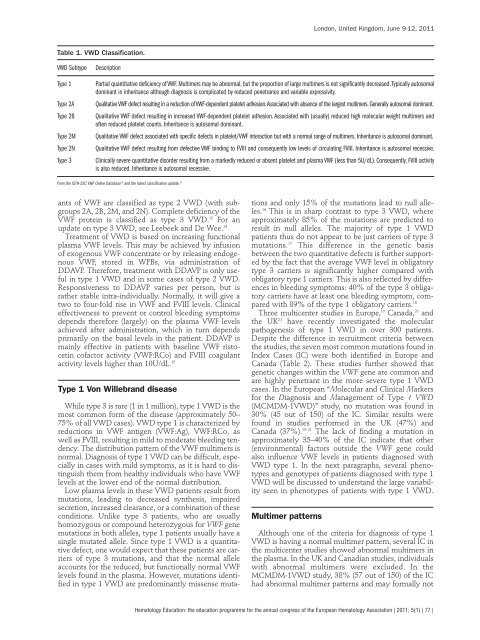
Table 1. VWD Classificat<strong>io</strong>n.<br />
VWD Subtype Descript<strong>io</strong>n<br />
ants of VWF are classified as type 2 VWD (with subgroups<br />
2A, 2B, 2M, and 2N). Complete deficiency of the<br />
VWF protein is classified as type 3 VWD. 13 For an<br />
update on type 3 VWD, see Leebeek and De Wee. 14<br />
Treatment of VWD is based on increasing funct<strong>io</strong>nal<br />
plasma VWF levels. This may be achieved by infus<strong>io</strong>n<br />
of exogenous VWF concentrate or by releasing endogenous<br />
VWF, stored in WPBs, via administrat<strong>io</strong>n of<br />
DDAVP. Therefore, treatment with DDAVP is only useful<br />
in type 1 VWD and in some cases of type 2 VWD.<br />
Responsiveness to DDAVP varies per person, but is<br />
rather stable intra-individually. Normally, it will give a<br />
two to four-fold rise in VWF and FVIII levels. Clinical<br />
effectiveness to prevent or control bleeding symptoms<br />
depends therefore (largely) on the plasma VWF levels<br />
achieved after administrat<strong>io</strong>n, which in turn depends<br />
primarily on the basal levels in the patient. DDAVP is<br />
mainly effective in patients with baseline VWF ristocetin<br />
cofactor activity (VWF:RCo) and FVIII coagulant<br />
activity levels higher than 10U/dL. 15<br />
Type 1 Von Willebrand disease<br />
While type 3 is rare (1 in 1 mill<strong>io</strong>n), type 1 VWD is the<br />
most common form of the disease (approximately 50–<br />
75% of all VWD cases). VWD type 1 is characterized by<br />
reduct<strong>io</strong>ns in VWF antigen (VWF:Ag), VWF:RCo, as<br />
well as FVIII, resulting in mild to moderate bleeding tendency.<br />
The distribut<strong>io</strong>n pattern of the VWF multimers is<br />
normal. Diagnosis of type 1 VWD can be difficult, especially<br />
in cases with mild symptoms, as it is hard to distinguish<br />
them from healthy individuals who have VWF<br />
levels at the <strong>lo</strong>wer end of the normal distribut<strong>io</strong>n.<br />
Low plasma levels in these VWD patients result from<br />
mutat<strong>io</strong>ns, leading to decreased synthesis, impaired<br />
secret<strong>io</strong>n, increased clearance, or a combinat<strong>io</strong>n of these<br />
condit<strong>io</strong>ns. Unlike type 3 patients, who are usually<br />
homozygous or compound heterozygous for VWF gene<br />
mutat<strong>io</strong>ns in both alleles, type 1 patients usually have a<br />
single mutated allele. Since type 1 VWD is a quantitative<br />
defect, one would expect that these patients are carriers<br />
of type 3 mutat<strong>io</strong>ns, and that the normal allele<br />
accounts for the reduced, but funct<strong>io</strong>nally normal VWF<br />
levels found in the plasma. However, mutat<strong>io</strong>ns identified<br />
in type 1 VWD are predominantly missense muta-<br />
t<strong>io</strong>ns and only 15% of the mutat<strong>io</strong>ns lead to null alleles.<br />
16 This is in sharp contrast to type 3 VWD, where<br />
approximately 85% of the mutat<strong>io</strong>ns are predicted to<br />
result in null alleles. The majority of type 1 VWD<br />
patients thus do not appear to be just carriers of type 3<br />
mutat<strong>io</strong>ns. 17 This difference in the genetic basis<br />
between the two quantitative defects is further supported<br />
by the fact that the average VWF level in obligatory<br />
type 3 carriers is significantly higher compared with<br />
obligatory type 1 carriers. This is also reflected by differences<br />
in bleeding symptoms: 40% of the type 3 obligatory<br />
carriers have at least one bleeding symptom, compared<br />
with 89% of the type 1 obligatory carriers. 18<br />
Three multicenter studies in Europe, 19 Canada, 20 and<br />
the UK 21 have recently investigated the molecular<br />
pathogenesis of type 1 VWD in over 300 patients.<br />
Despite the difference in recruitment criteria between<br />
the studies, the seven most common mutat<strong>io</strong>ns found in<br />
Index Cases (IC) were both identified in Europe and<br />
Canada (Table 2). These studies further showed that<br />
genetic changes within the VWF gene are common and<br />
are highly penetrant in the more severe type 1 VWD<br />
cases. In the <strong>European</strong> “Molecular and Clinical Markers<br />
for the Diagnosis and Management of Type 1 VWD<br />
(MCMDM-1VWD)” study, no mutat<strong>io</strong>n was found in<br />
30% (45 out of 150) of the IC. Similar results were<br />
found in studies performed in the UK (47%) and<br />
Canada (37%). 19–21 The lack of finding a mutat<strong>io</strong>n in<br />
approximately 35–40% of the IC indicate that other<br />
(environmental) factors outside the VWF gene could<br />
also influence VWF levels in patients diagnosed with<br />
VWD type 1. In the next paragraphs, several phenotypes<br />
and genotypes of patients diagnosed with type 1<br />
VWD will be discussed to understand the large variability<br />
seen in phenotypes of patients with type 1 VWD.<br />
Multimer patterns<br />
London, United Kingdom, June 9-12, 2011<br />
Type 1 Partial quantitative deficiency of VWF. Multimers may be abnormal, but the proport<strong>io</strong>n of large multimers is not significantly decreased. Typically autosomal<br />
dominant in inheritance although diagnosis is complicated by reduced penetrance and variable expressivity.<br />
Type 2A Qualitative VWF defect resulting in a reduct<strong>io</strong>n of VWF-dependent platelet adhes<strong>io</strong>n. Associated with absence of the largest multimers. Generally autosomal dominant.<br />
Type 2B Qualitative VWF defect resulting in increased VWF-dependent platelet adhes<strong>io</strong>n. Associated with (usually) reduced high molecular weight multimers and<br />
often reduced platelet counts. Inheritance is autosomal dominant.<br />
Type 2M Qualitative VWF defect associated with specific defects in platelet/VWF interact<strong>io</strong>n but with a normal range of multimers. Inheritance is autosomal dominant.<br />
Type 2N Qualitative VWF defect resulting from defective VWF binding to FVIII and consequently <strong>lo</strong>w levels of circulating FVIII. Inheritance is autosomal recessive.<br />
Type 3 Clinically severe quantitative disorder resulting from a markedly reduced or absent platelet and plasma VWF (less than 5U/dL). Consequently, FVIII activity<br />
is also reduced. Inheritance is autosomal recessive.<br />
From the ISTH-SSC VWF Online Database 16 and the latest classificat<strong>io</strong>n update. 13<br />
Although one of the criteria for diagnosis of type 1<br />
VWD is having a normal multimer pattern, several IC in<br />
the multicenter studies showed abnormal multimers in<br />
the plasma. In the UK and Canadian studies, individuals<br />
with abnormal multimers were excluded. In the<br />
MCMDM-1VWD study, 38% (57 out of 150) of the IC<br />
had abnormal multimer patterns and may formally not<br />
Hemato<strong>lo</strong>gy Educat<strong>io</strong>n: the educat<strong>io</strong>n programme for the annual congress of the <strong>European</strong> Hemato<strong>lo</strong>gy Associat<strong>io</strong>n | 2011; 5(1) | 77 |