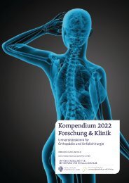
Kompendium 2020 Forschung & Klinik
Das Kompendium 2020 der Universitätsklinik für Orthopädie und Unfallchirurgie von MedUni Wien und AKH Wien (o. Univ.-Prof. R. Windhager) stellt einen umfassenden Überblick über die medizinsichen Leistungen und auch die umfangreichen Forschungsfelder dar. Die Veröffentlichungen zeigen die klinische Relevanz und innovative Ansätze der einzelnen Forschungsrichtungen. Herausgeber: Universitätsklinik für Orthopädie und Unfallchirurgie MedUni Wien und AKH Wien Prof. Dr. R. Windhager ISBN 978-3-200-07715-7
Das Kompendium 2020 der Universitätsklinik für Orthopädie und Unfallchirurgie von MedUni Wien und AKH Wien (o. Univ.-Prof. R. Windhager) stellt einen umfassenden Überblick über die medizinsichen Leistungen und auch die umfangreichen Forschungsfelder dar. Die Veröffentlichungen zeigen die klinische Relevanz und innovative Ansätze der einzelnen Forschungsrichtungen.
Herausgeber: Universitätsklinik für Orthopädie und Unfallchirurgie
MedUni Wien und AKH Wien
Prof. Dr. R. Windhager
ISBN 978-3-200-07715-7

Create successful ePaper yourself
Turn your PDF publications into a flip-book with our unique Google optimized e-Paper software.
TOP-Studien<br />
23<br />
defects (≤ International cartilage repair Society (ICRS) grade 2) at baseline<br />
and no surgical or invasive treatment during the follow up (4.0 ± 1.6 years).<br />
To ensure the technical consistency especially of the T2 mapping, hardware,<br />
including the MR scanner and the knee coil, as well as the MR sequence<br />
protocol, was identical for all subjects at both time points.<br />
Following the ICRS grading system, morphological cartilage changes over<br />
time were subdivided into a progression group, a non-progression group and<br />
regression group. Quantitative analysis of cartilage defects was performed<br />
by means of global and zonal T2 mapping (deep and superficial cartilage T2<br />
values) at both time points.<br />
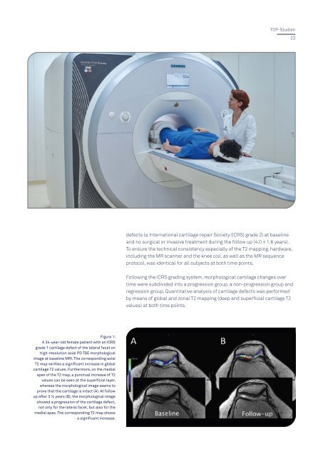
Figure 1:<br />
A 34-year-old female patient with an ICRS<br />
grade 1 cartilage defect of the lateral facet on<br />
high-resolution axial PD TSE morphological<br />
image at baseline MRI. The corresponding axial<br />
T2 map verifies a significant increase in global<br />
cartilage T2 values. Furthermore, on the medial<br />
apex of the T2 map, a punctual increase of T2<br />
values can be seen at the superficial layer,<br />
whereas the morphological image seems to<br />
prove that the cartilage is intact (A). At follow<br />
up after 3 ½ years (B), the morphological image<br />
showed a progression of the cartilage defect,<br />
not only for the lateral facet, but also for the<br />
medial apex. The corresponding T2 map shows<br />
a significant increase.