Kompendium 2020 Forschung & Klinik
Das Kompendium 2020 der Universitätsklinik für Orthopädie und Unfallchirurgie von MedUni Wien und AKH Wien (o. Univ.-Prof. R. Windhager) stellt einen umfassenden Überblick über die medizinsichen Leistungen und auch die umfangreichen Forschungsfelder dar. Die Veröffentlichungen zeigen die klinische Relevanz und innovative Ansätze der einzelnen Forschungsrichtungen. Herausgeber: Universitätsklinik für Orthopädie und Unfallchirurgie MedUni Wien und AKH Wien Prof. Dr. R. Windhager ISBN 978-3-200-07715-7
Das Kompendium 2020 der Universitätsklinik für Orthopädie und Unfallchirurgie von MedUni Wien und AKH Wien (o. Univ.-Prof. R. Windhager) stellt einen umfassenden Überblick über die medizinsichen Leistungen und auch die umfangreichen Forschungsfelder dar. Die Veröffentlichungen zeigen die klinische Relevanz und innovative Ansätze der einzelnen Forschungsrichtungen.
Herausgeber: Universitätsklinik für Orthopädie und Unfallchirurgie
MedUni Wien und AKH Wien
Prof. Dr. R. Windhager
ISBN 978-3-200-07715-7

Create successful ePaper yourself
Turn your PDF publications into a flip-book with our unique Google optimized e-Paper software.
TOP-Studien<br />
66<br />
Figure 1: Illustration of the position of the<br />
two 3D spatial position sensors: spatial<br />
position sensor epicutaneously centrally<br />
dorsally over the os sacrum for recording<br />
the pelvic position (white arrow (A and B)),<br />
transducer with 3D-printed adapter and<br />
spatial position sensor (white star (A)).<br />
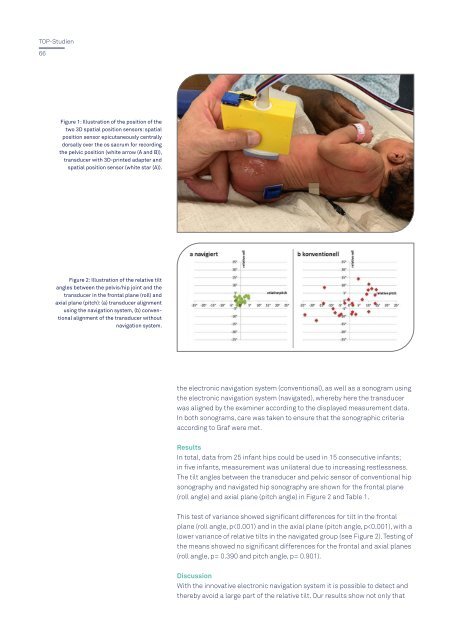
Figure 2: Illustration of the relative tilt<br />
angles between the pelvis/hip joint and the<br />
transducer in the frontal plane (roll) and<br />
axial plane (pitch): (a) transducer alignment<br />
using the navigation system, (b) conventional<br />
alignment of the transducer without<br />
navigation system.<br />
the electronic navigation system (conventional), as well as a sonogram using<br />
the electronic navigation system (navigated), whereby here the transducer<br />
was aligned by the examiner according to the displayed measurement data.<br />
In both sonograms, care was taken to ensure that the sonographic criteria<br />
according to Graf were met.<br />
Results<br />
In total, data from 25 infant hips could be used in 15 consecutive infants;<br />
in five infants, measurement was unilateral due to increasing restlessness.<br />
The tilt angles between the transducer and pelvic sensor of conventional hip<br />
sonography and navigated hip sonography are shown for the frontal plane<br />
(roll angle) and axial plane (pitch angle) in Figure 2 and Table 1.<br />
This test of variance showed significant differences for tilt in the frontal<br />
plane (roll angle, p