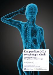
Kompendium 2020 Forschung & Klinik
Das Kompendium 2020 der Universitätsklinik für Orthopädie und Unfallchirurgie von MedUni Wien und AKH Wien (o. Univ.-Prof. R. Windhager) stellt einen umfassenden Überblick über die medizinsichen Leistungen und auch die umfangreichen Forschungsfelder dar. Die Veröffentlichungen zeigen die klinische Relevanz und innovative Ansätze der einzelnen Forschungsrichtungen. Herausgeber: Universitätsklinik für Orthopädie und Unfallchirurgie MedUni Wien und AKH Wien Prof. Dr. R. Windhager ISBN 978-3-200-07715-7
Das Kompendium 2020 der Universitätsklinik für Orthopädie und Unfallchirurgie von MedUni Wien und AKH Wien (o. Univ.-Prof. R. Windhager) stellt einen umfassenden Überblick über die medizinsichen Leistungen und auch die umfangreichen Forschungsfelder dar. Die Veröffentlichungen zeigen die klinische Relevanz und innovative Ansätze der einzelnen Forschungsrichtungen.
Herausgeber: Universitätsklinik für Orthopädie und Unfallchirurgie
MedUni Wien und AKH Wien
Prof. Dr. R. Windhager
ISBN 978-3-200-07715-7

You also want an ePaper? Increase the reach of your titles
YUMPU automatically turns print PDFs into web optimized ePapers that Google loves.
TOP-Studien<br />
47<br />
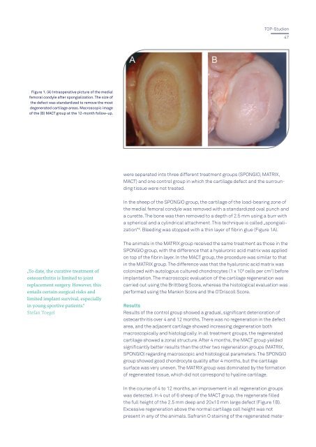
Figure 1. (A) Intraoperative picture of the medial<br />
femoral condyle after spongialization. The size of<br />
the defect was standardized to remove the most<br />
degenerated cartilage areas. Macroscopic image<br />
of the (B) MACT group at the 12-month follow-up.<br />
were separated into three different treatment groups (SPONGIO, MATRIX,<br />
MACT) and one control group in which the cartilage defect and the surrounding<br />
tissue were not treated.<br />
In the sheep of the SPONGIO group, the cartilage of the load-bearing zone of<br />
the medial femoral condyle was removed with a standardized oval punch and<br />
a curette. The bone was then removed to a depth of 2.5 mm using a burr with<br />
a spherical and a cylindrical attachment. This technique is called „spongialization“<br />
6 . Bleeding was stopped with a thin layer of fibrin glue (Figure 1A).<br />
„To date, the curative treatment of<br />
osteoarthritis is limited to joint<br />
replacement surgery. However, this<br />
entails certain surgical risks and<br />
limited implant survival, especially<br />
in young sportive patients.“<br />
Stefan Toegel<br />
The animals in the MATRIX group received the same treatment as those in the<br />
SPONGIO group, with the difference that a hyaluronic acid matrix was applied<br />
on top of the fibrin layer. In the MACT group, the procedure was similar to that<br />
in the MATRIX group. The difference was that the hyaluronic acid matrix was<br />
colonized with autologous cultured chondrocytes (1 x 10 6 cells per cm 2 ) before<br />
implantation. The macroscopic evaluation of the cartilage regeneration was<br />
carried out using the Brittberg Score, whereas the histological evaluation was<br />
performed using the Mankin Score and the O’Driscoll Score.<br />
Results<br />
Results of the control group showed a gradual, significant deterioration of<br />
osteoarthritis over 4 and 12 months. There was no regeneration in the defect<br />
area, and the adjacent cartilage showed increasing degeneration both<br />
macroscopically and histologically. In all treatment groups, the regenerated<br />
cartilage showed a zonal structure. After 4 months, the MACT group yielded<br />
significantly better results than the other two regeneration groups (MATRIX,<br />
SPONGIO) regarding macroscopic and histological parameters. The SPONGIO<br />
group showed good chondrocyte quality after 4 months, but the cartilage<br />
surface was very uneven. The MATRIX group was dominated by the formation<br />
of regenerated tissue, which did not correspond to hyaline cartilage.<br />
In the course of 4 to 12 months, an improvement in all regeneration groups<br />
was detected. In 4 out of 6 sheep of the MACT group, the regenerate filled<br />
the full height of the 2.5 mm deep and 20x10 mm large defect (Figure 1B).<br />
Excessive regeneration above the normal cartilage cell height was not<br />
present in any of the animals. Safranin O staining of the regenerated mate-