Kompendium 2020 Forschung & Klinik
Das Kompendium 2020 der Universitätsklinik für Orthopädie und Unfallchirurgie von MedUni Wien und AKH Wien (o. Univ.-Prof. R. Windhager) stellt einen umfassenden Überblick über die medizinsichen Leistungen und auch die umfangreichen Forschungsfelder dar. Die Veröffentlichungen zeigen die klinische Relevanz und innovative Ansätze der einzelnen Forschungsrichtungen. Herausgeber: Universitätsklinik für Orthopädie und Unfallchirurgie MedUni Wien und AKH Wien Prof. Dr. R. Windhager ISBN 978-3-200-07715-7
Das Kompendium 2020 der Universitätsklinik für Orthopädie und Unfallchirurgie von MedUni Wien und AKH Wien (o. Univ.-Prof. R. Windhager) stellt einen umfassenden Überblick über die medizinsichen Leistungen und auch die umfangreichen Forschungsfelder dar. Die Veröffentlichungen zeigen die klinische Relevanz und innovative Ansätze der einzelnen Forschungsrichtungen.
Herausgeber: Universitätsklinik für Orthopädie und Unfallchirurgie
MedUni Wien und AKH Wien
Prof. Dr. R. Windhager
ISBN 978-3-200-07715-7

You also want an ePaper? Increase the reach of your titles
YUMPU automatically turns print PDFs into web optimized ePapers that Google loves.
TOP-Studien<br />
24<br />
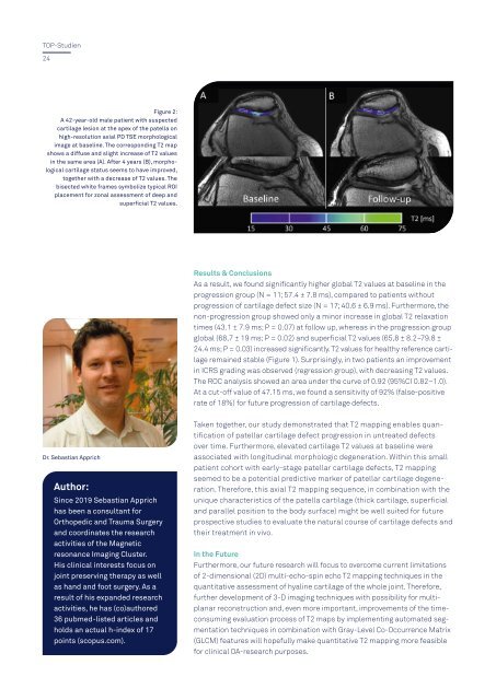
Figure 2:<br />
A 42-year-old male patient with suspected<br />
cartilage lesion at the apex of the patella on<br />
high-resolution axial PD TSE morphological<br />
image at baseline. The corresponding T2 map<br />
shows a diffuse and slight increase of T2 values<br />
in the same area (A). After 4 years (B), morphological<br />
cartilage status seems to have improved,<br />
together with a decrease of T2 values. The<br />
bisected white frames symbolize typical ROI<br />
placement for zonal assessment of deep and<br />
superficial T2 values.<br />
Results & Conclusions<br />
As a result, we found significantly higher global T2 values at baseline in the<br />
progression group (N = 11; 57.4 ± 7.8 ms), compared to patients without<br />
progression of cartilage defect size (N = 17; 40.6 ± 6.9 ms). Furthermore, the<br />
non-progression group showed only a minor increase in global T2 relaxation<br />
times (43.1 ± 7.9 ms; P = 0.07) at follow up, whereas in the progression group<br />
global (68,7 ± 19 ms; P = 0.02) and superficial T2 values (65,8 ± 8.2–79.8 ±<br />
24.4 ms; P = 0.03) increased significantly. T2 values for healthy reference cartilage<br />
remained stable (Figure 1). Surprisingly, in two patients an improvement<br />
in ICRS grading was observed (regression group), with decreasing T2 values.<br />
The ROC analysis showed an area under the curve of 0.92 (95%CI 0.82–1.0).<br />
At a cut-off value of 47.15 ms, we found a sensitivity of 92% (false-positive<br />
rate of 18%) for future progression of cartilage defects.<br />
Dr. Sebastian Apprich<br />
Author:<br />
Since 2019 Sebastian Apprich<br />
has been a consultant for<br />
Orthopedic and Trauma Surgery<br />
and coordinates the research<br />
activities of the Magnetic<br />
resonance Imaging Cluster.<br />
His clinical interests focus on<br />
joint preserving therapy as well<br />
as hand and foot surgery. As a<br />
result of his expanded research<br />
activities, he has (co)authored<br />
36 pubmed-listed articles and<br />
holds an actual h-index of 17<br />
points (scopus.com).<br />
Taken together, our study demonstrated that T2 mapping enables quantification<br />
of patellar cartilage defect progression in untreated defects<br />
over time. Furthermore, elevated cartilage T2 values at baseline were<br />
associated with longitudinal morphologic degeneration. Within this small<br />
patient cohort with early-stage patellar cartilage defects, T2 mapping<br />
seemed to be a potential predictive marker of patellar cartilage degeneration.<br />
Therefore, this axial T2 mapping sequence, in combination with the<br />
unique characteristics of the patella cartilage (thick cartilage, superficial<br />
and parallel position to the body surface) might be well suited for future<br />
prospective studies to evaluate the natural course of cartilage defects and<br />
their treatment in vivo.<br />
In the Future<br />
Furthermore, our future research will focus to overcome current limitations<br />
of 2-dimensional (2D) multi-echo-spin echo T2 mapping techniques in the<br />
quantitative assessment of hyaline cartilage of the whole joint. Therefore,<br />
further development of 3-D imaging techniques with possibility for multiplanar<br />
reconstruction and, even more important, improvements of the timeconsuming<br />
evaluation process of T2 maps by implementing automated segmentation<br />
techniques in combination with Gray-Level Co-Occurrence Matrix<br />
(GLCM) features will hopefully make quantitative T2 mapping more feasible<br />
for clinical OA-research purposes.