Kompendium 2020 Forschung & Klinik
Das Kompendium 2020 der Universitätsklinik für Orthopädie und Unfallchirurgie von MedUni Wien und AKH Wien (o. Univ.-Prof. R. Windhager) stellt einen umfassenden Überblick über die medizinsichen Leistungen und auch die umfangreichen Forschungsfelder dar. Die Veröffentlichungen zeigen die klinische Relevanz und innovative Ansätze der einzelnen Forschungsrichtungen. Herausgeber: Universitätsklinik für Orthopädie und Unfallchirurgie MedUni Wien und AKH Wien Prof. Dr. R. Windhager ISBN 978-3-200-07715-7
Das Kompendium 2020 der Universitätsklinik für Orthopädie und Unfallchirurgie von MedUni Wien und AKH Wien (o. Univ.-Prof. R. Windhager) stellt einen umfassenden Überblick über die medizinsichen Leistungen und auch die umfangreichen Forschungsfelder dar. Die Veröffentlichungen zeigen die klinische Relevanz und innovative Ansätze der einzelnen Forschungsrichtungen.
Herausgeber: Universitätsklinik für Orthopädie und Unfallchirurgie
MedUni Wien und AKH Wien
Prof. Dr. R. Windhager
ISBN 978-3-200-07715-7

You also want an ePaper? Increase the reach of your titles
YUMPU automatically turns print PDFs into web optimized ePapers that Google loves.
TOP-Studien<br />
67<br />
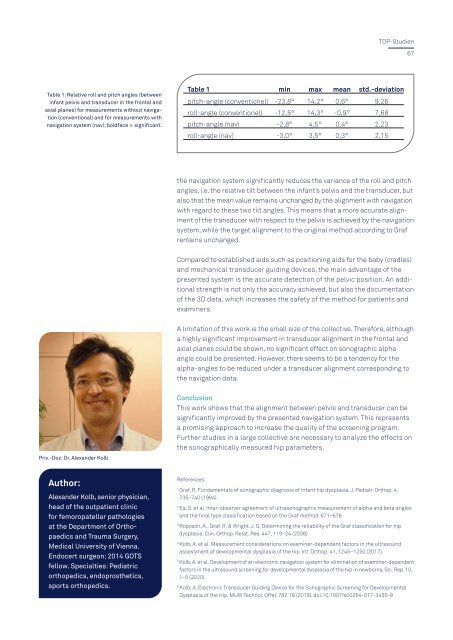
Table 1: Relative roll and pitch angles (between<br />
infant pelvis and transducer in the frontal and<br />
axial planes) for measurements without navigation<br />
(conventional) and for measurements with<br />
navigation system (nav); boldface = significant.<br />
Table 1 min max mean std.-deviation<br />
pitch-angle (conventionel) -23,8° 14,2° 0,6° 9,26<br />
roll-angle (conventionel) -12,5° 14,3° -0,9° 7,68<br />
pitch-angle (nav) -2,8° 4,5° 0,4° 2,23<br />
roll-angle (nav) -3,0° 3,5° 0,3° 2,15<br />
the navigation system significantly reduces the variance of the roll and pitch<br />
angles, i.e. the relative tilt between the infant’s pelvis and the transducer, but<br />
also that the mean value remains unchanged by the alignment with navigation<br />
with regard to these two tilt angles. This means that a more accurate alignment<br />
of the transducer with respect to the pelvis is achieved by the navigation<br />
system, while the target alignment to the original method according to Graf<br />
remains unchanged.<br />
Compared to established aids such as positioning aids for the baby (cradles)<br />
and mechanical transducer guiding devices, the main advantage of the<br />
presented system is the accurate detection of the pelvic position. An additional<br />
strength is not only the accuracy achieved, but also the documentation<br />
of the 3D data, which increases the safety of the method for patients and<br />
examiners.<br />
A limitation of this work is the small size of the collective. Therefore, although<br />
a highly significant improvement in transducer alignment in the frontal and<br />
axial planes could be shown, no significant effect on sonographic alpha<br />
angle could be presented. However, there seems to be a tendency for the<br />
alpha-angles to be reduced under a transducer alignment corresponding to<br />
the navigation data.<br />
Conclusion<br />
This work shows that the alignment between pelvis and transducer can be<br />
significantly improved by the presented navigation system. This represents<br />
a promising approach to increase the quality of the screening program.<br />
Further studies in a large collective are necessary to analyze the effects on<br />
the sonographically measured hip parameters.<br />
Priv.-Doz. Dr. Alexander Kolb<br />
Author:<br />
Alexander Kolb, senior physician,<br />
head of the outpatient clinic<br />
for femoropatellar pathologies<br />
at the Department of Orthopaedics<br />
and Trauma Surgery,<br />
Medical University of Vienna.<br />
Endocert surgeon; 2014 GOTS<br />
fellow. Specialties: Pediatric<br />
orthopedics, endoprosthetics,<br />
sports orthopedics.<br />
References:<br />
1 <br />
Graf, R. Fundamentals of sonographic diagnosis of infant hip dysplasia. J. Pediatr. Orthop. 4,<br />
735–740 (1984).<br />
2 <br />
Ea, S. et al. Inter-observer agreement of ultrasonographic measurement of alpha and beta angles<br />
and the final type classification based on the Graf method. 671–678<br />
3 <br />
Roposch, A., Graf, R. & Wright, J. G. Determining the reliability of the Graf classification for hip<br />
dysplasia. Clin. Orthop. Relat. Res. 447, 119–24 (2006).<br />
4 <br />
Kolb, A. et al. Measurement considerations on examiner-dependent factors in the ultrasound<br />
assessment of developmental dysplasia of the hip. Int. Orthop. 41, 1245–1250 (2017).<br />
5 <br />
Kolb, A. et al. Development of an electronic navigation system for elimination of examiner-dependent<br />
factors in the ultrasound screening for developmental dysplasia of the hip in newborns. Sci. Rep. 10,<br />
1–5 (<strong>2020</strong>).<br />
6 <br />
Kolb, A. Electronic Transducer Guiding Device for the Sonographic Screening for Developmental<br />
Dysplasia of the Hip. MUW Technol. Offer. 782.18 (2019). doi:10.1007/s00264-017-3455-9