

Remerciements Remerciements
Remerciements Remerciements
Remerciements Remerciements

Create successful ePaper yourself
Turn your PDF publications into a flip-book with our unique Google optimized e-Paper software.
tel-00769943, version 1 - 4 Jan 2013<br />
UNIVERSITE PARIS XI<br />
FACULTE DE MEDECINE PARIS-SUD<br />
ECOLE-DOCTORALE ED419: Signalisations et Réseaux Intégratifs en Biologie<br />
BIO-SigNE<br />
Année: 2011-2012 N°attribué par la bibliothèque<br />
THESE<br />
Pour obtenir le grade de<br />
DOCTEUR DE L’UNIVERSITE PARIS XI<br />
Champs disciplinaire: Médecine<br />
Spécialité: Biochimie, Biologie Cellulaire et Moléculaire<br />
Présentée et soutenue publiquement par<br />
Maya DEBBABI<br />
Le 16 Décembre 2011<br />
MECANISMES DE REGULATION DE LA NADPH OXYDASE<br />
NOX1 : ROLE DE LA PHOSPHORYLATION DE NOXA1 (NOX<br />
ACTIVATOR 1) ET DE NOXO1 (NOX ORGANIZER 1)<br />
Directrice de thèse: Dr. Pham-My chan DANG<br />
JURY<br />
Pr. Oliver NÜSSE Président<br />
Pr. Françoise MOREL Rapporteur<br />
Dr. Xavier de DEKEN Rapporteur<br />
Pr. Hervé Kovacic Examinateur<br />
Dr. Pham My-Chan DANG Directrice de thèse<br />
Pr. Marie-Anne GOUGEROT-POCIDALO Co-directrice de thèse<br />
1
tel-00769943, version 1 - 4 Jan 2013<br />
<strong>Remerciements</strong><br />
<strong>Remerciements</strong><br />
<strong>Remerciements</strong><br />
En préambule à ce mémoire, je souhaite adresser mes remerciements les plus<br />
sincères aux personnes qui m'ont apporté leur aide et qui ont contribué à<br />
l'élaboration de ce mémoire.<br />
Je remercie les rapporteurs de cette thèse le Pr Françoise MOREL et le Dr Xavier<br />
de DEKEN pour l’intérêt qu’ils ont porté à mon travail. Merci également aux autres<br />
membres du jury qui ont accepté de juger ce travail : le Pr Hervé Kovacic ainsi que<br />
le Pr Oliver NÜSSE qui a bien voulu présider le jury de ma thèse et qui a, en<br />
quelque sorte, fait partie de mon parcours universitaire.<br />
Je tiens à remercier ma directrice de thèse, le Dr My-chan DANG alias Michmich qui<br />
a dirigé cette thèse dans la continuité de mon stage de master. Tout au long de ces<br />
années, elle a su orienter mes recherches aux bons moments, elle a toujours été<br />
disponible pour d’intenses et rationnelles discussions. Pour tout cela, sa confiance,<br />
son soutien et tout le reste, je la remercie vivement. Mes remerciements vont<br />
également à Yolande KROVIARSKI alias YO pour la gentillesse, la disponibilité, la<br />
patience qu'elle a manifestées à mon égard durant cette thèse mais aussi pour tous<br />
ses conseils scientifiques et de tous les jours. Je remercie aussi le Dr Jamel EL<br />
BENNA qui m’a accueillie dans son laboratoire suite à la gentille demande de Mr<br />
Abdelhamid SAHLI. Un autre merci pour le Pr Marie-Anne GOUGEROT-<br />
POCIDALO qui a toujours fait preuve d’une grande gentillesse, disponibilité et qui a<br />
porté un intérêt important à notre bien être au laboratoire, je la remercie aussi de<br />
m’avoir aidé pour le concours de thèse et d’avoir relu mon manuscrit en si peu de<br />
temps.<br />
Je remercie aussi les autres membres de l’équipe : mes collègues et amis Karama,<br />
Pia, Sahra, Mahmoud, Loic, Riad, Michèle, Grégory, Pascal, Sanae, Eric, Imène,<br />
Margarita, Tarek sans oublier le « staff permanent de l’équipe » : Odile BOURNIER<br />
pour les manips de microscopie confocale mais aussi sa serviabilité et sa pêche,<br />
Jean claude MARIE d’avoir toujours apporté une pointe d’humour à notre quotidien<br />
au laboratoire, Françoise BRAUT pour sa gentillesse, Axel PERIANIN pour son<br />
esprit « rebelle », Eric CHASTRE et Larissa KOTELEVETS pour les discussions<br />
fructueuses lors des séminaires internes.<br />
2
tel-00769943, version 1 - 4 Jan 2013<br />
Je n’oublie pas Nicaise COMIN, Marie-Hélène CANOGA, Julien PUTHOD,<br />
Mathieu, Pascale et Margot SAUVADET qui ont facilité les démarches<br />
administratives mais aussi pour leurs sourires.<br />
Enfin, j'adresse mes plus sincères remerciements à tous mes proches qui m'ont toujours<br />
soutenue et encouragée. Un MERCI particulier à mes parents Coucoulinou et Nonny, qui<br />
ont été présents pour moi depuis le jour de ma naissance, qui m’ont inculqué mes valeurs et<br />
mes principes et sans lesquels je ne serai pas là aujourd’hui. Merci à mon frère Farrouss et<br />
ma sœur Diddy qui ont contribué à ma « maturation ». Merci également à mon très cher mari<br />
Wesswess qui a toujours su me faire rire mais aussi m’épauler et me rassurer quand j’en avais<br />
besoin. Un autre merci à ma ‘tite nièce chérie Bambi qui me fait garder mon âme d’enfant<br />
plus longtemps et à sa maman Syrriinne qui s’occupe très bien de mon frère et ma nièce et<br />
pour tous nos après-midi jeux. Une pensée pour mes Dia, Papy, Azizi et Ya.<br />
Et le plus important je me remercie d’avoir persévéré et d’être allée aussi loin dans<br />
mes études, je rigooooooooole.<br />
3
tel-00769943, version 1 - 4 Jan 2013<br />
Sommaire<br />
Liste des Abréviations.............................................................................................................. 6<br />
INTRODUCTION GENERALE ............................................................................................ 7<br />
I. NOX2, LE PROTOTYPE DES CELLULES PHAGOCYTAIRES............................... 11<br />
I.1- Bref rappel sur les Polynucléaires Neutrophiles (PN)................................................... 11<br />
I. 2- Structure de NOX2 ....................................................................................................... 14<br />
I.2.1- Les composants membranaires............................................................................... 15<br />
I.2.1.a- La gp91 PHOX / NOX2........................................................................................ 16<br />
I.2.1.b-La p22 PHOX ........................................................................................................ 18<br />
I.2.1.c- Processus de transfert des électrons par le cytochrome b558.......................... 21<br />
I. 2.2- Les composants cytosoliques................................................................................. 21<br />
I.2.2.a-La p47 PHOX ........................................................................................................ 22<br />
I.2.2.b-La p67 PHOX ........................................................................................................ 24<br />
I.2.2.c- La p40 PHOX ....................................................................................................... 25<br />
I.2.2.d- Rac 1/2............................................................................................................. 26<br />
I.3- Mécanismes d’activation de NOX2............................................................................... 28<br />
I.3.1- Importance de la phosphorylation des sous-unités de NOX2 dans l’activation de<br />
NOX2 ............................................................................................................................... 29<br />
I.3.1.a-Phosphorylation de la p47 PHOX ......................................................................... 30<br />
I.3.1.b-Phosphorylation de la p67 PHOX ......................................................................... 32<br />
I.3.1.c-Phosphorylation de la p40 PHOX ......................................................................... 33<br />
I.3.1.d-Phosphorylation de la gp91 PHOX / NOX2 ......................................................... 34<br />
I.3.1.e-Phosphorylation de la p22 PHOX ......................................................................... 34<br />
I.3.2- Rac2, un élément central de l’activation de NOX2 ................................................ 35<br />
I.3.3-Assemblage de NOX2: Importance des interactions protéines-protéines et<br />
protéines-lipides ............................................................................................................... 36<br />
II- LES HOMOLOGUES DE NOX2.................................................................................... 40<br />
II.1- NOX1 ........................................................................................................................... 42<br />
II.2- NOX3 ........................................................................................................................... 42<br />
II.3- NOX4 ........................................................................................................................... 43<br />
II.4- NOX5 ........................................................................................................................... 45<br />
II.5- DUOX1/DUOX2.......................................................................................................... 46<br />
III- DESCRIPTION DETAILLEE DE NOX1 .................................................................... 49<br />
III.1- Profil d’expression de NOX1...................................................................................... 50<br />
III.2- Modulation de l’expression de NOX1 ........................................................................ 51<br />
III.3- Localisation subcellulaire de la sous-unité NOX1...................................................... 52<br />
III.4- Composition du complexe NOX1............................................................................... 52<br />
III.4.1- La sous-unité catalytique NOX1.......................................................................... 54<br />
III.4.2- La sous-unité organisatrice NOXO1.................................................................... 54<br />
III.4.3- La sous-unité activatrice NOXA1........................................................................ 58<br />
III.5 - Assemblage et activation de NOX1........................................................................... 60<br />
III.6- Rôles physiologiques et physiopathologiques de NOX1............................................ 62<br />
III.6 .1- NOX1 dans le système vasculaire....................................................................... 63<br />
III.6.2- NOX1 au niveau du colon.................................................................................... 64<br />
IV. OBJECTIFS DU TRAVAIL ........................................................................................... 68<br />
4
tel-00769943, version 1 - 4 Jan 2013<br />
TRAVAUX DE RECHERCHE............................................................................................. 69<br />
Article 1: NOXA1 est phosphorylée et sa phosphorylation prévient l’hyperactivation de<br />
la NADPH oxydase NOX1 ..................................................................................................... 70<br />
Résultats supplémentaires: Etude de la phosphorylation de NOXA1: l’activation de<br />
NOX1 par le fMLF est associée à la déphosphorylation de NOXA1................................. 88<br />
Article 2: NOXO1β est phosphorylée et sa phosphorylation potentialise l’activité de la<br />
NADPH oxydase NOX1 ......................................................................................................... 94<br />
DISCUSSION GENERALE................................................................................................ 123<br />
REFERENCES BIBLIOGRAPHIQUES........................................................................... 136<br />
5
tel-00769943, version 1 - 4 Jan 2013<br />
Liste des Abréviations<br />
Aa Acide aminé<br />
AD Activation domain<br />
pcDNA Plasmide contenant l’Acide Désoxyribo Nucléique complémentaire<br />
AIR Auto-Inhibitory Region<br />
ARNm ARN messager<br />
ATP Adénosine TriPhosphate<br />
CGD Chronic Granulomatous Disease<br />
DPI DiPhenileneIodonium<br />
DUOX Dual Oxidase<br />
ERK Extracellular signal Regulated Kinase<br />
FAD Flavine Adénine Dinucléotide<br />
fMLF N-Formyl_methionyl-leucyl-phénylalanine<br />
fPR Récepteur du fMLF<br />
FRO Formes Réactives de l’Oxygène<br />
Fsk Forskoline<br />
GSH Glutathion<br />
GST Glutathion-S-Transférase<br />
H2O2<br />
Peroxyde d’hydrogène (eau oxygénée)<br />
HEK293 Human Embryonic Kidney cells<br />
IFNγ Interferon gamma<br />
IL Interleukine<br />
Kb kilobase<br />
kDa kiloDalton<br />
KO Knock Out<br />
LPS LipoPoliSaccharides<br />
MAPK Mitogene-Activated Protein Kinase<br />
MICI Maladies Inflamamtoires Chroniques de l’Intestin<br />
NADPH Nicotinamide Adénosine Dinucléotide PHosphate<br />
NFκB Nuclear Factor-kappa B<br />
NOX NADPH Oxydase<br />
NOXA1 NADPH Activator 1<br />
NOXO1 NADPH Organizer 1<br />
O2<br />
Oxygène moléculaire<br />
O2 -. Anion superoxyde<br />
PB1 Phox and Bem 1 domain<br />
Phox Phagocyte oxidase<br />
pI Point Isoélectrique<br />
PKA Protéine Kinase A<br />
PKC Protéine Kinase C<br />
PMA Phorbol-12-Myristate13-Acétate<br />
PN Polynucléaire Neutrophile<br />
PRR Proline Rich Region<br />
PtdIns Phosphatidylinositols<br />
PX Phox homology domain<br />
Rac Ras-related C3 botulinum toxin substrate<br />
Rho-GDI Rho-GDP-Dissociation Inhibitor<br />
SAXS Small Angle X-ray Scattering<br />
SH3 Src Homology domain<br />
TNFα Tumor Necrosis Factor α<br />
TPR TetratricoPeptide Repeat motif<br />
WT Wild Type<br />
6
tel-00769943, version 1 - 4 Jan 2013<br />
INTRODUCTION GENERALE<br />
7
tel-00769943, version 1 - 4 Jan 2013<br />
Les formes réactives de l’oxygène (FRO), telles que l’anion superoxyde (O2 °- ), le<br />
radical hydroxyl (OH°) et le peroxyde d’hydrogène ( H2O2) sont de petites molécules à<br />
forte potentialité oxydante longtemps considérées comme les produits secondaires<br />
de différents systèmes enzymatiques comme le cytochrome P450, la chaine<br />
respiratoire mitochondriale, la lipoxygénase ou encore la xanthine oxydase.<br />
La découverte de la NADPH oxydase phagocytaire il y a une trentaine d’années, a<br />
permis d’identifier pour la première fois un système enzymatique dont la fonction est<br />
uniquement dédiée à la production des FRO. La NADPH oxydase des phagocytes<br />
(polynucléaires neutrophiles, macrophages, monocytes), joue un rôle clé dans la<br />
défense anti-infectieuse de l’organisme contre divers agents pathogènes (bactéries<br />
ou levures principalement) en générant de façon rapide et massive les FRO<br />
hautement toxiques. Ce phénomène, connu aussi sous le nom d’explosion oxydative,<br />
a été identifié pour la première fois en 1933, bien avant la découverte même de<br />
l’identité moléculaire de la NADPH oxydase effectuée en 1978 par l’équipe de Segal.<br />
Pendant longtemps, il a été considéré que cette enzyme était restreinte aux<br />
phagocytes. Cependant dès 1991, Meier et Jones observent dans les fibroblastes<br />
une activité NADPH oxydase faible mais sensible au diphénylène iodonium (DPI),<br />
inhibiteur sélectif des flavoprotéines déshydrogénases dont fait partie la NADPH<br />
oxydase phagocytaire, suggérant ainsi l’existence de NADPH oxydases non<br />
phagocytaires. Ce n’est qu’à partir de 1999 que plusieurs homologues de la NADPH<br />
oxydase phagocytaire ont été décrits dans de nombreuses cellules au sein de<br />
différents organes. NOX1 (NOX, pour NADPH Oxidase), fortement exprimée dans<br />
les cellules épithéliales du colon, est le premier homologue à avoir été identifié. Par<br />
la suite, plusieurs autres homologues ont été clonés dans divers tissus par<br />
différentes équipes. Il s’agit de NOX3, NOX4, NOX5, DUOX1 et DUOX2 (DUOX,<br />
pour Dual Oxidase). La NADPH oxydase phagocytaire a été renommée NOX2. Les<br />
NOXs sont impliquées dans diverses fonctions biologiques telles que la signalisation<br />
intracellulaire, l’immunité innée, la prolifération cellulaire, l’apoptose, la régulation de<br />
la pression artérielle, la synthèse d’hormones. Cependant, en raison de leur haute<br />
réactivité avec les molécules biologiques telles que les lipides, les protéines et les<br />
acides nucléiques, les FRO peuvent représenter une arme à double tranchant. Ainsi,<br />
lorsqu’elles sont produites de façon inappropriée ou excessive, les FRO peuvent<br />
induire un état de stress oxydant responsable de lésions tissulaires contribuant à la<br />
8
tel-00769943, version 1 - 4 Jan 2013<br />
physiopathologie de nombreuses maladies inflammatoires, telles que la polyarthrite<br />
rhumatoïde ou encore les maladies inflammatoires chroniques de l’intestin (MICI).<br />
La haute toxicité et réactivité des FRO, impliquent que les NOXs soient soumises à<br />
une régulation très fine. Tel est le cas de NOX2. Dans les phagocytes au repos, les<br />
différentes sous-unités qui forment NOX2 sont dispersées entre le cytosol (p40 PHOX ,<br />
p47 PHOX , p67 PHOX et la petite protéine G Rac 1/2) et la membrane plasmique<br />
(gp91 PHOX /NOX2 et p22 PHOX formant le cytochrome b558) empêchant ainsi toute<br />
activation de l’enzyme. Un des événements majeurs de l’activation de NOX2 est la<br />
phosphorylation de certains composants cytosoliques tels que la p47 PHOX ou la<br />
p67 PHOX , conduisant à leur translocation vers le cytochrome b558 membranaire pour<br />
permettre la formation d’une enzyme catalytiquement active à l’origine de la<br />
production de FRO.<br />
Parmi les homologues de NOX2, NOX1 est structurellement et fonctionnellement la<br />
plus proche de NOX2. Elle interagit avec la p22 PHOX et son activation requiert<br />
également des protéines cytosoliques: NOX organizer 1 (NOXO1) et NOX activator<br />
1 (NOXA1), qui sont les homologues respectifs de la p47 PHOX et de la p67 PHOX . La<br />
petite protéine G, Rac1, est aussi nécessaire pour le fonctionnement de NOX1.<br />
NOX1, NOXO1 et NOXA1 sont abondamment exprimées dans les cellules<br />
épithéliales du colon et pourraient être impliquées dans la défense immunitaire innée<br />
de la muqueuse intestinale dans les conditions physiologiques. Cependant les<br />
mécanismes qui régulent l’activation du complexe NOX1 demeurent obscurs et la<br />
plupart du temps ont été déduits de l’homologie ou de la différence structurelle qui<br />
existent entre NOX2 et NOX1.<br />
Dans ce contexte, mon projet de thèse a eu pour objectif de déterminer les<br />
mécanismes moléculaires fondamentaux qui régulent l’activation de NOX1. Je me<br />
suis en particulier intéressée aux processus de phosphorylation, dont l’importance a<br />
été bien établie pour NOX2. Qu’en est-il pour NOX1?<br />
1) Dans un premier temps, j’ai étudié la phosphorylation de NOXA1. J’ai ainsi<br />
identifié les acides aminés phosphorylés sur NOXA1, déterminé les kinases<br />
impliquées et analysé les conséquences de ces phosphorylations sur<br />
l’assemblage et l’activation de NOX1.<br />
9
tel-00769943, version 1 - 4 Jan 2013<br />
2) Dans un deuxième temps, j’ai étudié la phosphorylation de NOXO1. Comme<br />
pour NOXA1, J’ai identifié les acides aminés phosphorylés sur NOXO1,<br />
déterminé les kinases impliquées et analysé les conséquences fonctionnelles<br />
de ces phosphorylations.<br />
Mes résultats montrent que:<br />
1) NOXA1, la sous-unité activatrice du complexe NOX1 est phosphorylée de<br />
façon constitutive dans les cellules au repos. Ces phosphorylations diminuent<br />
ses interactions avec la sous-unité catalytique NOX1 et la petite protéine G<br />
Rac1, ce qui a pour conséquence de prévenir l’hyperactivation de NOX1.<br />
2) NOXO1, la sous-unité organisatrice est phosphorylée par les PKC au cours de<br />
la stimulation des cellules par le Phorbol myristate acétate (PMA). Cette<br />
phosphorylation favorise les interactions de NOXO1 avec la sous-unité<br />
cytosolique NOXA1 et avec la sous-unité membranaire p22 PHOX , augmente sa<br />
localisation membranaire et entraîne une hyperactivation de NOX1.<br />
Dans ce manuscrit, je ferai d’abord un rappel des connaissances actuelles sur les<br />
différentes NADPH oxydases et leurs mécanismes de régulation, j’insisterai<br />
particulièrement sur NOX1. Je présenterai par la suite mes travaux de recherche et<br />
les résultats obtenus. Enfin, je terminerai par une discussion de mes résultats par<br />
rapport aux données de la littérature.<br />
10
tel-00769943, version 1 - 4 Jan 2013<br />
I. NOX2, LE PROTOTYPE DES CELLULES PHAGOCYTAIRES<br />
La NADPH oxydase NOX2 est le «patriarche» de la famille des NOXs et est un<br />
puissant système générateur de FRO dans les cellules phagocytaires à savoir les<br />
polynucléaires neutrophiles, les macrophages et les monocytes. Bien qu’il soit connu<br />
depuis 1933 que l’incubation des neutrophiles avec les bactéries entraîne une<br />
consommation accrue d’oxygène [Baldridge 1933] insensible au cyanide [Sbarra<br />
1959] et donc distincte de la respiration mitochondriale, ce n’est qu’en 1978 que<br />
l’enzyme impliquée dans ce processus, à savoir NOX2, a été découverte par l’équipe<br />
de Segal [Segal 1978].<br />
La production massive des FRO par NOX2 joue un rôle essentiel dans la<br />
bactéricidie. Ceci est bien illustré par la granulomatose septique chronique (CGD),<br />
une maladie génétique caractérisée par la déficience d’un des composants de<br />
NOX2, qui se traduit par l’absence de production de FRO et par l’incapacité à<br />
éliminer l’agent pathogène, ce qui aboutit à des infections bactériennes ou fongiques<br />
graves et répétées, dès le plus jeune âge [Heyworth 2003]. Bien que les monocytes,<br />
les macrophages et les lymphocytes B [Morel 1993] expriment également NOX2, les<br />
polynucléaires neutrophiles (PN) restent le système où l’enzyme est la mieux<br />
étudiée. Par ailleurs, la plupart de ce qui est connu de la structure et de la régulation<br />
des autres NOXs découlent directement de notre compréhension de NOX2. Après un<br />
bref rappel sur les PN, je détaillerai donc la structure et la régulation de NOX2, en<br />
développant tout particulièrement l’importance des phosphorylations dans cette<br />
première partie.<br />
I.1- Bref rappel sur les Polynucléaires Neutrophiles (PN)<br />
Les PN humains (Figure 1) jouent un rôle essentiel dans l’immunité innée en<br />
intervenant dans la défense anti-infectieuse de l’hôte contre les agents pathogènes<br />
(bactéries, levures, virus) [Witko-Sarsat 2000]. Cette cellule va acquérir, lors de sa<br />
maturation dans la moelle, des granulations azurophiles (primaires), des granulations<br />
spécifiques (secondaires), des granulations riches en gélatinase et des vésicules<br />
sécrétoires qui contiennent des peptides antimicrobiens et des enzymes<br />
protéolytiques. Après sa maturation, le PN reste 0 à 5 jours dans la moelle puis<br />
passe dans la circulation sanguine où sa demi-vie est classiquement de quelques<br />
heures. Sous l’influence de différents stimuli, les PN qui sont au repos dans le sang<br />
11
tel-00769943, version 1 - 4 Jan 2013<br />
circulant, vont adhérer à la paroi vasculaire, glisser entre les cellules endothéliales<br />
(diapédèse) puis migrer à travers les tissus vers le foyer infectieux en suivant un<br />
gradient de substances chimioattractantes (IL8, C5a, LTB4, PAF, fMLF,…) (Figure<br />
2). A ce niveau, l’adhérence à la cible suivie de son englobement aboutissent à la<br />
formation du phagolysosome et vont engager le PN dans la réponse bactéricide. Le<br />
PN utilise de façon coopérative deux systèmes pour détruire l’agent infectieux: le<br />
premier système qui est indépendant de l’oxygène, fait intervenir la libération, dans le<br />
phagosome, de peptides anti-microbiens et d’enzymes lytiques initialement contenus<br />
dans les granulations. Le deuxième système est dépendant de l’oxygène et fait<br />
intervenir une production rapide et massive des formes réactives de l’oxygène<br />
(FRO), appelée explosion oxydative. Les FRO ont une activité microbicide et<br />
cytotoxique. Ce sont des molécules à potentialité oxydante qui contribuent à la<br />
destruction des agents pathogènes en induisant des altérations des lipides, protéines<br />
et acides nucléiques.<br />
12
tel-00769943, version 1 - 4 Jan 2013<br />
Figure 1: Le polynucléaire neutrophile (PN)<br />
Figure 2: Migration du PN vers le site infectieux [Witko-Sarsat 2000]<br />
13
tel-00769943, version 1 - 4 Jan 2013<br />
I. 2- Structure de NOX2<br />
Comme mentionné précédemment, le système générateur des FRO dans les<br />
phagocytes est la NADPH oxydase NOX2. Ce système est étudié depuis quelques<br />
décennies. Dès 1961, les travaux de Lyer et coll. montrent que l’explosion oxydative<br />
entraîne une production de peroxyde d’hydrogène (H202) [Lyer 1961]. Rossi et Zatti,<br />
en 1964, observent que cette réaction est accompagnée de l’oxydation du NADPH<br />
(Nicotinamide Adénine Dinucléotide Phosphate) [Rossi 1964]. En 1973, Babior et<br />
coll. démontrent que le produit initial généré est en fait l’anion superoxyde O2 -° et non<br />
l’H2O2 [Babior 1973]. La somme de ces données renseigne sur l’origine de<br />
l’appellation NOX, pour NADPH oxydase. Il est maintenant bien établi que sous<br />
l’influence de différents stimuli, cette enzyme catalyse la formation des anions<br />
superoxydes (O2 °- ) par la réduction monovalente de l’oxygène moléculaire aux<br />
dépens du NADPH cellulaire provenant de la voie des hexoses monophosphates. La<br />
production des FRO se fait sur la face externe de la membrane plasmique qui<br />
devient la face interne de la membrane du phagosome lors des processus de<br />
phagocytose des microorganismes. La réaction conduisant à la production de l’anion<br />
superoxyde est la suivante:<br />
NADPH + 2O2 2O2 °- + NADP + + H +<br />
Les radicaux O2 °- sont les précurseurs d’autres molécules oxydantes hautement<br />
réactives qui sont le radical hydroxyle (OH ° ), l’oxygène singulet (O2 1 ), le peroxyde<br />
d’hydrogène (H2O2) et l’acide hypochloreux (HOCl) qui est formé en présence de<br />
chlorure (Cl - ).<br />
La NADPH oxydase NOX2 est un complexe multimoléculaire et les facteurs<br />
essentiels à son fonctionnement ont été mis en évidence grâce à la granulomatose<br />
septique chronique liée à un dysfonctionnement de l’enzyme mais également grâce<br />
à des études de reconstitution de l’activité enzymatique en système acellulaire<br />
(notion détaillée plus loin). La NADPH oxydase NOX2 est constituée de plusieurs<br />
protéines qui sont réparties entre les fractions membranaires et cytosoliques dans<br />
les PN au repos, évitant ainsi toute activation basale de l’enzyme [Chanock 1994,<br />
Babior 1999, El Benna 2005]. Les composants membranaires sont la gp91 PHOX ,<br />
renommée NOX2, et la p22 PHOX (PHOX pour phagocyte Oxydase); ils forment le<br />
flavocytochrome b558.<br />
14
tel-00769943, version 1 - 4 Jan 2013<br />
Les composants cytosoliques sont la p47 PHOX , la p67 PHOX , la p40 PHOX et la petite<br />
protéine G Rac2 (ou Rac1) (Figure 3).<br />
Membrane NOX2<br />
Cytosol<br />
Rho-GDI Rac2<br />
Figure 3: La NADPH oxydase au repos<br />
I.2.1- Les composants membranaires<br />
p67PHOX p67PHOX p22PHOX p22PHOX p22PHOX Cytochrome b558<br />
p40PHOX p40PHOX p47PHOX p47PHOX Le flavocytochrome b558 qui est intégré à la membrane représente le composant<br />
ultime de la chaine de transfert des électrons et doit son nom à son spectre<br />
d’absorption (bande α à 558nm) [Cross 1984]. Il a été identifié en 1978 par Segal et<br />
Jones qui ont noté son absence dans les PN de nombreux patients atteints de CGD<br />
[3]. Le cytochrome b558 est une hémoflavoprotéine composée de deux sous-unités:<br />
la p22 PHOX (sous-unité α) et la gp91 PHOX (sous-unité β ou NOX2 qui a donné son<br />
nom à l’enzyme), associées de façon non covalente. Les études de densitométrie et<br />
de «Cross-linking» effectuées sur des phagocytes ont montré un rapport<br />
stœchiométrique de 1:1 entre NOX2 et la p22 PHOX [Huang 1995, Wallach 1996].<br />
Cette hémoflavoprotéine a un potentiel d’oxydoréduction très bas (-245 mV à pH 7)<br />
ce qui lui permet de réduire directement l’oxygène de l’air [Rotrosen 1992]. Au<br />
repos, le cytochrome b558 est présent à 90% au niveau des membranes des<br />
granulations spécifiques et à 10% dans la membrane plasmique [Ginsel 1990]. Lors<br />
15
tel-00769943, version 1 - 4 Jan 2013<br />
de l’activation du système oxydasique, le cytochrome b558 contenu dans les<br />
granulations spécifiques est transféré vers les membranes plasmique et<br />
phagosomale [DeLeo 1996]. Le cytochrome b558 est également le point d’ancrage<br />
des autres composants de la NADPH oxydase après stimulation. Dans le PN stimulé,<br />
il intervient dans l’organisation du complexe actif par interaction de ses domaines<br />
cytosoliques avec les sous-unités cytosoliques en particulier la p47 PHOX [Rotrosen<br />
1990], la p67 PHOX [Dang 2001a, Dang 2002] et Rac 2 [Babior 2002].<br />
I.2.1.a- La gp91 PHOX / NOX2<br />
La gp91 PHOX / NOX2 est la sous-unité catalytique du complexe enzymatique NOX2.<br />
Le gène codant pour la gp91 PHOX a été cloné par Royer-Pokora [Royer-Pokora<br />
1986] et Teahan [Teahan 1987] vers la fin des années 80. Ce gène, CYBB<br />
(Cytochrome b558 Beta chain), de 30kb et formé de 13 exons est situé sur le<br />
chromosome X dans la région Xp21.1. L’ARN messager (ARNm) qui lui est<br />
spécifique fait 5kb. Le produit de cet ARNm est une protéine de 570 acides aminés<br />
ayant un point isoélectrique (PI) égal à 9.<br />
La protéine NOX2 est d’abord synthétisée comme une protéine de 58 kDa puis en<br />
tant que précurseur glycosylé de 65kDa dans le réticulum endoplasmique, appelé<br />
gp65. Seule une partie de la gp65 synthétisée est transformée en gp91 PHOX ou NOX2<br />
mature. La synthèse de NOX2 et son association avec la p22 PHOX , constituent une<br />
étape importante de la maturation du cytochrome b558 dans les cellules<br />
phagocytaires [Zhen 1993, Yu 1997]. Par ailleurs, il est connu que l’absence de l’une<br />
(NOX2 ou la p22 PHOX ) chez les patients atteints de CGD entraine l’absence de<br />
l’autre, suggérant un rôle stabilisateur des deux protéines [Yu 1998].<br />
L’alignement de la séquence de la gp91 PHOX /NOX2 avec les membres de la famille<br />
des ferrédoxines réductases prédit une structure topologique comprenant 6 hélices α<br />
transmembranaires notées de I à VI dans la partie N-terminale et une région<br />
cytoplasmique contenant les sites de fixation pour la Flavine Adénine Dinucléotide<br />
(FAD) [Babior 1977, Sumimoto 1992, Nisimoto 1995] et le NADPH [Doussiere<br />
1993] dans la partie C-terminale (Figure 4). Les sites de liaison potentiels du FAD<br />
sont localisés au niveau des acides aminés 338 HPFTLSA 342 et 354 IRIVGD 359<br />
[Rotrosen 1992, Segal 1992, Sumimoto 1992] alors que ceux du NADPH sont<br />
16
tel-00769943, version 1 - 4 Jan 2013<br />
situés au niveau des séquences 405 MLVGAGIGVTPF 416 , 442 YWLCR 446 , 504 GLKQ 507 et<br />
537 CG 538 [Segal 1992, Sumimoto 1992].<br />
Comme l’indiquent les deux lettres gp (gp: pour glycoprotéine), la gp91 PHOX /NOX2<br />
est une protéine fortement glycosylée [Harper 1985, Wallach 1997], qui sur gel de<br />
SDS-PAGE (Sodium Dodecyl Sulfate Polyacrylamide Gel), migre sous la forme d’une<br />
trainée de masse moléculaire apparente allant de 70 à 91kDa [Kleinberg 1989], ce<br />
qui reflète l’hétérogénéité de la glycosylation: la partie glucidique représente environ<br />
33% du poids moléculaire. La glycosylation a lieu sur les résidus asparagines 132,<br />
149 et 240 situés dans les seconde et troisième boucles extracellulaires [Wallach<br />
1997].<br />
Par ailleurs, la gp91 PHOX /NOX2 contient deux hèmes non identiques avec des<br />
potentiels d’oxydo-réduction de -225mV et de -265 mV [Cross 1995b]. Ces hèmes<br />
se lient à deux paires d’histidines, His 101-209 et His 115-222, localisées dans les<br />
hélices α transmembranaires III (His 101 et 115) et V (His 209 et 222) [Biberstine-<br />
Kinkade 2001]. La mutation de ces histidines en leucines empêche la maturation et<br />
l’adressage vers la membrane du cytochrome b558 et de plus entraîne une perte de<br />
la capacité à générer l’anion superoxyde [Biberstine-Kinkade 2001]. Bien que la<br />
glycosylation de la gp91 PHOX /NOX2 ne soit pas nécessaire à la formation de<br />
l’hétérodimère gp91 PHOX -p22 PHOX , l’acquisition de l’hème l’est impérativement [Yu<br />
1997, Yu 1999]. L’hétérodimère serait plus stable après insertion de l’hème.<br />
La gp91 PHOX /NOX2 contient donc tous les éléments nécessaires au transfert des<br />
électrons du NADPH vers l’oxygène moléculaire à savoir deux hèmes, des sites<br />
consensus de liaison pour la FAD et le NADPH. Elle catalyse ce transfert selon la<br />
séquence suivante:<br />
NADPH → FAD → Hème 1 → Hème 2 → O2<br />
En plus de son rôle catalytique, il a été montré que la gp91 PHOX / NOX2 possède<br />
plusieurs sites d’ancrage potentiel pour les protéines cytosoliques après stimulation<br />
des PN. Ainsi, plusieurs études ont suggéré que la séquence 559 RGVHFIF 565 de<br />
NOX2 est importante pour son interaction avec la p47 PHOX [Kleinberg 1990, DeLeo,<br />
1995], de même la leucine 505 semble jouer un rôle important dans son interaction<br />
avec la p67 PHOX [Li 2007]. De plus, l’équipe de Bokoch a récemment démontré que<br />
17
tel-00769943, version 1 - 4 Jan 2013<br />
la séquence 419 ILKSVWYKYCNN 430 de NOX2 intervient directement dans l’interaction<br />
avec Rac afin de promouvoir l’activation de la NADPH oxydase. Ce site est conservé<br />
dans les NOXs régulées par les petites protéines G Rac, à savoir NOX1-2-3, mais<br />
pas dans les autres membres de la famille NOX dont la régulation ne nécessite pas<br />
Rac (NOX4, 5 et DUOX1/2) [Kao 2008].<br />
Il est à noter que 2/3 des cas de CGD sont dus à des mutations dans le gène CYBB<br />
[Dinauer 1987, Rotrosen 1992, Roos 1996, Morel 2007]. Ces mutations,<br />
hétérogènes sans génotype prédominant [Jurkowska 2004] se traduisent dans 90%<br />
des cas par l’absence du cytochrome b558 [Segal 1987]. Certaines formes rares de<br />
ces CGD, dites liées à l’X, expriment une protéine NOX2 non fonctionnelle [Cross<br />
1995a].<br />
I.2.1.b-La p22 PHOX<br />
La p22 PHOX , protéine transmembranaire partenaire de la gp91 PHOX , a été identifiée en<br />
1987 par l’équipe de Segal qui a démontré que le cytochrome b558 était en fait<br />
composé de deux polypeptides de 91 et 22 kDa [Segal 1987]. Il s’agit d’une protéine<br />
de 195 acides aminés et de PI égal à 10. Comme l’indique son nom, elle migre sur<br />
gel de SDS-PAGE à un poids moléculaire apparent de 22kDa [Roos 1994]. C’est le<br />
produit du gène CYBA (Cytochrome b558 Alpha chain) localisé sur le chromosome<br />
16, dans la région 16q24 et faisant 8.5kb de taille [Dinauer 1990]. Ce gène formé de<br />
6 exons est transcrit en un ARNm de 0.8kb [Roos 2003]. Contrairement à NOX2 qui<br />
est surtout exprimée dans les cellules myéloïdes, l’ARNm de la p22 PHOX est exprimé<br />
de façon ubiquitaire [Bedard 2007].<br />
La p22 PHOX est une protéine non glycosylée [Parkos 1987] dont la structure<br />
topologique comprend une extrémité N-terminale cytosolique, deux hélices α<br />
transmembranaires et une longue queue C-terminale cytosolique contenant une<br />
Région Riche en Proline (PRR) de type PXXP (Figure 5) [Campion 2009]. Cette<br />
région PRR constitue le point de liaison aux domaines SH3 (Src Homology domain 3)<br />
des sous-unités cytosoliques [Sumimoto 1994, Leto 1994] notamment la p47 PHOX<br />
[Sumimoto 1996]. L’interaction p22 PHOX -p47 PHOX semble être critique pour<br />
l’activation de la NADPH oxydase car une mutation de la proline 156 (présente sur le<br />
18
tel-00769943, version 1 - 4 Jan 2013<br />
motif 156 PPRPP 160 ) de p22 PHOX bloque la fixation de la protéine p47 PHOX entrainant<br />
une absence d'activité NADPH oxydase [Dinauer 1991].<br />
La p22 PHOX , qui lie la gp91 PHOX avec un rapport 1:1 [Huang 1995, Wallach 1996] est<br />
essentielle à la maturation et à la stabilité de son partenaire permettant ainsi la<br />
formation d’un complexe NOX2 actif [DeLeo 2000]. En effet, la gp91 PHOX est absente<br />
chez les patients atteints de CGD due à une mutation de la p22 PHOX [Parkos 1989,<br />
Stasia 2002].<br />
19
tel-00769943, version 1 - 4 Jan 2013<br />
Les Sous-unités Membranaires de NOX2<br />
Figure 4: Structure de la gp91 PHOX [El-Benna 2010]<br />
Figure 5: Structure de la p22 PHOX [El-Benna 2010]<br />
20
tel-00769943, version 1 - 4 Jan 2013<br />
I.2.1.c- Processus de transfert des électrons par le cytochrome b558<br />
Le transfert d’électrons est un processus qui se fait en sept étapes (Figure 6): Tout<br />
d’abord, il y a transfert de deux électrons du NADPH cytosolique vers le FAD oxydé,<br />
le FAD est alors réduit en FADH2. Ce FADH2 va par la suite transférer les électrons<br />
un à un à l’hème interne, étant donné que l’hème ne peut en accepter qu’un à la fois.<br />
A l’issue du transfert du premier électron, le FADH2 est transformé en FAD<br />
semiquinonique (FAD . ). L’hème interne qui a reçu l’électron l’utilise pour réduire<br />
l’hème externe qui à son tour va le transférer à l’oxygène moléculaire et ainsi<br />
produire le premier anion superoxyde. Au cours des étapes cinq, six et sept, le<br />
deuxième électron est transféré du FAD . vers l’hème interne puis vers l’hème externe<br />
et finalement à l’oxygène moléculaire [Cross 2004]. Il y a alors génération de deux<br />
anions superoxydes.<br />
Figure 6: Chaine de transfert des électrons au sein du cytochrome b<br />
[Cross 2004]<br />
I. 2.2- Les composants cytosoliques<br />
L’implication de facteurs cytosoliques en tant qu’éléments entrant dans la<br />
composition de la NADPH oxydase phagocytaire a d’abord été suggérée par<br />
l’existence de certaines formes de CGD où la bande d’absorption α du cytochrome<br />
b558 est toujours présente [Borregaard 1983]. Le développement du système<br />
acellulaire permettant la reconstitution de l’activité NADPH oxydase grâce à<br />
21
tel-00769943, version 1 - 4 Jan 2013<br />
l’incubation de fractions membranaires avec des fractions cytosoliques de PN<br />
[Bromberg 1985] en présence d’agents activateurs comme l’acide arachidonique ou<br />
le Sodium Dodécyl Sulfate (SDS) a conduit à la découverte de la p67 PHOX et de la<br />
p47 PHOX . Ainsi, contrairement aux cytosols de PNs d’individus sains, les cytosols<br />
provenant de PNs de malades atteints de formes autosomales (non liées au<br />
chromosome X) de CGD n’induisent pas d’activité de production de FRO malgré la<br />
fonctionnalité des fractions membranaires qui leur sont associées. Ce défaut<br />
d’activité est lié à l’absence de l’une ou l’autre des protéines cytosoliques de NOX2<br />
[Nunoi1988, Volpp 1988] à savoir la p47 PHOX ou la p67 PHOX .<br />
I.2.2.a-La p47 PHOX<br />
La p47 PHOX est le produit du gène NCF-1 (Neutrophil Cytosolic Factor 1) de taille 17-<br />
18kb, formé de 9 exons et situé dans la région 7q11.23 du chromosome 7. Le<br />
transcrit de ce gène est un ARNm de 1.4kb exprimé essentiellement dans les cellules<br />
phagocytaires.<br />
La p47 PHOX est formée de 390 acides aminés, a un poids moléculaire apparent de<br />
47kDa et possède un PI avoisinant les 10. A l’état de repos du neutrophile, elle est<br />
présente dans le cytosol sous deux formes: une forme libre et une forme complexée<br />
avec la p67 PHOX et la p40 PHOX (à un rapport 1:1:1) [Park 1992, Lyer 1994, Lapouge<br />
2002].<br />
Cette protéine basique est composée d’un domaine PX, pour Phox Homology,<br />
(séquence conservée d’environ 120 acides aminés intervenant dans la liaison aux<br />
phospholipides) qui lui permet de lier les phosphoinositides [Ago 2001] notamment<br />
les phosphatidylinositols(3,4)-bisphosphates (PtdIns(3,4)P2) produits par la<br />
phosphatidylinositol-3-kinase (PI3K) suite à la stimulation des PNs [Kanai 2001,<br />
Karathanassis 2002], d’un tandem de domaines SH3 (SH3: domaine d’environ 60<br />
acides aminés assurant les liaisons protéine-protéine par interaction avec les<br />
séquences PRR) qui est impliqué dans l’interaction avec le domaine PRR de la<br />
p22 PHOX [Leto 1994, Leusen, 1994, Groemping 2003], d’une région auto-inhibitrice<br />
(AIR) qui prévient l’interaction avec la p22 PHOX tant que la p47 PHOX n’est pas<br />
phosphorylée [Ago 2003, Peng 2003] et enfin d’un domaine PRR qui lui permet<br />
d’interagir avec son partenaire: la p67 PHOX [Finan 1994] (Figure 7).<br />
22
tel-00769943, version 1 - 4 Jan 2013<br />
Les Sous-unités Cytosoliques composant NOX2<br />
4 121 159 214 229 284 292 340 363 368<br />
p47 PX<br />
PHOX p47 PX<br />
SH3A PHOX SH3 A<br />
p67PHOX p67PHOX 1<br />
1 SH3 B<br />
TPR 1 TPR 2 TPR 3<br />
Figure 7: Structure de la p47 PHOX<br />
TPR 4<br />
AD<br />
PRR<br />
SH3<br />
Figure 8: Structure de la p67 PHOX<br />
Figure 9: Structure de la p40 PHOX<br />
Figure 10: Structure de Rac 2<br />
AIR PRR<br />
6 36 70 104 120 154 199 212 240 299 345 428 457 518<br />
PB1<br />
p40 PX<br />
PHOX p40 PX<br />
SH3 PB1<br />
PHOX SH3 PB1<br />
SH3<br />
18 136 173 228 237<br />
329<br />
1 339<br />
Rac 2 N t I II Insert Eff<br />
GDP<br />
C t<br />
Rho<br />
GDI<br />
390<br />
526<br />
23
tel-00769943, version 1 - 4 Jan 2013<br />
La p47 PHOX est dite sous-unité “organisatrice” car c’est elle qui est responsable de la<br />
translocation du complexe protéique cytosolique (p47 PHOX -p67 PHOX -p40 PHOX ) vers la<br />
membrane et ce grâce à ses interactions: avec les lipides membranaires et avec la<br />
p22 PHOX d’une part et avec la p67 PHOX d’autre part. En effet, les PN de patients CGD<br />
déficients en p47 PHOX montrent une absence de translocation de la p67 PHOX et de la<br />
p40 PHOX vers la membrane [Heyworth 1991, Dusi 1996].<br />
I.2.2.b-La p67 PHOX<br />
La p67 PHOX est le produit du gène NCF-2 (Neutrophil Cytosolic Factor 2) situé dans la<br />
région 1q25 du chromosome1. Ce gène fait 40kb, contient 16 exons et est transcrit<br />
en un ARNm de 2.4kb codant pour une protéine non glycosylée de 526 acides<br />
aminés [franke 1990] et ayant un poids moléculaire apparent de 67kDa. La p67 PHOX<br />
a un PI d’environ 6.<br />
Elle possède 4 motifs TPR (TetratricoPeptide Repeats) [Ponting 1996], un domaine<br />
d’activation AD (AD pour Activation Domain), une région PRR, un domaine PB1, pour<br />
Phox and Bem1, et deux domaines SH3 localisés dans la partie centrale et C-<br />
terminale de la protéine (Figure 8). Ces domaines lui permettent notamment<br />
d’interagir avec les autres sous-unités composant NOX2.<br />
Les 4 motifs TPR (TPR: séquence d’une trentaine d’acides aminés où le profil des aa<br />
hydrophobes est conservé, ce qui constitue une poche pour les interactions protéine-<br />
protéine) de la p67 PHOX sont nécessaires à l’interaction avec la petite protéine G Rac<br />
1 / 2; notamment le résidu arginine 102 localisé dans le troisième motif semble<br />
important car il permet d’établir une liaison ionique avec Rac1/2 [Koga 1999]. Le<br />
domaine AD est une séquence de 11 acides aminés (résidus 199 - 210) qui est<br />
requise pour l’activation de NOX2 [Han 1998] et qui a été impliquée dans la<br />
régulation du transfert des électrons par le cytochrome b558 [Nisimoto 1999]. En<br />
effet la mutation en alanine des résidus localisés dans ce domaine, notamment la<br />
valine 204, altère le transfert des électrons du NADPH vers le FAD diminuant ainsi la<br />
production de FRO. En plus de cette séquence, il a été montré récemment que les<br />
acides aminés situés juste en amont du domaine AD semblent aussi important pour<br />
la régulation de l’activité de NOX2 [Maehara 2010]. De par ce rôle dans l’activation<br />
de la NADPH oxydase, la p67 PHOX est considérée comme la sous-unité «activatrice»<br />
du complexe NOX2. Par la technique du système double hybride et des essais<br />
d’interaction in vitro, il a été démontré que le domaine PB1 (qui est un module<br />
24
tel-00769943, version 1 - 4 Jan 2013<br />
d’interaction protéique conservé) assure l’interaction de la p67 PHOX avec la p40 PHOX<br />
[Dusi 1996, Nakamura 1998]. Quant au domaine SH3 C-terminal, il permet<br />
l’interaction de la p67 PHOX avec la région PRR de la p47 PHOX [Finan 1994, Leto<br />
1994] et en conséquence la translocation de la protéine vers la membrane.<br />
Les fonctions des domaines PRR et SH3 N-terminal de la p67 PHOX sont encore mal<br />
connues. Cependant, l’analyse structurelle de la p67 PHOX par SAXS (Small Angle X-<br />
ray Scattering) a récemment suggéré l’existence d’une interaction intramoléculaire<br />
engageant probablement le domaine SH3 N-terminal avec le domaine TPR [Durand<br />
2010]. Ceci est appuyé par l’existence de sites cryptiques de phosphorylation dans la<br />
partie C-terminale de la p67 PHOX qui ne sont détectés que lorsque la région TPR de la<br />
protéine est délétée [Dang 2003]. Une étude récente de Mahera parue en 2009<br />
[Maehara 2009] montre que la délétion du domaine SH3 N-terminal a pour effet de<br />
diminuer la capacité de la p67 PHOX à activer NOX2 indiquant un rôle possible de ce<br />
domaine dans la régulation du fonctionnement de la p67 PHOX . De nouveaux<br />
partenaires de la p67 PHOX ont récemment été découverts: il s’agit de la sous-unité αi<br />
des protéines G et de Vav1, une isoforme de la famille des Rho/Rac GEF. Les<br />
interactions directes de ces deux protéines avec la p67 PHOX vont promouvoir ses<br />
interactions respectives avec la p47 PHOX et Rac activée [Marty 2006, Ming 2007].<br />
I.2.2.c- La p40 PHOX<br />
La p40 PHOX est la dernière sous-unité de la NADPH oxydase à avoir été identifiée. Sa<br />
découverte s’est faite suite à sa co-immunoprécipitation et co-purification avec la<br />
p47 PHOX et la p67 PHOX [Someya 1993, Wientjes 1993].<br />
La p40 PHOX est le produit du gène NCF-4 (Neutrophil Cytosolic Factor 4) localisé<br />
dans la région 22q13.1 du chromosome 22; c’est un gène de 18kb qui comprend 10<br />
exons. L’expression de l’ARNm relatif à ce gène est limitée aux cellules<br />
hématopoïétiques à l’exception des cellules érythrocytaires [zhan 1996].<br />
La p40 PHOX est une protéine de 339 acides aminés ayant un poids moléculaire<br />
avoisinant les 40kDa et un PI de 7.28 [Cross 2004]. Elle est composée d’un<br />
domaine PX capable de lier les phosphoinositides membranaires [Kanai 2001] mais<br />
également capable de lier les éléments du cytosquelette tels que la coronine, la<br />
moésine et l’actine [Grogan 1997, Wientjes 2001, Shao 2010]. Elle possède aussi<br />
un domaine SH3 et un domaine PB1 (Figure 9) qui permettent les interactions avec<br />
25
tel-00769943, version 1 - 4 Jan 2013<br />
la p47 PHOX et la p67 PHOX respectivement [Fuchs 1995, Ito 1996, Fuchs 1996,<br />
Wientjes 1996] selon un rapport de 1:1:1 [Lapouge 2002]. L’interaction de la<br />
p40 PHOX à la p67 PHOX la stabilise fortement; en effet, l’expression de la p40 PHOX est<br />
réduite chez les patients CGD déficients en p67 PHOX [Tsunawaki 1994].<br />
Initialement, il a été montré que la p40 PHOX , par l’intermédiaire de son domaine SH3,<br />
régulait négativement l’activité de NOX2 [Sathyamoorthy 1997]. Par la suite<br />
plusieurs données ont indiqué un rôle de la p40 PHOX dans la régulation positive de<br />
NOX2. Ainsi, il a été montré que: i) la p40 PHOX participe à l’activation de NOX2 en<br />
augmentant l’affinité de la p47 PHOX pour le cytochrome b558 [Cross 2000], ii)<br />
L’interaction de la p40 PHOX avec la p67 PHOX favorise la translocation des facteurs<br />
cytosoliques p47 PHOX et p67 PHOX à la membrane [Kuribayashi 2002] ce qui a pour<br />
conséquence d’augmenter l’activité du complexe, iii) la liaison de la p40 PHOX avec le<br />
phosphatidyliniositol-3 phosphate (PtdIns(3)P) potentialise la production des FRO<br />
dans un système acellulaire [Ellson 2001], iv) le domaine PX de la p40 PHOX lie le<br />
cytosquelette d’actine pour stabiliser le complexe NOX2 dans les cellules au repos,<br />
alors qu’il lie le PtdIns(3)P dans les cellules stimulées par le PMA et le fMLF pour<br />
potentialiser la production d’O2 -. [Chen 2007]. En accord avec ces données, un<br />
déficit de production de FRO en réponse à des agents particulaires phagocytés a été<br />
récemment décrit dans un cas de CGD où la p40 PHOX est mutée au niveau du<br />
domaine PX (R58A) [Matute 2009]. La structure cristallographique de la p40 PHOX a<br />
été résolue [Honbou 2007] et révèle l’existence d’une interaction intramoléculaire<br />
entre le domaine PB1 et le domaine PX. L’interface du domaine PB1 impliquée dans<br />
cette interaction se situe à l’opposé de celle impliquée dans l’interaction avec la<br />
p67 PHOX et n’empêche donc pas la liaison de la p40 PHOX à cette dernière.<br />
I.2.2.d- Rac 1/2<br />
Une autre protéine cytosolique clé impliquée dans la régulation de l’activité de NOX2<br />
est la petite protéine G Rac1/2. L’importance de Rac 1/2 dans la régulation de<br />
l’activation de l’oxydase a été mise en évidence en 1991 [Knaus 1991, Abo1991].<br />
Rac 1 et Rac 2 sont des petites protéines G monomériques appartenant à la famille<br />
des GTPases de la sous-famille Rho. Contrairement à Rac 1 qui est exprimée de<br />
façon ubiquitaire [Moll 1991], Rac 2 est plus spécifique des cellules<br />
hématopoïétiques [Didsbury 1989, Moll 1991]. Il existe une troisième isoforme, Rac<br />
26
tel-00769943, version 1 - 4 Jan 2013<br />
3, qui est retrouvée au niveau du cœur, du placenta, du cerveau et du pancréas<br />
[Haataja 1997]. Rac 2 est composée de 192 acides aminés, a un poids moléculaire<br />
de 21kDa et un PI de 7.87 [Cross 2004]. Le gène dont elle est le produit est Rac 2<br />
situé dans la région 22q13.1 du chromosome 22. Outre la régulation de NOX2,<br />
Rac1/2 jouent un rôle essentiel dans une variété de processus cellulaires parmi<br />
lesquels l’organisation du cytosquelette d’actine [Cassimeris 1992] assurant ainsi la<br />
motilité et la contraction, la migration et la croissance cellulaires mais aussi<br />
l’inflammation et le cancer [Jaffe 2002]. Rac 1 et Rac 2 ont 92% d’identité de<br />
structure [Didsbury 1989]. Elles sont toutes les deux essentielles pour la formation<br />
d’un complexe NADPH oxydase actif [Abo 1991, Knaus 1991, Gabig 1995]: Rac 2<br />
est la plus abondante dans le PN humain [Knaus 1991, Heyworth 1993] et c’est elle<br />
qui est impliquée dans l’activation de NOX2 dans ces cellules, en revanche, c’est<br />
Rac 1 qui est mise en jeu lors de l’activation de NOX2 dans les monocytes [Zhao<br />
2003]. Récemment, il a été montré que Rac 3 pouvait également activer NOX2 dans<br />
les cellules de rein fœtal humain HEK293 où le complexe a été reconstitué par<br />
transfection des différentes sous-unités [Miyano 2009].<br />
Rac 2 est composée d’une région «effectrice Ras-like» en N-terminal (aa 20-40) qui<br />
assure les interactions avec la p67 PHOX [Freeman 1994, Nisimoto 1997]. Cette<br />
région chevauche le domaine appelé «switch I» (aa 30-38), domaine qui subit un<br />
changement conformationnel important lors de l’activation de Rac. Le domaine<br />
dénommé «switch II» (aa 60-78) semble intervenir dans l’activation de l’oxydase<br />
puisque la mutation T75D résulte en un défaut d’activité [Freeman 1996]. Elle<br />
possède aussi une région appelée «insert region» (aa 124-135) qui serait<br />
indispensable à l’activation de NOX2, en assurant la liaison au cytochrome b et en<br />
régulant le transfert des électrons [Diebold 2001]. Cependant, plusieurs études ont<br />
montré que l’ «insert region» n’est pas indispensable à l’activation de NOX2 [Alloul<br />
2001, Gorzalczany 2002, Miyano 2009]. Enfin, l’extrémité C-terminale est impliquée<br />
dans l’association de Rac2 à la membrane, via le motif CAAX isoprénylé qui assure<br />
l’ancrage lipidique. La délétion de cette extrémité bloque l’activation du complexe<br />
oxydatif [Kreck 1996] indiquant son rôle dans l’activation de NOX2 (Figure 10).<br />
L’importance de Rac 2 dans le neutrophile est illustrée par un cas de<br />
dysfonctionnement congénital à l’origine d’infections bactériennes répétées: les<br />
neutrophiles du patient présentent un défaut de polarisation, de chimiotactisme, de<br />
27
tel-00769943, version 1 - 4 Jan 2013<br />
dégranulation azurophile et de production d’O2 -. . L’origine de ce dysfonctionnement<br />
est la mutation D57N de Rac2, ce mutant ne lie que la forme GDP (Guanosine<br />
diphosphate) et par conséquent il n’y a pas d’activation de l’oxydase bien que toutes<br />
les autres sous-unités du complexe soient présentes [Ambruso 2000].<br />
I.3- Mécanismes d’activation de NOX2<br />
En raison de leur haute réactivité et de leur production massive, les FRO générées<br />
par la NADPH oxydase phagocytaire lors de la réponse inflammatoire pourraient<br />
entraîner de nombreux dommages tissulaires si elles étaient produites de façon<br />
constitutive. Fort heureusement, l’activation de NOX2 est très finement régulée de<br />
manière spatio-temporelle; en effet elle n’a lieu qu’après stimulation du phagocyte et,<br />
en situation normale, uniquement dans l’enceinte du phagosome.<br />
En l’absence d’agent pathogène, le PN est au repos et NOX2 est inactive: les<br />
protéines qui la composent sont dispersées entre la membrane plasmique et le<br />
cytosol.<br />
La stimulation de la NADPH-oxydase peut être induite par un grand nombre de<br />
facteurs solubles et particulaires. Les facteurs solubles sont le fragment du<br />
complément C5a, le Leucotriène B4 (LTB4), le «platelet activating factor» (PAF), les<br />
N-formyl peptides d’origine bactérienne dont le prototype est le formyl-méthionyl-<br />
leucyl-phénylalanine (fMLF), les endotoxines bactériennes (LPS pour<br />
lipopolisaccharides) [Forehand 1989, Casatella 1990, DeLeo 1998], les agonistes<br />
pharmacologiques comme les ionophores calciques (Ionomycine, A23187) ou les<br />
activateurs de PKC de type esters de phorbol (PMA et PdBU) [McPhail 1979, Cohen<br />
1981]. Les agonistes particulaires qui stimulent la production des FRO sont les<br />
agents pathogènes, le zymosan (complexe carbohydrate-protéine issu de la paroi<br />
cellulaire de levure) ou des particules de latex plus ou moins opsonisées. La<br />
conjonction de différents facteurs comme les cytokines pro-inflammatoires (TNFα,<br />
GM-CSF, IL-8) exerçant un effet potentialisateur (“priming”) envers un autre agoniste<br />
appliqué ultérieurement comme le fMLF joue également un rôle important dans<br />
l’activation de NOX2 [Khawaja 1992, Kodama 1999].<br />
Ces agents stimulants vont activer des cascades de signalisation intracellulaire<br />
distinctes faisant intervenir des protéines G (trimériques et monomériques), des<br />
kinases (protéines Kinases C (PKC), mitogene activated protein kinases (MAPK),<br />
28
tel-00769943, version 1 - 4 Jan 2013<br />
protéines Kinases A (PKA), PI3K) des phospholipases (PLC, PLD, PLA2) qui<br />
aboutissent à une activation plus ou moins importante de NOX2.<br />
Trois évènements majeurs accompagnent l’activation de NOX2: 1) la phosphorylation<br />
des composants cytosoliques, p47 PHOX , p67 PHOX et p40 PHOX . La phosphorylation de la<br />
p47 PHOX est très intense en comparaison avec la phosphorylation de la p67 PHOX et de<br />
la p40 PHOX [El Benna 1994, Dang 2001b, Dang 2003], 2) l’activation de la petite<br />
protéine G, Rac2, 3) la translocation des composants cytosoliques vers la membrane<br />
plasmique où ils se regroupent avec le cytochrome b558 pour former la NADPH<br />
oxydase active responsable de la production des FRO [Babior 1999, Dang 2002,<br />
Fontayne 2002]. Cet assemblage au niveau de la membrane fait intervenir des<br />
interactions protéines-protéines et protéines-lipides impliquant les domaines SH3, les<br />
domaines riches en proline, les motifs TPR et les domaines PX [Babior 1999]. La<br />
compréhension de la régulation de NOX2 est primordiale pour la compréhension du<br />
fonctionnement des NADPH oxydases non phagocytaires. Cette section développera<br />
donc ces trois aspects majeurs de la régulation de NOX2.<br />
I.3.1- Importance de la phosphorylation des sous-unités de NOX2 dans<br />
l’activation de NOX2<br />
Dès 1981, il a été montré que la stimulation des PN, chargés au préalable<br />
avec l’acide orthophosphorique radioactif ( 32 P H3PO4), par le PMA ou le fMLF induit<br />
un changement au niveau des taux de phosphorylations d’un grand nombre de<br />
phosphoprotéines [Schneider 1981]. La cinétique d’ajout de groupements<br />
phosphates sur ces protéines est en étroite corrélation avec la cinétique de<br />
production de l’anion superoxyde (O2 -. ). Cette relation phosphorylation des protéines<br />
/ production d’O2 - a été confirmée par des essais de cinétique et des études de<br />
phosphorylation faits dans les PN de malades atteints de CGD [Babior 1988]. Un<br />
lien entre la phosphorylation de ces protéines et leur translocation à la membrane<br />
suite à la stimulation des PN a été ensuite mis en évidence par Clark et Nauseef au<br />
début des années 90 [Clark 1990, Nauseef 1991]. Bien qu’elles ne constituent pas<br />
le seul mécanisme impliqué dans l’activation de NOX2, ces phosphorylations<br />
tiennent toutefois une place primordiale.<br />
29
tel-00769943, version 1 - 4 Jan 2013<br />
I.3.1.a-Phosphorylation de la p47 PHOX<br />
Avant même l’identification de la p47 PHOX , il a été observé un défaut de<br />
phosphorylation d’une protéine de 47 kDa associé à une anomalie de production de<br />
FRO dans les PN de patients atteints de la forme autosomale de CGD [Segal 1985,<br />
Kramer 1987]. La p47 PHOX est la sous-unité la plus largement phosphorylée avec un<br />
total de 10 à 11 sites phosphorylés situés dans l’extrémité C-terminale (dans les<br />
domaines AIR et PRR), allant de la Sérine 303 à la Sérine 379, dans les PN stimulés<br />
[El Benna 1994, Faust 1995, El Benna 1996] (Figure 11).<br />
Figure 11: Sites de phosphorylation de la p47 PHOX [El Benna 2009]<br />
La protéine étant phosphorylée de façon séquentielle, différents états de<br />
phosphorylations sont observés. Au début, survient une phosphorylation primaire de<br />
certains résidus sérines dont les sérines S345, S359, S370 [Johnson 1998], puis des<br />
phosphorylations supplémentaires se produisent sur les sérines S303, S304, S328 et<br />
S379.<br />
La phosphorylation de la p47 PHOX est essentielle pour la translocation membranaire<br />
de l’hétérotrimère p40 PHOX -p47 PHOX -p67 PHOX et l’activation de l’oxydase [Faust 1995,<br />
El Benna 2009]: la mutation individuelle de la S379 en alanine abolit totalement<br />
l’activité de NOX2 [Faust 1995] alors que celles des S303, S304, S328, S359 et<br />
S370 diminuent (pour chacune d’entre elles) l’activité du complexe de moitié.<br />
Parmi les protéines kinases impliquées dans ces phosphorylations, les protéines<br />
kinases C (PKC) ont été identifiées comme indispensables à l’activité de la NOX2 du<br />
neutrophile humain [Dang 2001b, Fontayne 2002, El Benna 2008, El Benna 2009].<br />
Par ailleurs les MAPK, ERK1/2 et p38MAPK, sont aussi impliquées dans la<br />
phosphorylation de la p47 PHOX en réponse aux cytokines pro-inflammatoires telles<br />
que le GM-CSF (Granulocyte Macrophage Colony Stimulating Factor) et le TNFα<br />
(Tumor necrosis factor α) [Dang 1999, Dewas 2003].<br />
30
tel-00769943, version 1 - 4 Jan 2013<br />
Les mécanismes par lesquels la phosphorylation de la p47 PHOX sur ces différents<br />
sites aboutit à l’activation de NOX2 a fait l’objet de nombreuses études. Ainsi notre<br />
équipe a démontré que la phosphorylation de la S345 induite par le GM-CSF ou le<br />
TNFα entraîne un changement de la conformation de la p47 PHOX afin de faciliter sa<br />
phosphorylation subséquente par les PKC au niveau des autres sites (S315, S320 et<br />
S328 entre autres), et donc, l’assemblage et l’activation de la NADPH oxydase. Ce<br />
changement de conformation est catalysé par la proline isomérase Pin-1 qui<br />
n’interagit avec la S345 que si cette dernière est phosphorylée [Dang 2006, El<br />
Benna 2008, Boussetta 2010].<br />
Par ailleurs il a été montré que la phosphorylation de la p47 PHOX au niveau de<br />
certaines sérines ciblées par les PKC, entraîne un changement conformationnel de la<br />
protéine, changement qui lui permet d’interagir avec ses partenaires membranaires.<br />
En effet, dans les cellules au repos, la p47 PHOX existe sous une conformation auto-<br />
inhibée où le domaine PX et le tandem SH3 sont masqués par une interaction<br />
intramoléculaire impliquant la région AIR C-terminale [Sumimoto 1994, Leto 1994,<br />
de Mendez 1997, Hiroaki 2001, Durand 2006, Marcoux 2010] (Figure 12). Cette<br />
interaction intramoléculaire prévient la liaison de ces domaines avec leurs cibles<br />
membranaires que sont les phosphoinositides membranaires et la région PRR de la<br />
p22 PHOX . Il a été montré que la phosphorylation simultanée des S303, S304 et S328,<br />
situées dans le domaine AIR, était cruciale pour rompre cette interaction<br />
intramoléculaire [Ago 1999, Marcoux 2009] exposant ainsi le domaine PX et le<br />
tandem SH3 de la p47 PHOX ce qui permet leurs liaisons subséquentes aux PtdIns 3-4<br />
bisphosphates et à la p22 PHOX . Ceci a pour conséquence d’initier l’assemblage de<br />
NOX2. Ce modèle a été consolidé par des études structurales de la partie de la<br />
p47 PHOX (aa 156-340) impliquée dans l’auto-inhibition [Groemping 2003, Yuzawa<br />
2004a, Yuzawa 2004b, Groemping 2005].<br />
Quant à la phosphorylation de la S379, elle est impliquée dans la translocation de la<br />
p47 PHOX ; en effet, la mutation de ce site en alanine inhibe la translocation de la<br />
p47 PHOX à la membrane [Faust 1995] ce qui pourrait expliquer la perte d’activité<br />
totale observée suite à la seule mutation de cette sérine. Cependant, des études ont<br />
également suggéré que la phosphorylation de la S379 de la p47 PHOX , déstabilisait<br />
l’interaction avec le domaine SH3 de la p67 PHOX avec pour conséquence une<br />
diminution de l’activité de NOX2 [Massenet 2005, Mizuki, 2005].<br />
31
tel-00769943, version 1 - 4 Jan 2013<br />
La figure 12 résume les différentes étapes du changement de conformation que subit<br />
la p47 PHOX suite à l’activation de NOX2: de la conformation auto-inhibée à la<br />
conformation finale où la protéine est complètement dépliée et capable d’interagir<br />
avec ses partenaires.<br />
Figure 12: Changement conformationnel de la p47 PHOX durant l’activation de<br />
NOX2 [Marcoux 2010]<br />
I.3.1.b-Phosphorylation de la p67 PHOX<br />
C’est en 1993 que les travaux de Dusi et Rossi ont permis de montrer, pour la<br />
première fois, que la p67 PHOX était phosphorylée au cours de l’activation des PN<br />
[Dusi 1993b]. La cinétique de cette phosphorylation est similaire à celle de la<br />
p47 PHOX . Elle a lieu dans le cytosol et est suivie de la translocation de la p67 PHOX à la<br />
membrane [Dusi 1993b]. La phosphorylation de la p67 PHOX au cours de la<br />
stimulation des cellules est confortée par l’observation qu’aucune protéine de 67kD<br />
phosphorylée n’est détectée dans les PN de malades atteints de CGD liée à une<br />
déficience de la p67 PHOX [Dusi 1993b]. La phosphorylation et la translocation<br />
continues de la p67 PHOX permettent de maintenir soutenue l’activité oxydase [Dusi<br />
1993a]. Les travaux de notre équipe ont permis de montrer que la phosphorylation<br />
de la p67 PHOX impliquait une voie dépendante des PKC et une voie indépendante des<br />
PKC, ainsi le GF109203X, inhibiteur sélectif des PKC, diminue fortement la<br />
phosphorylation de la p67 PHOX sous PMA mais n’exerce aucun effet sous fMLF [El<br />
Benna 1997]. De plus, la phosphorylation de la p67 PHOX induite par le PMA a lieu sur<br />
des résidus sérines. L’utilisation d’inhibiteurs pharmacologiques nous a permis de<br />
32
tel-00769943, version 1 - 4 Jan 2013<br />
montrer que les voies des MAPK, ERK2 et p38MAPK, étaient impliquées dans la<br />
phosphorylation de la p67 PHOX au cours de l’activation des neutrophiles par le fMLF<br />
[Dang 2003]. Cette observation est appuyée par le fait qu’ERK2 et la p38MAPK sont<br />
capables de phosphoryler la p67 PHOX in vitro, sur des sites sélectifs. De plus la<br />
phosphorylation de la partie C-terminale de la p67 PHOX (aa 244-526) est inhibée par<br />
plusieurs constructions de la partie N-terminale de la protéine (aa 1-243, aa 1-210,<br />
aa 1-199) indiquant qu’un changement de conformation est nécessaire pour que la<br />
phosphorylation ait pleinement lieu. La thréonine 233 a été identifiée comme étant le<br />
site majeur de phosphorylation par les MAP kinases, elle se situe entre les motifs<br />
TPR et le domaine d’activation AD [Forbes 1999]. Nos récents travaux montrent<br />
également que la p67 PHOX est constitutivement phosphorylée dans les PN non<br />
stimulés et que cette phosphorylation constitutive est le résultat d’un cycle continu de<br />
phosphorylation / déphosphorylation qui est contrôlé d’une part par MEK1/2 et une<br />
tyrosine kinase et d’autre part par une sérine/thréonine phosphatase de type 1 /2A<br />
(PP1/2A) [Dang 2011].<br />
I.3.1.c-Phosphorylation de la p40 PHOX<br />
La phosphorylation de la p40 PHOX a été initialement mise en évidence par la<br />
technique d’IsoElectroFocalisation (IEF) (Si elle est phosphorylée, une protéine se<br />
déplace vers des points isoélectriques plus acides) et confirmée par<br />
immunoprécipitation de la p40 PHOX dans les cellules chargées avec l’acide<br />
orthophosphorique radioactif ( 32 P H3PO4) [Fuchs 1997]. Ces approches ont permis<br />
de montrer que dans les cellules HL60 différenciées en PNs, la p40 PHOX est<br />
phosphorylée au niveau basal et qu’elle subit des phosphorylations supplémentaires<br />
suite à la stimulation des cellules par le PMA ou le fMLF. De plus la cinétique de<br />
phosphorylation induite par chacun de ces agonistes est en étroite corrélation avec la<br />
cinétique de production des FRO [Fuchs 1997]. Les travaux de la même équipe ont<br />
montré que la p40 PHOX est phosphorylée sur les résidus sérine S315 et thréonine<br />
T154, et que cette phosphorylation implique les PKC [Bouin 1998].<br />
Le rôle de la phosphorylation de la p40 PHOX dans la régulation de l’activité de NOX2<br />
est controversé: en effet certaines données indiquent qu’en système acellulaire, la<br />
p40 PHOX phosphorylée inhibe l’activité de la NADPH oxydase en présence de PKC<br />
[Lopez 2004] alors que des études plus récentes mettent l’accent sur un rôle<br />
potentialisateur de la p40 PHOX phosphorylée: ainsi, les neutrophiles provenant de<br />
33
tel-00769943, version 1 - 4 Jan 2013<br />
souris dont la moelle osseuse a été reconstituée par des cellules progénitrices<br />
exprimant la p40 PHOX mutée au niveau de la thréonine 154 (T154A, mutant non<br />
phosphorylable) produisent significativement moins de FRO en réponse au fMLF<br />
(40% moins), aux globules rouges opsonisés par des IgG (67% moins) ou à S.aureus<br />
opsonisé par le sérum (63% moins) que ceux exprimant la p40 PHOX sauvage. Cette<br />
perte d’activité est associée à une diminution de la translocation de la p47 PHOX à la<br />
membrane suggérant que la phosphorylation de la thréonine 154 de la p40 PHOX est<br />
un signal physiologique important pour l’assemblage et l’activation de la NADPH<br />
oxydase du neutrophile [Chessa 2010].<br />
I.3.1.d-Phosphorylation de la gp91 PHOX / NOX2<br />
Dès 1988, l’analyse des protéines phosphorylées dans les neutrophiles normaux et<br />
les neutrophiles de patients atteints de CGD, autosomale ou liée à l’X, a suggéré que<br />
la gp91 PHOX était phosphorylée après la stimulation des PNs [Garcia 1988]. Mais ce<br />
n’est qu’en 2009 que cette phosphorylation a été clairement démontrée et<br />
caractérisée grâce à l’utilisation d’anticorps spécifiques. Notre équipe a ainsi mis en<br />
évidence la phosphorylation de la gp91 PHOX /NOX2 au cours de l’activation des PNs<br />
humains par le PMA, le fMLF ou le zymosan opsonisé (un dérivé de la paroi de la<br />
levure S.cerevisiae). Cette phosphorylation n’est pas détectée dans les neutrophiles<br />
des patients atteints de CGD liée à une déficience en gp91 PHOX confirmant ainsi<br />
l’identité de la protéine phosphorylée. La phosphorylation de la gp91 PHOX (ou NOX2)<br />
est médiée par les PKC qui ciblent des résidus sérines et thréonines localisés dans<br />
la partie cytosolique C-terminale de la protéine et a pour conséquences<br />
l’augmentation de l’interaction de NOX2 avec ses partenaires cytosoliques à savoir la<br />
p47 PHOX , la p67 PHOX et Rac2 mais aussi la potentialisation de l’activité diaphorase du<br />
complexe [Raad 2009]. Par conséquent, la phosphorylation de la sous unité<br />
catalytique Nox2 constitue un nouveau mécanisme de régulation de l’activité de la<br />
NADPH oxydase.<br />
I.3.1.e-Phosphorylation de la p22 PHOX<br />
L’étude de l’activation de la NADPH oxydase phagocytaire en système acellulaire a<br />
révélé que l’ajout d’acide phosphatidique dans le mélange réactionnel entraîne la<br />
phosphorylation de la p22 PHOX . Cette phosphorylation fait intervenir des PKC<br />
34
tel-00769943, version 1 - 4 Jan 2013<br />
conventionnelles et des kinases dépendantes de l’acide phosphatidique [Regier<br />
1999]. Il a été par la suite mis en évidence que la p22 PHOX est phosphorylée dans les<br />
PNs humains en réponse à différents stimuli comme le PMA, le zymosan opsonisé et<br />
le fMLF, et que cette phosphorylation est en corrélation avec l’activation de la<br />
NADPH oxydase [Regier 2000]. Cette phosphorylation qui touche des résidus<br />
thréonines met en jeu deux mécanismes: l’un est dépendant des phospholipases D<br />
(PLD), l’autre en est indépendant [Regier 1999]. Récemment, il a été montré que la<br />
phosphorylation de la Thr 147 est indispensable à la formation d’un complexe NOX2<br />
actif. En effet, la mutation de la T147 en alanine, résidu non phosphorylable, inhibe<br />
de près de 70% l’activité de NOX2 dans les cellules ovariennes d’hamster chinois<br />
CHO transfectées avec le système NOX2 complet (gp91 PHOX , p47 PHOX , p67 PHOX ) et<br />
empêche l’interaction de la p22 PHOX avec la p47 PHOX . De façon intéressante, la<br />
mutation de ce même résidu en acide aspartique, qui mime la phosphorylation,<br />
permet de restaurer cette interaction ainsi que l’activité du complexe [Lewis 2009].<br />
I.3.2- Rac2, un élément central de l’activation de NOX2<br />
Les petites protéines G sont des GTPases qui fonctionnent comme des interrupteurs<br />
de la signalisation cellulaire. Elles lient de façon réversible les nucléotides<br />
guaniliques et existent par conséquent sous deux conformations, active et inactive.<br />
Dans les PNs au repos, Rac 2 est inactif, lie le GDP et est maintenu dans le cytosol<br />
par l’inhibiteur Rho-GDI (Rho-Guanine nucléotide dissociation inhibitors) qui interagit<br />
avec sa région switch II et avec sa partie isoprénylée en C-terminal [Scheffzek<br />
2000]. Suite à l’activation des PNs, Rac 2 est libéré de Rho-GDI, et sous l’influence<br />
de facteurs d’échanges (GEFs) (essentiellement Vav1, PRex1 dans les PNs) et en<br />
présence du cofacteur Mg 2+ , va échanger son GDP contre un GTP (Guanosine<br />
triphosphate), acquérant ainsi une conformation active [Mizuno 1992, Welch 2002,<br />
Kim 2003, Bokoch 2006]. Une fois actif, Rac 2 transloque vers la membrane [Quinn<br />
1993, Abo 1994] indépendamment des autres sous-unités régulatrices [Heyworth<br />
1994, Dusi 1996] et lie ses protéines effectrices afin de réguler les diverses fonctions<br />
du PN et notamment l’activité NADPH oxydase. Une protéine GAP (GTPase<br />
Activating Protein) interagit ensuite avec la forme active pour stimuler l’activité<br />
GTPasique intrinsèque de Rac2 et permettre le retour à la conformation inactive liant<br />
le GDP. La GTPase Rac 2, sous sa conformation active, régule ainsi l’activation de la<br />
35
tel-00769943, version 1 - 4 Jan 2013<br />
NADPH oxydase à plusieurs niveaux: 1) sa région «effectrice Ras-like» en N-terminal<br />
(aa 20-40) interagit avec les domaines TPR de la p67 PHOX [Diekmann 1994, Koga<br />
1999] et aurait une fonction de protéine adaptatrice permettant la translocation de la<br />
p67 PHOX (en conjonction avec la p47 PHOX ) vers la membrane [Gorzalczany 2000]. A<br />
ce niveau, la p67 PHOX interagit directement avec le cytochrome b558 par<br />
l’intermédiaire de sa partie N-terminale (aa 1-199) [Dang 2001, Dang 2002], et Rac<br />
assurerait le bon positionnement du domaine d’activation (AD) de la p67 PHOX par<br />
rapport au cytochrome b558 pour la régulation du transfert d’électrons, [Gorzalczany<br />
2002]. 2) l’«insert region» (aa 124-135) de Rac 2 interagit directement avec le<br />
cytochrome b558 et stimulerait directement la première étape du transfert d’électrons<br />
(du NADPH vers le FAD), alors que l’interaction de Rac 2 avec la p67 PHOX , serait<br />
indispensable pour assurer la deuxième étape de ce transfert (du FAD vers l’hème<br />
puis vers l’oxygène moléculaire) [Diebold 2001]. Cependant plusieurs études ont<br />
également montré que la délétion de l’«insert region» n’empêche pas l’activation de<br />
NOX2 suggérant au contraire que cette région de Rac 2 ne serait donc pas<br />
indispensable à la régulation du transfert d’électrons [Alloul 2001, Gorzalczany<br />
2002, Miyano 2009].<br />
I.3.3-Assemblage de NOX2: Importance des interactions protéinesprotéines<br />
et protéines-lipides<br />
L’état de repos et l’état actif de NOX2 sont gouvernés par différentes combinaisons<br />
d’interactions protéines-protéines et protéines-lipides impliquant différents domaines<br />
structuraux.<br />
Dans les PN au repos, les composants protéiques de la NADPH oxydase<br />
phagocytaire NOX2 sont répartis entre la membrane plasmique et le cytosol. Au<br />
niveau de la membrane plasmique, la sous-unité catalytique gp91 PHOX ou NOX2 et la<br />
p22 PHOX sont associées entre elles. Dans le cytosol (Figure 13) sont présentes la<br />
p47 PHOX , la p67 PHOX , la p40 PHOX à l’état de trimère et la petite protéine G Rac 2 à<br />
l’état inactif car associée à son inhibiteur RhoGDI [Kwong 1993].<br />
Dans cet état basal, les interactions impliquées dans la formation du complexe<br />
trimèrique sont les suivantes: le domaine PRR de la p47 PHOX interagit avec le<br />
domaine SH3 situé en C–terminal de la p67 PHOX . La p67 PHOX et la p40 PHOX<br />
interagissent entre elles via leurs domaines PB1 respectifs [Lapouge 2002, Leto<br />
36
tel-00769943, version 1 - 4 Jan 2013<br />
1994]. Certaines données de la littérature suggèrent également que la p40 PHOX , via<br />
son domaine SH3, lie la p47 PHOX [Fuchs 1995, Ito 1996, Fuchs 1996, Wientjes<br />
1996] mais l’affinité de cette interaction est beaucoup plus faible que celle qui existe<br />
entre la p40 PHOX et la p67 PHOX [Groemping 2005]. Mis à part ces interactions<br />
protéine-protéine, il existe au sein de ces trois protéines des interactions<br />
intramoléculaires qui empêchent leurs liaisons aux protéines membranaires tant que<br />
la cellule n’est pas stimulée, maintenant ainsi le complexe NOX2 inactif.<br />
Ainsi, dans les cellules au repos, la p47 PHOX est repliée sur elle-même : sa région<br />
auto-inhibitrice AIR est associée au tandem SH3 [Sumimoto 1994, Leto 1994]<br />
formant alors une protéine de structure globulaire et compacte qui l’empêche de lier<br />
la p22 PHOX membranaire. Cette association prévient également la liaison du domaine<br />
PX aux phospholipides membranaires [Yuzawa 2004]. Parallèlement à cela, des<br />
expériences de SAXS ont mis en évidence l’existence d’une interaction<br />
intramoléculaire entre le domaine PX et le domaine SH3A de la p47 PHOX [Durand<br />
2006]. Cette donnée est confortée par les travaux de Marcoux, qui montre par<br />
spectrométrie de masse couplée à la détermination des échanges d’hydrogène que<br />
des résidus de ce domaine (SH3A) devenaient en effet accessibles après stimulation<br />
de l’oxydase [Marcoux 2009].<br />
La p67 PHOX se trouve également dans une conformation auto-inhibée qui l’empêche<br />
de lier Rac et NOX2; les domaines TPR situés en N terminal sont masqués par une<br />
liaison au SH3 situé à l’extrémité N-terminale [Durand 2010]. De plus, la<br />
phosphorylation de la partie C-terminale (aa 244-526) est inhibée par plusieurs<br />
constructions de la partie N-terminale [Dang 2003]. La p40 PHOX ne fait pas<br />
l’exception : son domaine PX est associé à son domaine PB1 [Honbou 2007], la<br />
privant de ce fait de toute liaison à la membrane, via le domaine PX, mais sans pour<br />
autant gêner son interaction à la p67 PHOX .<br />
Suite à la stimulation du phagocyte, la p47 PHOX est phosphorylée sur plusieurs<br />
résidus sérines, elle change alors de conformation et ses domaines SH3 en tandem<br />
se démasquent et interagissent avec le domaine riche en proline de la p22 PHOX<br />
[Huang 1999, Hata 1998, Groemping 2003] et son PX est alors capable de<br />
s’associer aux phosphatidylinositols (4,5) bisphosphates permettant ainsi la<br />
translocation de l’ensemble des protéines cytosoliques vers la membrane [Ago<br />
37
tel-00769943, version 1 - 4 Jan 2013<br />
2003] (Figure 14). Durant l’activation de NOX2, la p40 PHOX est aussi phosphorylée et<br />
sa phosphorylation au niveau de la T154 semble favoriser la translocation de la<br />
p47 PHOX à la membrane et par conséquent potentialiser l’activité de l’oxydase<br />
[Chessa 2010]. La protéine dépliée est aussi capable de s’associer aux<br />
phospholipides membranaires, via son PX démasqué. La p67 PHOX est également<br />
phosphorylée après stimulation des PNs. Cette phosphorylation entraine<br />
probablement un changement conformationnel de la protéine, lui permettant<br />
d’interagir, après translocation à la membrane, avec le cytochrome b pour ainsi<br />
réguler la première étape du transfert d’électrons [Nisimoto 1999]. Le domaine de la<br />
p67 PHOX impliqué dans cette interaction n’est pas clairement défini ; le rôle important<br />
du domaine AD dans l’activation de NOX2 suggère une interaction directe de ce<br />
domaine avec le cytochrome b bien que ceci n’ait pas été clairement démontré.<br />
D’autre part, il a été mis en évidence que le fragment [1-199] de la p67 PHOX , qui ne<br />
possède pas le domaine AD était suffisant pour permettre cette interaction [Dang<br />
2002]. Par ailleurs, la p67 PHOX interagit avec Rac GTP via ses motifs TPR pour<br />
assurer, en synergie avec la petite protéine G, la suite du mécanisme de transfert<br />
des électrons [Koga 1999].<br />
38
tel-00769943, version 1 - 4 Jan 2013<br />
AD AD AD<br />
PRR PRR PRR<br />
Figure 13 : Les sous-unités cytosoliques de NOX2 à l’état de repos du<br />
complexe<br />
NN tt<br />
CC tt<br />
GTP GTP GTP<br />
p40PHOX p40PHOX Rac 2<br />
NN tt<br />
TPR TPR TPR11 1<br />
Eff Eff Eff<br />
II II II II<br />
I<br />
TPR TPR TPR22 2<br />
SH3 SH3 SH3<br />
SH3 SH3 SH3<br />
TPR 3<br />
TPR TPR TPR44 4<br />
TPR TPR33 Insert<br />
TPR TPR TPR33 3<br />
TPR TPR22 TPR 2<br />
O 2<br />
TPR TPR TPR44 4<br />
AD AD AD<br />
p67PHOX p67PHOX NOX2<br />
Fe<br />
Fe<br />
C t<br />
PX PX PX<br />
TPR TPR TPR11 1<br />
PRR PRR PRR<br />
PB1 PB1 PB1<br />
O -. -.<br />
2<br />
FAD FAD FAD<br />
NADPH NADPH NADPH<br />
SH3 SH3 SH3<br />
N t<br />
PB1 PB1 PB1<br />
N t<br />
N t<br />
GDP<br />
C t<br />
p22PHOX p22PHOX PB1 PB1 PB1<br />
I II Insert<br />
PB1 PB1 PB1<br />
C t<br />
PRR<br />
SH3<br />
p67PHOX p67PHOX AIR AIR AIR<br />
SH3 SH3 SH3<br />
PX<br />
SH3a<br />
PRR PRR PRR<br />
Rac 2<br />
PRR<br />
SH3 SH3 SH3<br />
p47PHOX p47PHOX Figure 14 : Le complexe NOX2 à l’état activé<br />
C t<br />
C t<br />
AIR<br />
C t<br />
SH3b<br />
Eff<br />
PX PX PX<br />
N t<br />
SH3 SH3 SH3<br />
SH3 SH3<br />
p40PHOX p40PHOX N t<br />
C t<br />
Rho<br />
GDI<br />
p47PHOX p47PHOX N t<br />
PX PX<br />
39
tel-00769943, version 1 - 4 Jan 2013<br />
II- LES HOMOLOGUES DE NOX2<br />
En 1991, Meier et coll montrent que les fibroblastes humains possèdent une activité<br />
NADPH oxydase inhibable par le DPI, inhibiteur sélectif des flavoenzymes [Meier<br />
1991]. La même équipe a par la suite montré que cette activité était conservée dans<br />
les fibroblastes de patients souffrant de CGD, et donc n’exprimant pas NOX2, en<br />
réponse à divers stimuli (TNFα, IL-1, calcium ionophores) [Emmendorffer 1993],<br />
suggérant ainsi l’existence de NADPH oxydase non phagocytaire. C’est en 1999 que<br />
Lambeth et coll clonent le premier homologue, NOX1, qui est fortement exprimé dans<br />
les cellules épithéliales du colon. Cet homologue a été d’abord dénommé MOX1<br />
(Mitogene Oxidase 1) pour ses propriétés mitogéniques [Suh 1999]. En très peu de<br />
temps, cinq autres homologues ont été clonés par différentes équipes dans différents<br />
tissus, il s’agit de NOX3, NOX4, NOX5 et les DUOX1-2 [Lambeth 2004]. Ces<br />
homologues, au nombre de 7, ont été regroupés sous l’acronyme NOX pour NADPH<br />
Oxydases (NOX-1 à NOX-5) et DUOX pour Dual Oxydases (DUOX1 et DUOX2). La<br />
gp91 PHOX a été renommée NOX2.<br />
L’étude des profils d’expression des différentes NOX montre qu’elles sont exprimées<br />
de manière sélective dans certaines cellules et certains organes (Figure 15) où elles<br />
seraient impliquées dans de nombreux processus physiologiques et<br />
physiopathologiques.<br />
NOX3<br />
Inner ear, semicircular canal<br />
DUOX1/2<br />
NOX4 NOX2<br />
Kidney, vascular smooth<br />
muscle and endothelium,<br />
cardiomyocytes, bone, ovary,<br />
pancreas, skeletal muscle,<br />
testis, prostate<br />
NOX5 NOX1<br />
Lymphoid tissue,<br />
endothelium, testis, prostate<br />
Thyroid,<br />
pulmonary and gut mucosal<br />
epithelium<br />
Myeloid and lymphoid cells,<br />
Vascular tissue<br />
Colon and gastric pit<br />
epithelium,vascular smooth<br />
muscle, skin<br />
Figure 15: Localisations des NOX dans l’organisme [Quinn 2006]<br />
.<br />
40
tel-00769943, version 1 - 4 Jan 2013<br />
Les NOX/DUOX peuvent être classées en trois groupes selon la présence ou non de<br />
domaines additionnels aux domaines du prototype NOX2 (Figure 16). Le premier<br />
groupe comprend NOX1, NOX2, NOX3 et NOX4 qui sont formées de six domaines<br />
transmembranaires avec à l’extrémité C-terminale des motifs conservés pour la<br />
fixation du NADPH et du FAD (Flavine Adénine Dinucléotide), de plus les quatre<br />
histidines impliquées dans la liaison des deux groupements hèmes sont conservées.<br />
En plus de ces domaines, NOX5, l’unique membre du deuxième groupe, possède<br />
dans la partie N-terminale intra cytoplasmique quatre motifs appelés EF-Hand<br />
pouvant lier le calcium. Quant aux DUOX1 et DUOX2, qui font partie du troisième<br />
groupe, elles possèdent, dans la région N-terminale, un domaine dit «peroxydase<br />
like» en raison de sa forte homologie avec les peroxydases, suivi d’un septième<br />
passage transmembranaire et de deux motifs EF-Hand.<br />
N<br />
O 2<br />
Fe<br />
Fe<br />
NOX1<br />
NOX2<br />
NOX3<br />
NOX4<br />
O 2 ° -<br />
FAD<br />
NADPH<br />
C<br />
N<br />
Ca<br />
Ca Ca<br />
EF-Hands<br />
O 2 O 2 ° - N<br />
Ca<br />
Fe<br />
Fe<br />
FAD<br />
NADPH<br />
NOX5 DUOX1<br />
Figure 16: Structure des NOX<br />
C<br />
Peroxidase<br />
homology<br />
Ca Ca<br />
O 2<br />
EF-Hands<br />
Fe<br />
Fe<br />
DUOX2<br />
H 2 O 2<br />
FAD<br />
NADPH<br />
C<br />
41
tel-00769943, version 1 - 4 Jan 2013<br />
II.1- NOX1<br />
La NADPH oxydase NOX1, majoritairement exprimée au niveau des cellules<br />
épithéliales du colon, est composée, tout comme NOX2, de protéines membranaires<br />
et de protéines cytosoliques. Les composants membranaires sont la sous unité<br />
catalytique NOX1 et la p22 PHOX . Les composants cytosoliques sont NOXA1 pour<br />
«NOXActivator 1», NOXO1 pour «NOXOrganizer 1»et la petite protéine G, Rac1.<br />
La structure, la régulation ainsi que les fonctions de NOX1, homologue qui constitue<br />
l’objet de ma thèse, seront détaillées dans le prochain chapitre du manuscrit.<br />
II.2- NOX3<br />
Clonée en 2001, NOX3 est composée de 568 acides aminés et possède 56%<br />
d’identité de séquence avec la sous-unité catalytique NOX2 [Cheng 2001]. Elle est<br />
fortement exprimée au niveau de l’oreille interne [Banfi 2004b] où elle est impliquée<br />
dans la biosynthèse de l’otoconie: une structure constituée de bio-minéraux (cristaux<br />
de carbonate de calcium) nécessaires à la perception de la gravité et du mouvement.<br />
En effet des souris het (Head-tilt) qui ont un défaut de perception de la gravité et du<br />
mouvement associé à l’absence de la biosynthèse de l’otoconie, présentent une<br />
mutation au niveau du gène de NOX3 [Paffenholz 2004]. NOX3 est également<br />
exprimée dans les cellules endothéliales pulmonaires où son expression est régulée<br />
par les Toll-Like Receptors 4 (TLR4) [Zhang 2006] ainsi que dans la rate et le rein<br />
fœtaux.<br />
En ce qui concerne sa régulation, il a été montré que la sous-unité catalytique, tout<br />
comme NOX2, interagit avec la p22 PHOX qui la stabilise [Ueno 2005, Nakano 2007].<br />
De plus, lorsqu’il est exprimé dans des systèmes hétérologues, cet hétérodimère<br />
possède une forte activité basale qui est augmentée par les sous-unités<br />
organisatrices (p47 PHOX et NOXO1) et activatrices (p67 PHOX et NOXA1) . Cependant,<br />
le domaine d’activation AD de la p67 PHOX ne semble pas être impliqué dans<br />
l’activation de NOX3, puisque la mutation du résidu valine 204 en alanine n’affecte<br />
pas sa production de FRO [Cheng 2004b, Ueno 2005, Cheng 2004b]. Par ailleurs,<br />
NOXO1 peut induire l’activation de NOX3 en l’absence de NOXA1 [Cheng 2004b].<br />
Par ailleurs, la mutation de Rac1 ou sa délétion par des siRNA affecte l’activité de<br />
NOX3 indiquant l’importance de cette petite protéine G dans l’activation du complexe<br />
42
tel-00769943, version 1 - 4 Jan 2013<br />
[Ueyama 2006]. Le rôle de NOXO1 comme partenaire essentiel de NOX3 est<br />
souligné par le fait que les souris hslt (Head slant) qui portent une mutation au<br />
niveau du gène codant pour NOXO1, homologue de la p47 PHOX , présentent le même<br />
phénotype que les souris het .<br />
II.3- NOX4<br />
NOX4, initialement décrite dans le rein [Geiszt 2000, Shiose 2001], présente 39%<br />
d’identité de séquence avec NOX2. Il existe plusieurs transcrits de NOX4 résultant<br />
d’un épissage différentiel [Goyal 2005]: NOX4A code pour la forme entière de la<br />
protéine, NOX4B ne possède pas de site de liaison au NADPH, NOX4C est tronquée<br />
dans toute sa partie C-terminale, NOX4D est tronquée dans sa partie N-terminale et<br />
ne possède pas de région transmembranaire, et NOX4E est tronquée dans sa partie<br />
N-terminale et ne possède pas de site de liaison au NADPH. Seules NOX4A et<br />
NOX4D sont actives. Cependant, il reste à déterminer si ces transcrits sont traduits<br />
physiologiquement. Mise à part cette forte expression dans les cellules du cortex<br />
rénal, NOX4 est exprimée dans plusieurs types cellulaires dont les cellules du<br />
muscle lisse vasculaire [Hilenski 2004], les cellules endothéliales [Ago 2004], les<br />
fibroblastes [Cucoranu 2005], les cardiomyocytes [Li 2006], les ostéoclastes [Yang<br />
2001], les kératinocytes [Chamulitrat 2004], les neurones [Vallet 2005] et les<br />
hépatocytes [Carmona-Cuenca 2006].<br />
Dans le rein, il a été proposé que NOX4 ait le rôle de senseur de l’oxygène et assure<br />
le contrôle de l’expression de l’érythropoïétine mais cette fonction reste controversée<br />
[Geiszt 2000, Shiose 2001]. Les FRO produites par NOX4 interviennent dans<br />
d’autres processus physiologiques notamment la sénescence [Geiszt 2000, Shiose<br />
2001, Schilder 2009], l’apoptose [Pedruzzi 2004], la migration [Meng 2008,<br />
Pendyala 2009], la différenciation [Cucoranu 2005, Yang 2004, Li 2006, Clempus<br />
2007] et plus récemment l’immunité innée. En effet, les cellules HEK293 exprimant<br />
NOX4 produisent des FRO en réponse à un traitement au LPS et cette activité est<br />
accompagnée de l’activation de NFКB (Nuclear Factor-kappa B) connue pour son<br />
rôle dans la réponse inflammatoire [Park 2004]. Au niveau du poumon, NOX4 serait<br />
impliquée dans la fibrose pulmonaire: dans les modèles murins de fibrose<br />
pulmonaire, le H2O2 produit par NOX4 en réponse au TGFβ1 (Transforming Growth<br />
Factor β 1) stimule la différenciation des myofibroblastes et l’accumulation de la<br />
43
tel-00769943, version 1 - 4 Jan 2013<br />
matrice extracellulaire toutes deux marqueurs de la fibrose, mais cette FRO est<br />
également impliquée dans la migration des fibroblastes induite par le PDGF (Platelet<br />
Derived Growth Factor) [Hecker 2009, Amara 2010]. De plus, il y a inhibition de<br />
l’apoptose des cellules alvéolaires chez les souris où NOX4 a été invalidée<br />
[Carnesecchi 2011]. Les souris NOX4 KO sont protégées du stress oxydant et de la<br />
survenue d’accident vasculaire cérébral observée chez les souris NOX4 WT suite à<br />
l’ischémie reperfusion [Kleinschnitz 2010]; selon Kuroda et coll., les souris où le<br />
gène NOX4 a été invalidé de façon spécifique dans les cardiomyocytes ont une<br />
meilleure activité cardiaque et sont mieux protégées contre l’hypertrophie cardiaque<br />
et la fibrose interstitielle induites par la surcharge de pression [Kuroda 2010] mais<br />
de façon contradictoire, Zhang et coll., indiquent, par utilisation de souris NOX4 KO<br />
et de souris NOX4 surexprimant NOX4, que cette oxydase améliore les fonctions<br />
angiogéniques des cardiomyocytes en augmentant l’activité de HIF1 (Hypoxia<br />
Inducible Factor 1alpha) et l’expression du VEGF (Vascular Endothelial Growth<br />
Factor) d’où le fait que les souris où NOX4 est surexprimée sont mieux protégées<br />
contre les risques d’une attaque cardiaque [Zhang 2010].<br />
En termes de régulation, la p22 PHOX est indispensable pour la stabilité et l’activation<br />
de NOX4 [Ambasta 2004]. Le domaine de la p22 PHOX impliqué dans cette interaction<br />
comprend les acides aminés 6 à 11; cependant, la région C-terminale cytosolique de<br />
la p22 PHOX , notamment la région PRR ne semble pas nécessaire [Von Löhneysen<br />
2008]. Sa régulation ne requiert ni les sous unités organisatrices (p47 PHOX et<br />
NOXO1) ni les sous-unités activatrices (p67 PHOX et NOXA1). De même la petite<br />
protéine G, Rac, n’est pas nécessaire [Martyn 2006]. Contrairement à NOX1, NOX2<br />
et NOX3, NOX4 produit de l’H2O2 et non de l’O2 °- .Cette particularité est attribuée à la<br />
troisième boucle extracellulaire de NOX4 (boucle E) qui contient des acides aminés<br />
spécifiques de NOX4 et dont la délétion ou mutation oriente vers la production d’O2 °-<br />
[Takac 2011]. Notamment, cette boucle E contient un résidu histidine qui pourrait<br />
accélérer la dismutation spontanée de l’O2 °- en H2O2. Le complexe NOX4/ p22 PHOX a<br />
une forte activité constitutive qui peut être augmentée suite à l’induction de<br />
l’expression protéique de la sous-unité catalytique, notamment par le TGFβ dans les<br />
fibroblastes [Cucoranu 2005] et par l’insuline dans les adipocytes [Schroder 2009].<br />
L’activité constitutive de NOX4 a été imputée à sa partie C-terminale contenant le<br />
domaine déshydrogénase [Nisimoto 2010]. Une récente étude de l’équipe de<br />
Griendling a identifié un nouveau partenaire de la p22 PHOX dénommé Poldip2<br />
44
tel-00769943, version 1 - 4 Jan 2013<br />
(Polymerase delta-interacting protein 2) lequel en se liant à celle-ci augmente<br />
l’activité de NOX4 dans les cellules musculaires lisses vasculaires [Lyle 2009].<br />
II.4- NOX5<br />
NOX5, clonée en 2001 [Cheng 2001, Banfi 2001] possède 27% d’identité de<br />
séquence avec la NADPH oxydase phagocytaire. Elle est absente chez le rongeur<br />
[Kawahara 2007]. Il existe 5 transcrits de NOX5, résultant d’épissages alternatifs au<br />
niveau du domaine «EF-hand»: NOX5α, β, δ, γ et ε (ou NOX5-S) [Fulton 2009]. La<br />
signification fonctionnelle de ces différentes isoformes n’est pas clairement établie.<br />
NOX5 est exprimée dans plusieurs types cellulaires dont les cellules endothéliales<br />
[Belaiba 2007], les cellules musculaires lisses vasculaires [Jay 2008] et les cellules<br />
du cancer de la prostate [Brar 2003] mais également dans différents organes tels<br />
que la rate [Cheng 2001], les testicules [Banfi 2001], le placenta et les ovaires<br />
[Banfi 2001, Cheng 2001]. Certains auteurs pensent que NOX5 joue un rôle dans la<br />
prolifération cellulaire [Brar 2003, Krause 2004, Kamiguti 2005, Belaiba 2007, Si<br />
2007, Jay 2008]; cette hypothèse est confortée par la forte expression de la protéine<br />
dans les lignées cancéreuses [Brar 2003, Kamiguti 2005, Si 2007 ,Si 2008] mais<br />
aussi par la démonstration récente de la régulation positive de l‘activité de NOX5 par<br />
la kinase proto-oncogène c-Abl dans les K562, une lignée cellulaire de leucémie<br />
érythromyéloblastoide humaine [El Jamali 2008]. En ce qui concerne les organes<br />
reproducteurs mâles, la fonction de NOX5 n’est pas encore bien déterminée mais il<br />
est clairement établi que les FRO jouent un rôle primordial dans diverses fonctions<br />
vitales de la reproduction à savoir la maturation du sperme, la capacitation, la<br />
régulation du pH intracellulaire,… [Baker 2004, de Lamirande 2008, Ford 2004].<br />
L’expression abondante de NOX5 dans ces organes, suggère qu’elle soit la source<br />
de ces FRO et par conséquent impliquée dans ces événements.<br />
Contrairement aux autres NOX, NOX5 fonctionne en l’absence de tout autre facteur<br />
cytosolique ou membranaire. Son activation est dépendante du calcium [Banfi<br />
2001]: la liaison des ions calcium aux motifs EF-hand résulte en un changement<br />
conformationnel de ce domaine qui sera alors capable d’interagir avec le domaine<br />
catalytique, localisé en C-terminal, afin d’assurer le transfert d’électrons [Banfi<br />
2004a]. Par ailleurs, la partie C-terminale de NOX5 possède également un site de<br />
liaison pour la calmoduline et cette liaison augmente la sensibilité au calcium [Tirone<br />
2007, Tirone 2010]. Il a été également montré que l’interaction de NOX5 avec le<br />
45
tel-00769943, version 1 - 4 Jan 2013<br />
phosphatidyl-inositol(4,5)bisphosphate par l’intermédiaire d’un motif polybasique<br />
situé en aval de la région EF-hand module la localisation du complexe à la<br />
membrane plasmique [kawahara 2008]. Une autre étude a également mis en<br />
évidence le rôle de la phosphorylation de la T494 et de la S498, induite sous PMA,<br />
dans la régulation positive de l’activité de NOX5. Cette phosphorylation augmente la<br />
sensibilité des domaines EF-hand au Ca 2+ intracellulaire [Jagnandan 2007]. La<br />
même équipe a montré que la voie MAPK kinase est également capable de réguler<br />
l’activité de NOX5 en modulant la phosphorylation de la S498 [Pandey 2011]. Plus<br />
récemment, les travaux de l’équipe de Lambeth montrent qu’un processus d’homo-<br />
oligomérisation de protéines NOX5 conduit à la formation d’un tétramère fonctionnel ;<br />
les protéines NOX5 s’associant entre elles via les domaines déshydrogénases<br />
[Kawahara 2011].<br />
II.5- DUOX1/DUOX2<br />
Le rôle d’une NADPH oxydase, en conjonction avec la thyroperoxidase, dans la<br />
synthèse des hormones thyroïdiennes est connu depuis longtemps mais l’identité de<br />
cette oxydase au niveau des glandes thyroïdiennes n’a été mise en évidence que<br />
récemment, il s’agit des DUOX1 et DUOX2 [Dupuy 1999, De Deken 2000]. Les<br />
DUOX1 et DUOX2 sont des protéines respectivement de 1551 acides aminés et<br />
1548 acides aminés qui sont très fortement similaires entre elles (83% d’homologie<br />
de séquence). Leurs parties NADPH oxydases présentent 50% d’identité de<br />
séquence avec NOX2 [Geiszt 2006]. Outre la thyroïde, elles sont également<br />
exprimées dans l’épithélium pulmonaire, le long du tractus digestif (uniquement<br />
DUOX2) [Geiszt 2003c, El-Hassani 2005, Forteza 2005] et dans les lymphocytes T<br />
[Kwon 2010].<br />
L’identification de patients atteints d’hypothyroïdisme du à des mutations du gène<br />
codant pour DUOX2 a confirmé le rôle primordial de cette protéine dans la synthèse<br />
des hormones thyroïdiennes [Moreno 2002]. DUOX1 ne semble pas jouer de rôle<br />
prépondérant dans cette synthèse, en effet les souris déficientes en DUOX1 ne<br />
montrent pas de phénotype apparent et ont un niveau de thyroxine sérique normal<br />
[Donko 2010]. Au niveau de l’épithélium des voies respiratoires, l’augmentation de<br />
l’expression de l’ARNm de DUOX1 par les interleukines IL4 et IL13 ainsi que<br />
l’induction de DUOX2 par l’IFNγ (Interféron gamma) laissent à penser que DUOX1<br />
46
tel-00769943, version 1 - 4 Jan 2013<br />
est constitutivement exprimée en vue de maintenir les fonctions basales de<br />
l’épithélium alors que DUOX2 serait induite en réponse aux infections [Harper<br />
2005b]. Il a été également proposé que les DUOX constituent la source d’H2O2 qui<br />
est utilisé par la lactoperoxydase pour produire, en présence de thiocyanate (SCN - ),<br />
l’hypothiocyanite (OSCN - ), un oxydant à fort pouvoir microbicide [Geiszt 2003c,<br />
Moskwa 2007]. Ainsi l’hypothiocyanite permet l’élimination de Staphylococcus<br />
aureus et de Pseudomonas aeruginosa, mais n’est pas toxique pour l’hôte. Il se<br />
trouve également que l’épithélium des patients atteints de mucoviscidose, et donc<br />
déficients en CFTR (Cystic Fibrosis Transmembrane conductance Regulator), ne<br />
sécrète pas d’ion thiocyanate rendant ainsi le système inactif [Moskwa 2007]. Par<br />
ailleurs, il a été démontré que les FRO produites par les DUOX sont des effecteurs<br />
majeurs de l’immunité intestinale chez la drosophile. En effet, la délétion de dDUOX<br />
(la DUOX de la drosophile) chez la drosophile diminue la production de FRO et<br />
augmente la mortalité, en réponse à une charge bactérienne, au niveau de l’intestin<br />
[Ha 2005,]. Ce rôle chez l’homme n’a pas été clairement établi. Cependant, il a été<br />
montré dans la lignée du cancer colique humain Caco 2, que DUOX2 était impliquée<br />
dans la production des FRO en réponse à l’activation de NOD2 (Nucleotide<br />
Oligomerisation Domain containing protein) par les Muramyldipeptides (MDP),<br />
composants de la paroi bactérienne. De plus DUOX2, qui interagit et co-localise avec<br />
NOD2, est nécessaire pour les propriétés bactéricides de NOD2 contre Listeria<br />
monocytogenes [Lipinski 2009]. Par ailleurs, l’expression colique de DUOX2 est<br />
augmentée dans les colons de patients atteints de la maladie de Crohn en<br />
comparaison avec les colons de sujets sains [Csillag 2007]. Cette élévation pourrait<br />
être induite par l’INFγ connu pour son rôle dans cette maladie. Ces donnés reflètent<br />
une relation étroite entre les DUOX et la défense immunitaire innée mucosale de<br />
l’hôte [Allaoui 2009, Bae 2010], cependant aucune anomalie de l’immunité innée n’a<br />
jusqu’à présent été rapportée dans les cas d’hypothyroïdisme lié à une mutation de<br />
DUOX2.<br />
Pour être fonctionnelles, les DUOX ont besoin de leurs facteurs de maturation,<br />
DUOXA1 et DUOXA2, qui leur permettent d’acquérir la bonne conformation<br />
(modifications post-traductionnelles) et la localisation à la membrane plasmique<br />
[Grasberger 2006]. En plus de leur fonction dans la maturation, les DUOXA, forment<br />
des hétérodimères fonctionnels stables avec les DUOX [Morand 2009, Luxen 2009]<br />
47
tel-00769943, version 1 - 4 Jan 2013<br />
de façon similaire à l’interaction de la p22 PHOX avec la gp91 PHOX . Bien que la p22 PHOX<br />
co-immunoprécipite avec les DUOX dans les thyrocytes humains, son implication<br />
dans l’activation de ces dernières n’est pas connue [Wang 2005]. Par ailleurs, la<br />
protéine EFP1 (EF-Hands Binding Protein1) homologue de la thioredoxine, est un<br />
autre partenaire des DUOX dans les thyrocytes mais sa fonction reste à déterminer<br />
[Wang 2005]. La petite protéine G Rac, quant à elle, n’est pas impliquée dans la<br />
régulation des DUOX, [Fortemaison 2005], et les sous-unités organisatrices et<br />
activatrices ne semblent pas non plus nécessaires. Une étude a cependant montré<br />
que l’interaction de DUOX avec NOXA1 dans les cellules épithéliales pulmonaires<br />
interviendrait dans l’inhibition de l’activité de DUOX et que cette interaction peut être<br />
dissociée par le calcium [Pacquelet 2008]; les DUOX sont en effet régulées par le<br />
calcium [De Deken 2002, Ameziane-El Hassani 2005] via les motifs EF-Hands. De<br />
façon intéressante, la protéolyse partielle de Duox par l’α-chymotrypsine résulte en la<br />
formation d’une enzyme active en l’absence de calcium [Ameziane-El Hassani<br />
2005]. Les phosphorylations constituent un autre mécanisme par lesquels les DUOX<br />
sont régulées. Ainsi DUOX1 est phosphorylée, dans la thyroïde, par la PKA sous<br />
l’action de la forskoline (Fsk) et DUOX2 est phosphorylée par les PKC après<br />
stimulation par les esters de phorbol [Rigutto 2009]. La phosphorylation de DUOX1<br />
a lieu sur les S955 et S1217, mais alors que la phosphorylation de la S955 intervient<br />
dans la régulation positive de DUOX1, la phosphorylation de la S1217 semble au<br />
contraire inhiber son activation [Rigutto 2009].<br />
La fonction du domaine «peroxydase like» reste à élucider, certaines données<br />
montrent que ce domaine possède une activité peroxydase [Edens 2001] alors que<br />
d’autres études suggèrent que pendant le processus de maturation, ce domaine<br />
pourrait acquérir une conformation capable de promouvoir la dismutation de l’anion<br />
superoxyde. En effet les formes immatures de DUOX, localisées dans le réticulum<br />
endoplasmique sont capables de générer l’anion superoxyde de manière calcium<br />
dépendante [Ameziane El-Hassani 2005]. Cependant une étude récente a montré<br />
que le domaine «peroxydase like» ne possède ni activité peroxydase intrinsèque, ni<br />
activité superoxyde dismutase significative [Meitzler 2009].<br />
48
tel-00769943, version 1 - 4 Jan 2013<br />
III- DESCRIPTION DETAILLEE DE NOX1<br />
NOX1 est non seulement le premier homologue de la NADPH oxydase phagocytaire<br />
à avoir été identifié mais aussi la NOX la plus proche de NOX2 en termes de<br />
structure et de régulation. Elle a été clonée à partir de la lignée cancéreuse du colon,<br />
Caco-2 et présente 56% d’identité de séquence avec NOX2 [Suh 1999]. Comme<br />
pour NOX2, elle interagit avec la p22 PHOX et est régulée par les sous-unités<br />
organisatrices et activatrices ainsi que la petite protéine G Rac1 [Geiszt 2006]. La<br />
figure 17 indique que les gènes de NOX2 et de NOX1 sont tous deux localisés sur le<br />
chromosome X, tous deux constitués de treize exons et tous deux ont des tailles<br />
voisines (30 kb). Il existe plusieurs transcrits du gène de NOX1 résultant d’épissages<br />
alternatifs: NOX1α (ou NOH-1L) code pour la forme entière de 564 acides aminés,<br />
NOX1β (ou NOH-1Lv) ne possède pas l’exon 11 et code pour une protéine inactive<br />
(515 acides aminés) en raison de l’absence du domaine liant le NADPH. Cette<br />
variante est très abondante dans les cellules du colon [Geiszt 2004, Harper 2005a]<br />
mais aussi dans les lignées du cancer de colon à savoir HT29 et Caco2. Un autre<br />
transcrit court de NOX1, NOX1γ(ou NOH-1S) décrit par Banfi, en 2000 [Banfi 2000],<br />
s’est révélé être un artéfact résultant de la formation d’une boucle stable au sein de<br />
l’ARNm (de l’exon 6 à l’exon 12); de plus aucune séquence d’épissage consensus<br />
GT/AG n’est retrouvée dans l’exon 5 au niveau génomique [Geiszt 2004].<br />
49
tel-00769943, version 1 - 4 Jan 2013<br />
Figure 17: Comparaison des gènes codants NOX1 et NOX2 [Banfi 2000]<br />
III.1- Profil d’expression de NOX1<br />
L’ARNm de NOX1 est très abondant dans le colon [Suh 1999, Banfi 2000, Kikuchi<br />
2000] particulièrement le long de la surface luminale [Szanto 2005, Geiszt 2003b].<br />
Au niveau du colon, l’expression de NOX1 suit un gradient croissant allant du colon<br />
ascendant (ou proximal) vers le colon descendant (ou distal). Ce profil d’expression<br />
est en étroite corrélation avec la densité de la flore bactérienne [Glebov 2003,<br />
Szanto 2005]. Les cellules qui expriment NOX1 dans le colon sont les cellules<br />
épithéliales ; de plus l’ARNm de NOX1 est détecté dans plusieurs lignées de cellules<br />
épithéliales cancéreuses [Perner 2003, Sadok 2008, de Carvalho 2008].<br />
Cependant, NOX1 est aussi exprimée de façon constitutive ou inductible dans de<br />
nombreux autres organes et cellules mais de façon beaucoup plus faible que dans le<br />
colon. Il est possible de citer: les cellules épithéliales du poumon [Ranjan 2006], les<br />
cellules endothéliales [Goyal 2004, Mittal 2007, Carnesecchi 2009, Ago 2005], les<br />
neurones et la lignée neurale PC12 [Ibi 2006, Dai 2006, Cao 2007], les astrocytes et<br />
cellules microgliales [Sorce 2009], la prostate [Suh 1999, Banfi 2000, Kikuchi<br />
2000] l’utérus [Suh 1999], les ovaires [Cui 2006, Cui 2010]; les kératinocytes<br />
50
tel-00769943, version 1 - 4 Jan 2013<br />
[Chamulitrat 2003, chamulitrat 2004, Benedyk 2007, Valencia 2008], les<br />
ostéoclastes [Lee 2005] mais aussi le système cardiovasculaire [Griendling 2004]<br />
et en particulier les cellules musculaires lisses vasculaires (VSMC) [Lassègue<br />
2001].<br />
Plusieurs modèles cellulaires on été utilisés pour l’étude de l’expression et de<br />
l’activité de NOX1 parmi lesquels on retrouve les cellules épithéliales gastriques du<br />
cochon d’inde [Teshima 2000, Kawahara 2005a], les lignées cancéreuses<br />
épithéliales du colon: HT29, Caco2, DLD1 ou encore les T84 [Perner 2003, Clarck<br />
2004] et les cellules musculaires lisses vasculaires de l’aorte de rat [Griendling<br />
2004].<br />
III.2- Modulation de l’expression de NOX1<br />
De nombreux agonistes et agents peuvent induire l’expression de NOX1. Ainsi au<br />
niveau des cellules épithéliales coliques, l’expression de l’ARNm de NOX1 peut être<br />
augmentée par l’INFγ [Geiszt 2003b, Kawahara 2004,Kuwano 2006], la 1α,25-<br />
dihydroxyvitamine D3 [Geiszt 2003b] ou encore les PAMPs (Pathogen Associated<br />
Motif Patterns) dont la flagelline [Kawahara 2004] et au niveau des cellules<br />
épithéliales de l’estomac de cochon d’inde, son expression est augmentée par les<br />
LPS d’Helicobacter pylori et d’Escherichia coli [Teshima 1999, Kahwahara 2001a,<br />
Kawahara 2001b, Kawahara 2005a]. Dans les cellules musculaires lisses<br />
vasculaires, ce sont l’angiotensine II [Lassègue 2001, Wingler 2001], la<br />
prostaglandine F2 α (PGF2 α) via le récepteur à l’EGF (Endothelial Growth Factor)<br />
ou encore l’activation de la voie ERK1/2, PI3K, ATF1 (Activating Transcription Factor<br />
1) [Katsuyama 2002, Fan 2005, Katsuyama 2005] et le PDGF [Suh 1999] qui<br />
stimulent la transcription de NOX1.<br />
Dans les cellules épithéliales du colon, les facteurs de transcription impliqués dans la<br />
régulation de NOX1 sont GATA, Cdx1/2, HNF-1α (Hepatocyte Nuclear Factor-1<br />
alpha) et STAT1 (Signal Transducers and Activators of Transcription) [Brewer 2006,<br />
Valente 2008, Kuwano 2006]. De plus le niveau d’expression de NOX1 dans le<br />
colon coïncide avec le niveau d’expression de GATA, Cdx1/2 et HNF-1α [Brewer<br />
2006, Valente 2008]. Kuwano et Coll ont analysé la séquence du promoteur de<br />
NOX1 dans les cellules épithéliales coliques T84 et ont ainsi démontré la présence<br />
d’éléments de réponse à l’IFNγ localisés entre -4.3 et -2.6 kb ainsi qu’une séquence<br />
GAS (Gamma-Activated Sequence) située entre -3,818 et -3,810 bp. L’IFNγ induit la<br />
51
tel-00769943, version 1 - 4 Jan 2013<br />
phosphorylation de STAT1 et la formation d’un complexe STAT1-GAS responsable<br />
de l’induction de l’expression de NOX1 et par conséquent de l’augmentation de la<br />
production de FRO [Kuwano 2006].<br />
Dans les cellules musculaires lisses vasculaires, les facteurs de transcription<br />
impliqués dans la régulation de NOX1 sont ATF-1, MEF2B (Myocyte Enhancer<br />
Factor 2B) et JUNB [Katsuyama 2010]. Il est également intéressant de noter que,<br />
dans les cellules musculaires lisses vasculaires, sont exprimés deux autres ARNm<br />
de NOX1 dénommés c (c-type) et f (f-type) dont la transcription est régulée par une<br />
région promotrice différente selon le phénotype des cellules. Ces transcrits codent<br />
pour codent pour 28 acides aminés supplémentaires situés dans la partie N-<br />
terminale de la protéine [Arakawa 2006].<br />
III.3- Localisation subcellulaire de la sous-unité NOX1<br />
Il n’existe pas d’anticorps spécifiques dirigés contre NOX1 qui soient de qualité, ce<br />
qui rend les analyses de la distribution cellulaire de NOX1 particulièrement difficiles.<br />
Néanmoins, l’analyse immunocytochimique de NOX1 dans les kératinocytes indique<br />
une localisation intranucléaire prononcée doublée d’un marquage cytoplasmique<br />
[Chamulitrat 2003]. En ce qui concerne les VSMC, NOX1 a été détectée au niveau<br />
du réticulum endoplasmique [Janiszewski 2005], de la membrane plasmique<br />
[Hilenski 2004, Helmcke 2009], des cavéoles [Hilenski 2004] mais aussi des<br />
endosomes précoces où elle colocalise avec le canal chloride-3 (CLC-3) [Miller<br />
2007]. Dans les cellules microgliales du système nerveux central et par utilisation de<br />
l’anticorps anti–p22 PHOX , Chéret et coll montrent que l’hétérodimère NOX1- p22 PHOX<br />
localisé dans des compartiments intracellulaires est recruté au niveau des<br />
membranes phagosomales suite à la phagocytose [Chéret 2008]. La localisation<br />
subcellulaire de NOX1 dans les cellules épithéliales du colon n’est pas connue.<br />
III.4- Composition du complexe NOX1<br />
Dans les systèmes d’expression hétérologue, la transfection de NOX1 seule,<br />
n’entraîne qu’une production infime de FRO, conduisant ainsi à l’hypothèse que<br />
NOX1 nécessite des partenaires fonctionnels pour être pleinement active. Les<br />
travaux de Banfi et coll ont démontré que la co-transfection de la p47 PHOX et de la<br />
p67 PHOX avec NOX1 dans les cellules HEK293 permet la production de l’anion<br />
52
tel-00769943, version 1 - 4 Jan 2013<br />
superoxyde [Banfi 2003], indiquant ainsi une similitude fonctionnelle entre NOX1 et<br />
NOX2. L’expression de p47 PHOX et p67 PHOX étant essentiellement restreinte aux<br />
cellules myéloïdes, ceci a laissé suggérer l’existence de protéines homologues dans<br />
les cellules et tissus exprimant NOX1. En effet, les cDNAS de NOXO1 «NOX<br />
Organizer 1» et de NOXA1 «NOX Activator 1» homologues respectifs de la p47 PHOX<br />
et de la p67 PHOX ont été rapidement identifiés par trois équipes indépendantes à<br />
partir de la base de données génomiques [Banfi 2003, Geiszt 2003a, Takeya 2003].<br />
Des expériences de Northern Blot montrent très clairement une co-expression des<br />
ARNms de NOX1 et de NOXO1 dans différentes sections intestinales dont le<br />
cæcum, l’iléum et particulièrement le colon où l’expression est très élevée. De même<br />
l’ARNm de NOXA1 est très fortement exprimé dans le colon [Geiszt 2003a]. Lorsque<br />
NOXO1, NOXA1 et NOX1 sont co-exprimées dans les cellules HEK293, les Cos-7<br />
(cellules rénales du singe vert d’Afrique), les CHO ou encore les HeLa (cellules de<br />
cancer cervical), une activité constitutive du complexe est détectée, l’absence d’une<br />
des protéines est suffisante pour prévenir cette activité [Banfi 2003, Cheng 2004a].<br />
L’activité constitutive du complexe NOX1 peut être augmentée par un traitement au<br />
PMA [Geiszt 2003a, Takeya 2003].<br />
En plus de ces deux partenaires cytosoliques, NOX1 interagit, tout comme NOX2,<br />
avec la p22 PHOX membranaire [Takeya 2003, Ambasta 2004, Kawahara 2005b] qui<br />
la stabilise. Ainsi, la surexpression de la p22 PHOX dans les cellules CHO, qui<br />
n’expriment pas de p22 PHOX endogène, en conjonction avec NOX1, NOXA1 et<br />
NOXO1, potentialise fortement la production d’anion superoxyde par le complexe<br />
NOX1 en comparaison avec les cellules ne surexprimant pas la p22 PHOX [Takeya<br />
2003]. La p22 PHOX co-immunoprécipite avec NOX1 dans les cellules HEK293<br />
[Ambasta 2004]. Finalement, l’utilisation de mutants mais aussi d’ARN interférents<br />
de la p22 PHOX inhibent spécifiquement l’activité de NOX1 dans les HEK293<br />
[Kawahara 2005b].<br />
La petite protéine G Rac1 joue également un rôle essentiel dans l’activation de<br />
NOX1 [Quinn 2004]; en effet: i) Les mutants et les siRNA de Rac1 inhibent l’activité<br />
de NOX1 [Ueyama 2006], ii) Rac1 participe à la production d’anion superoxyde par<br />
NOX1 en réponse au LPS d’H.pylori [Kawahara 2005a], iii) des expériences de<br />
double hybride indiquent que NOXA1 interagit avec la forme constitutivement active<br />
de Rac1 (Rac1-Q61L) mais pas la forme dominante négative (Rac1-T17N) [Takeya<br />
53
tel-00769943, version 1 - 4 Jan 2013<br />
2003], iiii) lorsqu’on bloque la liaison NOXA1- Rac en mutant l’une ou l’autre, on note<br />
un défaut d’activation de l’oxydase [Miyano 2006].<br />
A la différence du colon, NOX1 du système vasculaire, notamment des cellules<br />
musculaires lisses vasculaires, requiert la sous-unité organisatrice p47 PHOX et non<br />
NOXO1 lors de son activation [Patterson 1999, Lavigne 2001, Barry-Lane 2001, Li<br />
2002, Brandes 2002, Landmesser 2002]. Ceci est conforté par le fait que la<br />
production de FRO par NOX1 est fortement inductible par l’angiotensine II ou le<br />
PDGF [Lassègue 2001].<br />
III.4.1- La sous-unité catalytique NOX1<br />
La sous-unité catalytique NOX1 a un poids moléculaire apparent d’environ 55-<br />
60kDa, ce qui suggère qu’elle n’est pas glycosylée malgré la présence de séquences<br />
consensus de glycosylation NXT/S dans les boucles extracellulaires. Tout comme<br />
NOX2, NOX1 est une protéine membranaire qui, associée à la p22 PHOX , transfère les<br />
électrons du NADPH vers l’oxygène moléculaire qu’elle réduit pour former l’anion<br />
superoxyde. C’est aussi une protéine à six passages transmembranaires; les<br />
passages III et V contiennent chacun deux histidines qui vont lier un hème.<br />
L’extrémité C-terminale comporte un domaine de liaison au NADPH et un domaine<br />
de liaison au FAD tout deux hautement conservés [Bedard 2007]. Cette sous unité<br />
catalytique a 1000 fois plus d’affinité pour le NADPH que pour le NADH [Yoshida<br />
2004].<br />
III.4.2- La sous-unité organisatrice NOXO1<br />
NOXO1, produit du gène situé dans la région 16p13.3 du chromosome 16, est une<br />
protéine de 370 acides aminés ayant un poids moléculaire apparent de 41kDa et<br />
27% d’identité de séquence avec la p47 PHOX . Il existe quatre variants de NOXO1: α,<br />
β, δ et γ issus d’épissages alternatifs de l’exon 3, exon qui code pour le domaine PX<br />
[Cheng 2005, Takeya 2006] (Figure 18). Les formes α et δ ont une délétion de la<br />
lysine 50; δ a en plus une insertion de 5 acides aminés. Les formes β et γ n'ont pas<br />
de délétion de la lysine 50; γ en plus a une insertion de 5 acides aminés [Cheng<br />
2005]. Les ARNm de NOXO1α et NOXO1δ sont faiblement exprimés dans les tissus<br />
et les cellules, de plus leurs protéines sont instables dans des systèmes d’expression<br />
hétérologue (E. Coli) [Cheng 2005]. Ces isoformes ne semblent donc pas avoir de<br />
54
tel-00769943, version 1 - 4 Jan 2013<br />
rôle biologique significatif. En revanche, les ARNm de NOXO1β et NOXO1γ sont très<br />
abondants dans le colon et les testicules respectivement, leurs protéines sont stables<br />
dans les systèmes d’expression hétérologue. Ces isoformes auraient donc un rôle<br />
biologique significatif [Cheng 2005]. L’expression de ces variants dans les cellules<br />
HEK293, montre une localisation subcellulaire différentielle: NOXO1α et NOXO1δ,<br />
sont retrouvées dans les vésicules et les agrégats cytoplasmiques, NOXO1β est<br />
exclusivement localisée à la membrane plasmique, et NOXO1γ est détectée dans la<br />
membrane plasmique et le noyau [Ueyama 2007]. Les isoformes α et δ ne sont pas<br />
capables d’activer NOX1, alors que isoformes β et γ le peuvent, l’isoforme β étant<br />
celle qui assure la meilleure activation [Cheng 2005, Ueyama 2007, Takeya 2006].<br />
Figure 18: Les différents transcrits issus de l’épissage alternatif du gène de<br />
NOXO1 [Cheng 2005]<br />
Hormis une expression prédominante dans le colon (parallèle au profil d’expression<br />
de la sous-unité catalytique), NOXO1 est également détectée au niveau du rein, du<br />
foie, du pancréas, de l’utérus, des testicules et de l’oreille interne [Banfi 2003,<br />
Cheng 2005, Takeya 2003, Kiss 2006]. Son expression peut être induite par<br />
différents acteurs de la réponse inflammatoire tels que le TNFα, l’INFγ ou encore le<br />
LPS. En effet, la stimulation des cellules gastriques du cochon d’inde par le LPS<br />
d’H.pylori [Kawahara 2005a] ou encore des cellules T84 (lignée de cancer colique<br />
humain) par le TNFα et l’INFγ, entraîne une augmentation de l’expression de NOXO1<br />
au niveau de l’ARNm et de la protéine [Kuwano 2006, Kuwano 2008, Kamizato<br />
55
tel-00769943, version 1 - 4 Jan 2013<br />
2009]. A cette expression corrèle une élévation de l’activité du complexe NOX1 qui<br />
cependant reste dépendante du PMA. De plus il a été montré que l’IL-10, une<br />
cytokine anti-inflammatoire, est capable de prévenir l’augmentation de l’expression<br />
de NOXO1 induite par le TNFα et l’INFγ dans les cellules T84 [Kamizato 2009].<br />
Dans les cellules transfectées, NOXO1 peut assurer l’activation de NOX1, NOX2 et<br />
NOX3 [Geiszt 2003a, Takeya 2003, Banfi 2004, Cheng 2005], cependant le niveau<br />
d’activation varie fortement selon la combinaison utilisée. NOXO1 active le plus<br />
efficacement NOX1 et est donc le partenaire privilégié pour NOX1. Il est probable<br />
que dans les différents tissus, cette combinaison dépend du profil d’expression des<br />
gènes codant pour ces différentes sous-unités. Lorsque NOXO1 est associée à<br />
NOX2, l’activité de NOX2 devient constitutive et n’est plus dépendante de la<br />
stimulation des cellules par les agonistes. En ce qui concerne NOX3, NOXO1 peut<br />
l’activer en l’absence de sous-unités activatrices (p67 PHOX ou NOXA1) suggérant un<br />
rôle plutôt dans l’activation directe de NOX3 que dans l’assemblage du complexe<br />
[Cheng 2004b, Ueno 2005, Kiss 2006]. L’importance de NOXO1 dans la régulation<br />
de NOX3 est illustrée par le fait que les souris ayant des mutations dans le gène de<br />
NOX3 (souris head tilt) ou de NOXO1 (souris head slant) présentent le même<br />
phénotype c’est à dire des problèmes d’équilibre et de perception du mouvement<br />
[Kiss 2006, Paffenholz 2004].<br />
Malgré une faible identité de séquence avec la p47 PHOX , NOXO1 présente plusieurs<br />
motifs structuraux similaires (Figure 19): Ainsi elle possède un domaine PX situé en<br />
N-terminale qui intervient dans la liaison aux phosphoinositides membranaires [Sato<br />
2001] et plus spécifiquement aux PtdIns (4)P, PtdIns (5)P et PtdIns(3,5)P2 [Cheng<br />
2004a]. Deux domaines SH3 en tandem permettent l’interaction à la p22 PHOX<br />
[Takeya 2003] et une région riche en proline (PRR) en C-terminale assure la liaison<br />
à la sous-unité activatrice, NOXA1 [Takeya 2003]. Il est intéressant de noter que la<br />
région AIR, pour «Autoinhibitory region» est absente dans NOXO1. Dans la p47 PHOX ,<br />
cette région est responsable de l’auto-inhibition de la protéine et est le siège des<br />
phosphorylations qui sont primordiales pour l’activation du complexe NOX2.<br />
56
tel-00769943, version 1 - 4 Jan 2013<br />
NOXO1<br />
4 121 159 214 229 284 292 340 363 368<br />
N ter ter<br />
SH3 B AIR<br />
p47 PX<br />
PHOX p47 PX<br />
SH3A PHOX SH3 A<br />
2 130 158 219 232<br />
N ter<br />
SH3 A SH3 B<br />
PRR<br />
PX<br />
Figure 19: Comparaison des structures et interactions des sous-unités<br />
organisatrices<br />
L’absence du domaine AIR dans la séquence de NOXO1 laisse à penser que cette<br />
protéine, contrairement à la p47 PHOX , existe sous une conformation ouverte qui lui<br />
permet d’interagir, de façon constitutive avec la p22 PHOX d’une part (via le tandem<br />
SH3 non masqué) et avec les phosphoinositides membranaires d’autre part (par<br />
l’intermédiaire du domaine PX). Cette conformation ouverte et ces interactions<br />
pourraient expliquer la localisation constitutive à la membrane de NOXO1 ainsi que<br />
l’activité constitutive du complexe NOX1, observées dans les cellules transfectées<br />
[Geiszt 2003a, Takeya 2003, Cheng 2005]. De ce fait, il est couramment admis<br />
qu’une phosphorylation de NOXO1 n’est pas nécessaire pour induire une production<br />
de FRO par NOX1. Mais ceci n’a pas été clairement démontré, d’autant plus que<br />
l’activité constitutive du complexe peut être potentialisée par différents agonistes dont<br />
le PMA et l’angiotensine II [Geiszt 2003a, Takeya 2003, Choi 2008]. D’autre part,<br />
par des approches de «pull-down» et de titration calorimétrique, utilisant différentes<br />
constructions de NOXO1, deux équipes différentes ont récemment démontré que<br />
malgré l’absence de domaine AIR, une interaction intramoléculaire impliquant le<br />
tandem SH3 et le domaine PRR existe au sein de NOXO1 [Yamamoto 2007, Dutta<br />
2010] et préviendrait de ce fait la liaison à la p22 PHOX .<br />
AIR PRR<br />
290 322 330<br />
390<br />
C ter<br />
Il est important de spécifier l’existence d’autres homologues de la p47 PHOX et de<br />
NOXO1, les Tks 4 et 5 (Tks pour Tyrosine kinase substrate), qui sont impliqués dans<br />
l’activation de NOX1 mais aussi de NOX3 [Gianni 2009]. Ces Tks sont formés d’un<br />
domaine PX en N-terminal suivi de plusieurs domaines SH3, (quatre pour Tks4 et<br />
cinq pour Tks5) (Figure 20), ils interagissent également avec NOXA1 par<br />
l’intermédiaire de leurs domaines SH3 qui reconnaitraient une région PRR située en<br />
C ter ter<br />
57
tel-00769943, version 1 - 4 Jan 2013<br />
N-terminal (aa 34-37) de la sous-unité activatrice [Gianni 2011], de plus le rôle de<br />
ces Tks dans la régulation des NOX est dépendant de Rac [Gianni 2009]. Les TKs 4<br />
et 5 seraient importants pour la formation d’un complexe NOX1 actif au niveau des<br />
invadopodes [Gianni 2009], structures invasives riches en phosphotyrosines<br />
impliquées dans la dégradation de la matrice extracellulaire, dégradation qui est<br />
observée au cours de la cancérogenèse [Seals 2005, Blouw 2008, Buschmann<br />
2009]. Les FRO produites par NOX1 seraient nécessaires à la formation et<br />
stabilisation des invadopodes [Diaz 2009].<br />
Figure 20: Représentation schématique des membres de la superfamille de la<br />
p47 PHOX [Gianni 2009]<br />
III.4.3- La sous-unité activatrice NOXA1<br />
NOXA1 est le produit du gène situé dans la région 9q34.3 du chromosome 9. Cette<br />
protéine formée de 476 aa a un poids moléculaire de 51kDa [Takeya 2003] et<br />
présente 38% d’identité de séquence avec la p67 PHOX . L’ARNm de NOXA1 est<br />
détecté dans plusieurs tissus et organes où l’ARNm de NOX1 est également présent<br />
tels que le colon, l’utérus, la prostate, l’intestin grêle et l’estomac [Suh 1999,<br />
Teshima 2000]. Cependant l’ARNm de NOXA1 est aussi détecté dans d’autres<br />
tissus tels que la thyroïde, les poumons, les glandes salivaires, le foie et les reins<br />
[Geiszt 2003a]. Le colon reste le tissu qui exprime le plus abondamment NOXA1. La<br />
protéine est également exprimée dans les cellules épithéliales primaires bronchiques<br />
humaines (NHBEC) [Lavigne 2005] et dans le système vasculaire notamment dans<br />
les cellules musculaires lisses vasculaires [Ambasta 2006, Lassègue 2006]. Dans<br />
58
tel-00769943, version 1 - 4 Jan 2013<br />
les cellules endothéliales HUVEC, il a été montré que l’expression de NOXA1 peut<br />
être modulée par les LDL oxydés (LDL pour Low Density Lipoprotein), l’augmentation<br />
de l’expression étant en corrélation avec la capacité des LDL oxydés à induire la<br />
production des FRO [Honjo 2008].<br />
NOXA1 a une structure similaire à la p67 PHOX (Figure 21). Elle comprend quatre<br />
motifs TPR localisés dans la partie N-terminale et nécessaires à l’interaction avec<br />
Rac, un domaine d’activation (AD) dont le contact avec la sous-unité catalytique va<br />
induire un changement conformationnel de cette dernière la rendant capable de<br />
transférer les électrons, un domaine PB1 qui au contraire de la p67 PHOX n’interagit<br />
pas avec la p40 PHOX ; ceci serait du à l’absence d’une lysine qui normalement est<br />
conservée dans les domaines PB1 (Lys-355 dans la p67 PHOX ). Enfin, à la différence<br />
de la p67 PHOX qui possède deux domaines SH3, NOXA1 n’en contient qu’un seul,<br />
localisé dans la partie C-terminale. Ce domaine, par analogie avec la p67 PHOX , serait<br />
impliqué dans l’interaction avec la région PRR de la sous-unité organisatrice<br />
(NOXO1 ou p47 PHOX ).<br />
p67PHOX p67 N ter TPR SH3<br />
SH3<br />
1 TPR2 TPR3 TPR4 PB1<br />
C ter<br />
PHOX N ter TPR SH3<br />
SH3<br />
1 TPR TPR2 TPR3 TPR SH3<br />
4<br />
PB1<br />
SH3<br />
2 TPR 3 TPR 4<br />
C ter ter<br />
NOXA1<br />
Figure 21: Comparaison des structures et interactions des sous-unités<br />
organisatrices<br />
NOXA1 peut participer à la formation d’un complexe actif avec NOX1 et NOX2 dans<br />
les cellules transfectées, mais il est clair que NOXA1 active NOX1 de façon<br />
beaucoup plus efficace [Banfi 2003, Takeya 2003]. Par ailleurs, son implication dans<br />
la régulation de la NOX3 humaine reste controversée: certaines études montrent que<br />
NOXA1 potentialise l’effet activateur direct de NOXO1 sur NOX3 [Cheng 2004b]<br />
alors que d’autres indiquent que NOXA1 a un effet inhibiteur [Ueno 2006]. Dans les<br />
cellules musculaires lisses vasculaires, NOXA1 interagit avec la p47 PHOX et non<br />
NOXO1 pour participer à la formation d’un complexe NOX1 actif [Ambasta 2006,<br />
Niu 2010].<br />
6 36 70 104 120 154 199 212 240 299 345 428 457 518<br />
N ter<br />
4 105 122 155 200 212 313 392<br />
TPR 1 TPR 2 TPR 3<br />
AD<br />
TPR 4<br />
PRR<br />
AD<br />
PB1<br />
457 468<br />
SH3<br />
C ter<br />
59
tel-00769943, version 1 - 4 Jan 2013<br />
La phosphorylation de NOXA1 apparaît comme un processus important dans la<br />
modulation de l’activité du complexe NOX1. Ainsi Kim et coll., par mutagénèse<br />
dirigée et utilisation d’anticorps dirigés contre les sites consensus de phosphorylation<br />
reconnus par la PKA, ont montré que cette kinase pouvait phosphoryler NOXA1<br />
aussi bien in vitro que dans les cellules HEK293, sur les S172 et S461 [Kim 2007].<br />
Cette étude montre que la phosphorylation de NOXA1 par la PKA dans la cellule<br />
exerce un effet inhibiteur sur l’activité de NOX1. La phosphorylation de NOXA1<br />
permet en effet son interaction avec la protéine 14-3-3, interaction qui maintient<br />
NOXA1 dans le cytosol et prévient la formation d’un complexe NOX1 actif au niveau<br />
de la membrane. D’autres travaux de la même équipe, montrent que les toxines de<br />
l’anthrax inhibent l’activité de NOX1 dans les cellules HT-29 par ce processus [Kim<br />
2009]. Une autre étude a montré, au contraire, que la phosphorylation de NOXA1 sur<br />
la tyrosine 110 par la tyrosine kinase c-src peut potentialiser l’activité de NOX1 en<br />
permettant son interaction avec la Tks4 (sous sa forme phosphorylée) dans la lignée<br />
épithéliale DLD1 du colon ce qui contribue ainsi à la formation des invadopodes<br />
[Gianni 2010] et donc au processus cancéreux.<br />
D’autre part des souris homozygotes invalidées pour le gène NOXA1 ont été<br />
récemment générées, cependant le phénotype de ces souris ainsi que la fonction de<br />
NOXA1 dans chaque tissu n’ont pas encore été déterminés [Flaherty 2010].<br />
III.5 - Assemblage et activation de NOX1<br />
Les interactions protéines-protéines et protéine-lipides qui gouvernent l’assemblage<br />
d’un complexe NOX1 actif seraient très similaires à NOX2 (Figure 22). Ainsi les<br />
travaux de Takeya et coll. ont montré par système double hybride et co-précipitation<br />
que: 1) Le tandem SH3 de NOXO1 (aa 154-292) interagit avec la partie C-terminale<br />
de la p22 PHOX (aa 132-195) contenant la région riche en proline 2) NOXA1 par<br />
l’intermédiaire de son domaine SH3 C-terminal lie la partie C-terminale de NOXO1<br />
(aa 293-371) contenant la région riche en proline 3) la forme constitutivement active<br />
de Rac1 (Rac1-Q61L) interagit avec la partie N-terminale de NOXA1 (aa 1-224)<br />
contenant les régions TPR, ce qui n’est pas le cas du dominant négatif de Rac<br />
1(Rac1-T17N) [Takeya 2003]. D’autre part, les travaux de Cheng et coll. ont montré<br />
que la partie N-terminale de NOXO1 (1-147) contenant le domaine PX, était capable<br />
60
tel-00769943, version 1 - 4 Jan 2013<br />
d’interagir avec les PtdIns(4)P, PtdIns(5)P et les PtdIns(3,5)P2 par la technique du<br />
«Dot-blot» [Cheng 2004a].<br />
Les mécanismes qui régulent l’activité de NOX1 quant à eux, restent très mal<br />
connus. Il a été montré que le complexe NOX1 possède une activité constitutive<br />
dans plusieurs systèmes d’expression hétérologue (HEK293, COS-7, NIH-3T3 :<br />
lignées de fibroblastes embryonnaires de souris) [Geiszt 2003a, Banfi, 2003,<br />
Takeya 2003]. Ceci a été attribué à l’absence du domaine AIR dans NOXO1 qui<br />
permettrait son interaction constitutive à la p22 PHOX et aux phosphoinositides<br />
membranaires et donc d’initier l’assemblage du complexe NOX1. Ceci semble<br />
conforté par les observations suivantes: 1) par système double hybride, il a été<br />
montré que la forme entière de NOXO1 est capable de lier constitutivement la partie<br />
C-terminale cytosolique de la p22 PHOX (aa 132-195) contenant la région riche en<br />
proline [Takeya 2003] 2) Dans les cellules transfectées NOXO1 localise à la<br />
membrane plasmique [Cheng 2004a, Ueyama 2006]. Dans ce cas de figure le seul<br />
facteur limitant pour la formation d’un complexe NOX1 actif serait l’activation de Rac<br />
1. Dans la plupart des modèles d’études pour l’activation de NOX1, un faible<br />
pourcentage de Rac1-GTP endogène est en fait déjà détecté au niveau<br />
membranaire [Cheng 2006, Nisimoto 2008]. Cependant l’observation que l’activité<br />
de NOX1 puisse être augmentée par le PMA [Geiszt 2003a, Takeya 2003] suggère<br />
que d’autres signaux sont nécessaires pour l’activation optimale du complexe.<br />
Excepté la phosphorylation de NOXA1, l’implication des évènements de<br />
phosphorylation dans la régulation de l’activité de NOX1 reste très mal connue.<br />
61
tel-00769943, version 1 - 4 Jan 2013<br />
Figure 22: Interactions protéiques au sein de NOX1 [Takeya 2006]<br />
III.6- Rôles physiologiques et physiopathologiques de NOX1<br />
De par la génération des FRO, qui sont des acteurs de la signalisation intracellulaire,<br />
NOX1 régule de nombreuses fonctions cellulaires dont la prolifération, l’apoptose, la<br />
migration cellulaire et la transcription génique [Brown 2009, Jiang 2011]. Cette<br />
signalisation met en jeu la modification de l’activité de certaines kinases ou<br />
phosphatases dont les MAP Kinases (JNK, ERK1/2, p38MAPK) [Lassègue 2001,<br />
Ranjan 2006] et les protéines phosphatases (PTEN, PTP1B, slingshot<br />
phosphatases) [Kwon 2004, Sharma 2008, Maheswaranathan 2011]. Cependant<br />
NOX1 possède aussi des fonctions bien spécialisées, notamment au niveau du<br />
système vasculaire où son rôle a été bien établi par l’utilisation de souris déficientes<br />
en NOX1. D’autre part au niveau du colon, l’expression élevée de tous les éléments<br />
du complexe suggère que NOX1 pourrait exercer un rôle physiologique important,<br />
bien que ceci n’ait pas été encore clairement démontré. Le dysfonctionnement ou<br />
l’expression anormale de NOX1 au niveau de ces tissus pourraient également<br />
contribuer à des processus pathologiques.<br />
62
tel-00769943, version 1 - 4 Jan 2013<br />
III.6 .1- NOX1 dans le système vasculaire<br />
Dans le système vasculaire, NOX1 est faiblement exprimée. Cependant son<br />
importance dans les fonctions vasculaires, notamment la régulation de la pression<br />
artérielle a été mise en évidence grâce à l’utilisation de souris pour lesquelles le gène<br />
de NOX1 a été invalidé. Ainsi l’augmentation de la pression artérielle induite par<br />
l’angiotensine II est inhibée chez les souris déficientes en NOX1 [Matsuno 2005], de<br />
plus chez ces souris, la pression artérielle basale est également diminuée [Gavazzi<br />
2006]. Ces données indiquent donc que NOX1 est impliquée dans le maintien de la<br />
pression artérielle basale mais aussi dans son augmentation en réponse à<br />
l’angiotensine II. Cependant dans un modèle transgénique d’hypertension murin où<br />
la rénine d’origine humaine est surexprimée (TTRhRen), il a été montré que<br />
l’invalidation du gène de NOX1 n’avait aucun effet sur la pression artérielle bien que<br />
la production des FRO soit diminuée [Yogi 2008]. Il est possible que ce modèle<br />
d’hypertension soit trop robuste pour permettre l’observation d’un effet modulateur de<br />
NOX1. Les mécanismes par lesquels NOX1 module la pression artérielle pourraient<br />
impliquer: i) une diminution de la biodisponibilité du monoxyde d’azote (NO) dont le<br />
rôle dans la vasorelaxation est bien connu [Matsuno 2005]. ii) une relocalisation des<br />
récepteurs de l’angiotensine II à la surface membranaire au niveau des cavéoles, en<br />
effet chez les souris déficientes en NOX1 le récepteur de l’angiotensine II est retenu<br />
dans les vésicules en raison d’un défaut de la phosphorylation de la cavéoline et<br />
empêche ainsi la signalisation de l’angiotensine II [Basset 2009]. iii) une<br />
accumulation de la matrice extracellulaire qui contribue à l’épaississement de la<br />
média et à une rigidité vasculaire [Gavazzi 2006]. iv) finalement, NOX1 en inactivant<br />
la tyrosine phosphatase SHP-2 favorise la signalisation de l’angiotensine II; en effet<br />
les siRNA (small interfering RNA) de NOX1 préviennent l’inactivation de SHP-2 et<br />
diminuent la phosphorylation d’Akt (ou encore protéine kinase B) induite par<br />
l’angiotensine II dans les cellules musculaires lisses vasculaires de rat [Tabet 2008].<br />
Outre ce rôle dans la régulation de la pression artérielle, NOX1 a été impliquée dans<br />
le remodelage vasculaire. Ainsi, chez les souris déficientes en NOX1 la formation de<br />
la néointima suite à une lésion mécanique est altérée; ceci est associé à une<br />
diminution de la prolifération et de la migration des cellules musculaires lisses<br />
vasculaires [Lee 2009]. Ces résultats indiquent donc que NOX1 joue un rôle clé dans<br />
63
tel-00769943, version 1 - 4 Jan 2013<br />
la formation de la néointima en facilitant la migration et la prolifération des cellules<br />
musculaires lisses vasculaires.<br />
Plusieurs données indiquent que NOX1 pourrait aussi contribuer au développement<br />
de situations physiopathologiques au niveau vasculaire, telles que l’hypertension ou<br />
l’athérosclérose. Ainsi des souris transgéniques surexprimant NOX1 dans les cellules<br />
musculaires lisses vasculaires ont une réponse exagérée à l’angiotensine II qui se<br />
traduit par une augmentation de l’hypertension et une hypertrophie vasculaire<br />
[Dikalova 2005]. En ce qui concerne l’athérosclérose, il a été montré récemment que<br />
l’invalidation du gène de NOX1 dans un modèle murin d’athérosclérose (souris ApoE -<br />
/- x NOX1 -/y soumises à un régime riche. APOE pour Apolipoprotein E) diminue de<br />
façon significative les lésions aortiques, la production des FRO et l’infiltration des<br />
macrophages [Sheehan 2011], ce qui suggère un rôle de NOX1 dans le<br />
développement de cette pathologie. Ceci est appuyée par l’observation que<br />
l’expression de la sous-unité activatrice NOXA1 est augmentée dans l’aorte et au<br />
niveau des lésions athérosclérotiques des souris ApoE -/- . D’autre part, NOXA1 est<br />
aussi détectée dans les cellules musculaires lisses vasculaires de l’intima et de la<br />
média au niveau des lésions athérosclérotiques de la carotide humaine [Niu 2010].<br />
Les études concernant NOX1 dans le système vasculaire ont été, pour la plupart,<br />
effectuées dans des modèles murins. L’implication de NOX1 dans les fonctions<br />
physiologiques et physiopathologiques humaines reste donc à éclaircir. Il se trouve<br />
en effet que l’expression de NOX1 n’est pas détectée dans les artères humaines<br />
[Touyz 2002]; de plus la production des FRO induite par l’angiotensine II dans les<br />
cellules musculaires lisses vasculaires humaines est inhibée par les siRNA de NOX2<br />
mais pas par ceux de NOX1 [Chose 2008].<br />
III.6.2- NOX1 au niveau du colon<br />
Le colon reste le tissu qui exprime le plus abondamment tous les composants du<br />
complexe NOX1 (NOX1, NOXA1 et NOXO1) ce qui laisse suggérer que NOX1<br />
pourrait y jouer un rôle physiologique important. Cependant le rôle précis de NOX1<br />
au sein de ce tissu reste à éclaircir. Deux fonctions ont été proposées: la défense<br />
immunitaire innée et la prolifération cellulaire.<br />
Plusieurs arguments appuient l’hypothèse d’un rôle dans la réponse immunitaire<br />
innée. Ainsi, il existe une corrélation étroite entre le niveau d’expression de NOX1 et<br />
64
tel-00769943, version 1 - 4 Jan 2013<br />
la densité de la flore bactérienne dans le colon, l’expression étant la plus élevée<br />
dans les zones où la flore est la plus abondante [Geiszt 2003b, Glebov 2003,<br />
Szanto 2005]. D’autre part, les cytokines pro-inflammatoires et anti-inflammatoires<br />
sont capables de moduler l’expression de certaines sous-unités de NOX1:i) l’INF γ,<br />
l’IL-1 β et le TNFα, cytokines pro-inflammatoires, augmentent l’expression de NOX1<br />
au niveau de l’ARNm et de la protéine dans la lignée colique épithéliale T84. L’INFγ<br />
et le TNFα sont également capables d’induire l’expression de NOXO1 dans ces<br />
mêmes cellules. L’augmentation de l’expression de ces sous-unités est en corrélation<br />
avec l’augmentation de la production des FRO [Kuwano 2006, Kuwano 2008,<br />
Kamizato 2009]. ii) L’IL-10, une cytokine anti-inflammatoire, prévient l’augmentation<br />
de l’expression de NOX1 et de NOXO1 ainsi que la production des FRO induites par<br />
l’INFγ et le TNFα dans les cellules T84. In vivo, chez la souris, cette cytokine inhibe<br />
l’augmentation de l’expression de NOX1 liée au vieillissement. En effet l’invalidation<br />
du gène de l’IL-10 chez la souris augmente l’expression de NOX1 en relation avec<br />
l’augmentation de l’INFγ [Kamizato 2009]. iii) De façon paradoxale l’IL-13, une autre<br />
cytokine anti-inflammatoire, active NOX1 dans la lignée colique HT-29 [Mandal<br />
2010]. Par ailleurs, les PAMPs régulent aussi l’expression de NOX1. Ainsi, la<br />
flagelline de S.enteritidis lie le TLR5, induit l’expression et l’activation de NOX1 dans<br />
les cellules épithéliales primaires du colon de cochon d’inde et la lignée colique T84<br />
[Kawahara 2004]. L’augmentation des FRO dans ces cellules conduit à une<br />
augmentation de la synthèse de la cytokine chimioattractante, IL-8. Il faut noter que<br />
dans les cellules de la muqueuse gastrique du cochon d’inde, les LPS d’H.pylori et<br />
d’E.coli induisent également la transcription de NOX1 et de NOXO1 et la production<br />
des FRO [Teshima 1999, Kawahara 2001a, Kawahara 2001b]. Finalement, au<br />
niveau des lésions inflammatoires du colon des patients atteints de Crohn ou de<br />
colite ulcérative, une forte expression de l’ARNm de NOX1 est détectée dans les<br />
lymphocytes. Il est intéressant de noter que la maladie de Crohn est associée à la<br />
surproduction d’INFγ et de TNFα [Bouma 2003].<br />
En plus de cette fonction dans l’immunité innée, les premières études concernant les<br />
effets d’une expression hétérologue de NOX1 dans les cellules NIH3T3 ont suggéré<br />
un rôle de cette oxydase dans la prolifération cellulaire et la tumorigénèse. En effet,<br />
lorsqu’elles sont transfectées avec NOX1, ces cellules ont une prolifération accrue,<br />
développent un phénotype transformé se caractérisant par une élongation et une<br />
65
tel-00769943, version 1 - 4 Jan 2013<br />
perte de l’inhibition de la prolifération de contact [Suh 1999]; de plus, elles forment<br />
des tumeurs dans les souris athymiques nude d’où la première dénomination de<br />
Mox1 [Suh 1999, Arnold 2001]. Cependant cette première fonction a été remise en<br />
cause, du fait de la présence du proto-oncogène K-Ras dans les cellules NIH3T3<br />
utilisées pour ces études [Lambeth 2004]. La fonction proliférative de NOX1 prête<br />
donc à débat. En faveur d’un rôle dans la prolifération, il a été démontré que la<br />
production des FRO est beaucoup plus élevée dans les lignées coliques HT-29 et<br />
Caco-2 subconfluentes que confluentes; les cellules subconfluentes étant<br />
caractérisées par un taux de prolifération plus élevé [Perner 2003]. D’autre part,<br />
l’utilisation d’inhibiteurs sélectifs, a montré que la voie phospholipase-A2/12-<br />
lipoxygénase/NOX1 contrôle la prolifération de la lignée HT-29-D4 [De Carvalho<br />
2008]. Par ailleurs, il a été montré que NOX1 est en fait un médiateur critique de la<br />
transformation oncogénique de Ras incluant la prolifération cellulaire. Ainsi,<br />
l’oncogène K-Ras entraîne une augmentation de l’expression de NOX1 et les ARNs<br />
interférents de NOX1 suppriment le phénotype transformé dont la prolifération<br />
cellulaire [Mitsushita 2004]. A l’encontre d’un rôle dans la prolifération, il a été<br />
montré que la suppression de NOX1 dans la lignée cancéreuse du colon HT-29<br />
n’altère pas la croissance cellulaire; au contraire les promoteurs de la différenciation<br />
cellulaire tel que le métabolite 1α, 25(OH)2D3 de la vitamine D3, connus pour inhiber<br />
la prolifération des cellules cancéreuses du colon [Thomas 1992], augmentent<br />
fortement l’expression de l’ARNm de NOX1 dans les cellules HT-29 et Caco2 [Geiszt<br />
2003b]. Cette observation est appuyée par le fait que l’expression de NOX1 est<br />
élevée dans les tumeurs du colon bien différenciées et faible dans les tumeurs du<br />
colon peu différenciées. Ainsi la surexpression de NOX1 dans les tumeurs bien<br />
différenciées corrèle négativement avec le marqueur de prolifération Ki67<br />
[Fukuyama 2005]. Indépendamment ou non de son rôle dans la prolifération, il<br />
semble cependant clair que l’expression de NOX1 est augmentée dans les<br />
adénocarcinomes du colon humain [Fukuyama 2005, Laurent 2008]. Cela concerne<br />
NOX1 mais également des sous-unités NOXA1 et NOXO1 [Juhasz 2009]. Dans les<br />
cancers du colon humain, l’augmentation de l’expression de NOX1 pourrait être la<br />
conséquence directe des mutations activatrices au sein du proto-oncogène K-Ras.<br />
En effet, non seulement NOX1 est fonctionnellement nécessaire pour la<br />
transformation cellulaire médiée par K-ras [Mitsushita 2004], mais également la<br />
surexpression de NOX1 dans les cancers du colon humain est en étroite corrélation<br />
66
tel-00769943, version 1 - 4 Jan 2013<br />
avec les mutations activatrices de K-Ras [Laurent 2008]. Outre son influence<br />
potentielle sur la prolifération cellulaire, lorsqu’elle est surexprimée dans le colon,<br />
NOX1 peut participer au développement du processus cancéreux par plusieurs<br />
autres mécanismes, i) elle favorise l’angiogénèse des cellules cancéreuses du colon<br />
en régulant positivement l’expression du VEGF via l’activation du facteur de<br />
transcription Sp1 [Komatsu 2008]. ii) elle stimule les voies anti-apoptotiques<br />
dépendantes de NF-κB dans les cellules d’adénocarcinomes du colon [Fukuyama<br />
2005]. iii) elle contrôle la migration des cellules d’adénocarcinomes du colon en<br />
affectant le turnover des intégrines [Sadok 2008], cette régulation est liée à l’activité<br />
de RhoA [Sadok 2009] iv) elle induirait une inflammation chronique via la production<br />
soutenue et prolongée de FRO et entraîne ainsi des dommages au niveau de l’ADN<br />
qui contribuent directement à la carcinogénèse dans le colon [Meira 2008].<br />
67
tel-00769943, version 1 - 4 Jan 2013<br />
IV. OBJECTIFS DU TRAVAIL<br />
Comme cela a été décrit dans les paragraphes précédents, NOX1 est<br />
structurellement et fonctionnellement la NOX la plus proche de NOX2. Dans les<br />
conditions physiologiques, NOX1 pourrait être impliquée dans la défense immunitaire<br />
innée de la muqueuse intestinale. Cependant, tout comme NOX2, en cas de<br />
dysfonctionnement, elle pourrait contribuer au développement de pathologies<br />
inflammatoires, notamment les maladies inflammatoires chroniques de l’intestin<br />
(MICI) qui prédisposent aux cancers colorectaux. Cependant les mécanismes<br />
moléculaires qui régulent son activation demeurent très mal connus.<br />
Dans ce contexte, mes travaux de thèse ont eu pour objectif de mieux<br />
comprendre les mécanismes moléculaires fondamentaux qui régulent l’activation de<br />
NOX1. Je me suis en particulier intéressée aux processus de phosphorylations, dont<br />
l’importance a été bien établie pour NOX2 mais dont le rôle dans la régulation de<br />
NOX1 reste à éclaircir. Dans un premier temps, je me suis intéressée à la sous-unité<br />
activatrice NOXA1 et dans un deuxième temps à la sous-unité organisatrice NOXO1.<br />
Pour ces deux sous-unités, j’ai entrepris d’identifier les acides aminés phosphorylés,<br />
de déterminer les kinases impliquées dans ces phosphorylations et d’analyser les<br />
conséquences de ces phosphorylations sur l’assemblage et l’activation de NOX1.<br />
La compréhension précise des mécanismes impliqués dans la régulation de<br />
NOX1 permettrait à plus long terme d’identifier de nouvelles cibles pharmacologiques<br />
potentielles pour le développement d’inhibiteurs spécifiques de NOX1. Ceci<br />
permettrait de limiter la production des FRO au cours des maladies inflammatoires<br />
telles que les maladies inflammatoires intestinales.<br />
68
tel-00769943, version 1 - 4 Jan 2013<br />
TRAVAUX DE RECHERCHE<br />
69
tel-00769943, version 1 - 4 Jan 2013<br />
Article 1: NOXA1 est phosphorylée et sa phosphorylation prévient<br />
l’hyperactivation de la NADPH oxydase NOX1<br />
La sous-unité activatrice, NOXA1, possède des domaines structuraux<br />
organisés de façon très similaire à son homologue phagocytaire, la p67 PHOX . Les<br />
travaux de notre équipe ont montré que la p67 PHOX était faiblement phosphorylée<br />
dans les neutrophiles au repos et que cette phosphorylation était augmentée au<br />
cours de l’activation des cellules par le PMA (activateur direct des PKC) et le fMLF<br />
[El-Benna 1997, Dang 2003]. Cette phosphorylation induite par le PMA et le fMLF<br />
implique les voies PKC et MAPK kinases respectivement. Qu’en est-il pour<br />
NOXA1? Pour répondre à cette question, nous avons, dans une première étape,<br />
étudié la phosphorylation de NOXA1 recombinante par différentes kinases in vitro et<br />
identifié les sites phosphorylés par mutagenèse dirigée et cartes<br />
phosphopeptidiques. Dans une deuxième étape nous avons déterminé si NOXA1<br />
était phosphorylée, sur les mêmes sites dans les cellules intactes en reconstituant le<br />
système NOX1 complet dans les cellules HEK293. Finalement dans une troisième<br />
étape, nous avons tenté d’évaluer les conséquences de ces phosphorylations sur<br />
l’assemblage et l’activation de NOX1.<br />
Notre étude a permis de montrer que NOXA1 recombinante pouvait être<br />
phosphorylée par la PKA, la PKC, ERK2 et p38MAPK, in vitro. Par mutagenèse<br />
dirigée et cartes phosphopeptidiques nous avons montré que: la PKA cible les<br />
sérines 172 et 461, la PKC cible la sérine 172, la p38MAPK cible les sérines 282 et<br />
239, ERK2 cible la sérine 282. Cependant, dans les cellules HEK293, nous avons<br />
montré que seules les sérines 172 et 282 de NOXA1 étaient phosphorylées. Ces<br />
deux sites sont phosphorylés à l’état basal. L’utilisation d’inhibiteurs<br />
pharmacologiques montre que cette phosphorylation basale implique la PKC et ERK<br />
et dans une moindre mesure la PKA. La phosphorylation basale de ces sites diminue<br />
l’activité constitutive de NOX1. En effet, les simples mutants de NOXA1 où les<br />
résidus sérines 172 ou 282 ont été mutés en alanines, résidus non phosphorylables<br />
(S172A et S282A respectivement), augmentent significativement l’activité constitutive<br />
de NOX1 par rapport à la protéine sauvage et le double mutant S172A/S282A<br />
l’augmente encore plus. Enfin des expériences de co-précipitation indiquent que la<br />
phosphorylation de NOXA1 sur les sérines 172 et 282 diminue son interaction avec<br />
ses partenaires, la sous-unité catalytique NOX1 et la petite protéine G Rac1.<br />
70
tel-00769943, version 1 - 4 Jan 2013<br />
En conclusion, ce travail démontre clairement que la phosphorylation de NOXA1,<br />
médiée par les PKC et la PKA au niveau de la S172 et par les MAPK au niveau de la<br />
S282 constitue un mécanisme permettant de prévenir l’hyperactivation de NOX1.<br />
Ces travaux ont fait l’objet de la publication suivante:<br />
Kroviarski Y*, Debbabi M*, Bachoual R, Périanin A, Gougerot-Pocidalo MA, El-Benna J and<br />
Dang PM. Phosphorylation of NADPH oxidase activator 1 (NOXA1) on serine 282 by MAP<br />
kinases and on serine 172 by protein kinase C and protein kinase A prevents NOX1<br />
hyperactivation. FASEB J. 2010 Jun, 24 (6):2077-2092.<br />
* : Les deux premiers auteurs ont contribué de façon équivalente au travail.<br />
71
tel-00769943, version 1 - 4 Jan 2013<br />
The FASEB Journal • Research Communication<br />
Phosphorylation of NADPH oxidase activator 1<br />
(NOXA1) on serine 282 by MAP kinases and on<br />
serine 172 by protein kinase C and protein kinase A<br />
prevents NOX1 hyperactivation<br />
Yolande Kroviarski,* ,†,‡,1 Maya Debbabi,* ,†,1 Rafik Bachoual,* Axel Périanin, §<br />
Marie-Anne Gougerot-Pocidalo* ,†,‡ Jamel El-Benna* ,† and Pham My-Chan Dang* ,†,2<br />
*INSERM, U773, Centre de Recherche Biomédicale Bichat Beaujon (CRB3), Paris, France;<br />
† Université Paris 7 Denis Diderot, Paris, France; ‡ Assistance Publique Hôpitaux de Paris, Centre<br />
Hospitalier Universitaire Xavier Bichat, Centre d’Investigation Biomédical (CIB) Phenogen, Paris,<br />
France; and § Institut Cochin, INSERM U567, Centre National de la Recherche Scientifique (CNRS)<br />
UMR 8104, Paris, France<br />
ABSTRACT NADPH oxidase activator 1 (NOXA1)<br />
together with NADPH oxidase organizer 1 (NOXO1) are<br />
key regulatory subunits of the NADPH oxidase NOX1.<br />
NOX1 is expressed mainly in colon epithelial cells and<br />
could be involved in mucosal innate immunity by producing<br />
reactive oxygen species (ROS). Contrary to its phagocyte<br />
counterpart NOX2, the mechanisms involved in<br />
NOX1 activation and regulation remain unclear. Here we<br />
report that NOX1 activity is regulated through MAP<br />
kinase (MAPK), protein kinase C (PKC), and protein<br />
kinase A (PKA)-dependent phosphorylation of NOXA1.<br />
We identified Ser-282 as target of MAPK and Ser-172 as<br />
target of PKC and PKA in vitro and in a transfected human<br />
embryonic kidney 293 (HEK293) cell model using site directed<br />
mutagenesis and phosphopeptide mapping analysis.<br />
In HEK293 cells, phosphorylation of these sites occurred at<br />
a basal level and down-regulated constitutive NOX1 activity.<br />
Indeed, S172A and S282A single mutants of NOXA1 significantly<br />
up-regulated constitutive NOX1-derived ROS production,<br />
and S172A/S282A double mutant further increased<br />
it, as compared to wild-type NOXA1. Furthermore, phosphorylation<br />
of NOXA1 on Ser-282 and Ser-172 decreased its<br />
binding to NOX1 and Rac1. These results demonstrated a<br />
critical role of NOXA1 phosphorylation on Ser-282 and<br />
Ser-172 in preventing NOX1 hyperactivation through the<br />
decrease of NOXA1 interaction to NOX1 and Rac1.—<br />
Kroviarski, Y., Debbabi, M., Bachoual, R., Périanin, A.,<br />
Gougerot-Pocidalo, M.-A., El-Benna, J., Dang, P. M. Phosphorylation<br />
of NADPH oxidase activator 1 (NOXA1) on<br />
serine 282 by MAP kinases and on serine 172 by protein<br />
kinase C and protein kinase A prevents NOX1 hyperactivation.<br />
FASEB J. 24, 2077–2092 (2010). www.fasebj.org<br />
Key Words: reactive oxygen species innate immunity inflammation<br />
The nicotinamide adenine dinucleotide phosphate<br />
(NADPH) oxidases (or NOX) are a family of enzymes,<br />
0892-6638/10/0024-2077 © FASEB<br />
the function of which is dedicated to the production of<br />
reactive oxygen species (ROS), such as superoxide<br />
anions (O 2 ) and hydrogen peroxide (H2O 2), by<br />
means of the single-electron reduction of molecular<br />
oxygen using NADPH as the electron donor (1). This<br />
family has been defined based on the structural homology<br />
of their members with gp91 PHOX (now renamed<br />
NOX2), the catalytic subunit of the phagocyte NADPH<br />
oxidase. In phagocytes, such as neutrophils and monocytes/macrophages,<br />
this enzyme system plays a critical<br />
role in antimicrobial host defenses through the robust<br />
production of highly cytotoxic ROS in a phenomenon<br />
known as the “respiratory burst” (2). The enzymatic<br />
activity of the phagocyte NADPH oxidase has been<br />
studied for 30 yr, and its structure and regulation<br />
have been studied extensively over the past decade (3,<br />
4). The phagocyte NADPH oxidase is a multicomponent<br />
enzyme complex constituted of several cytosolic<br />
and membrane-bound components: in the cytosol,<br />
p47 PHOX (phox for phagocyte oxidase), p67 PHOX ,<br />
p40 PHOX , and a small GTPase Rac1 or Rac2; and in the<br />
membrane, gp91 PHOX and p22 PHOX , which together<br />
comprise cytochrome b558. The catalytic core of the<br />
enzyme gp91 PHOX contains both flavin and heme<br />
groups, which are necessary for electron transfer from<br />
NADPH to oxygen. It is only recently that several<br />
homologs of gp91 PHOX have been described in many<br />
nonphagocytic cells within various tissues. This new<br />
family of NADPH oxidases (NOX) encompasses 7<br />
members: NOX1, NOX2 (gp91 PHOX ), NOX3, NOX4,<br />
NOX5, DUOX1, and DUOX2 (5–9). They have been<br />
shown to mediate various biological functions, including<br />
innate immunity, angiogenesis, cell growth and<br />
1 These authors contributed equally to this work.<br />
2 Correspondence: INSERM U773, Faculté de Médecine<br />
Xavier Bichat, 16 rue Henri Huchard, 75018, Paris, France.<br />
E-mail: my-chan.dang@inserm.fr<br />
doi: 10.1096/fj.09-147629<br />
2077
tel-00769943, version 1 - 4 Jan 2013<br />
apoptosis, blood pressure regulation, otoconia biosynthesis,<br />
and hormone biosynthesis (10). However, contrary<br />
to their phagocyte counterpart, NOX2, the molecular<br />
mechanisms involved in their activation and<br />
regulation remain poorly defined.<br />
As inappropriate or excessive ROS production can<br />
damage surrounding tissues and participate in inflammatory<br />
disorders, the activity of NOX2 is tightly regulated.<br />
Thus, NOX2 is inactive unless 3 major events<br />
occur during cell stimulation: 1) phosphorylation of<br />
the cytosolic components; 2) small GTPase activation;<br />
and 3) translocation of cytosolic components to the<br />
plasma membrane, where they assemble with cytochrome<br />
b558 to form the active complex (11, 12).<br />
Phosphorylation of p47 PHOX on 7 to 8 sites in the AIR<br />
(autoinhibitory region) (13), located in its C terminus,<br />
allows the disruption of the autoinhibitory internal<br />
interactions that exist within the protein (14, 15). This<br />
conformational change leads to subsequent binding of<br />
p47 PHOX with p22 PHOX , which is mediated by the SH3<br />
(Src homology 3) and PRR (proline-rich region) domains<br />
present, respectively, on p47 PHOX and p22 PHOX<br />
(16). Phosphorylation of p47 PHOX also releases its PX<br />
domain in the N terminus and allows its interaction<br />
with membrane phosphoinositides (17). Both interactions<br />
are essential for targeting p47 PHOX to cytochrome<br />
b558. Simultaneously, p47 PHOX binds to p67 PHOX and by<br />
this mean recruits p67 PHOX to the membrane-bound<br />
cytochrome b558; hence, the concept that p47 PHOX is<br />
the “organizer” protein of the enzyme complex (18). At<br />
the membrane, p67 PHOX through its activation domain<br />
(AD) together with Rac activates NOX2; therefore,<br />
p67 PHOX is considered to be the “activator” protein of<br />
the enzyme complex (19). P67 PHOX has also been<br />
reported to be phosphorylated by PKC, p38MAPK, and<br />
ERK during phagocyte NADPH oxidase activation, although<br />
the functional significance of this phosphorylation<br />
remains to be determined (20, 21).<br />
Among the new NADPH oxidase homologs, NOX1 is<br />
the most structurally and functionally related to NOX2.<br />
It interacts with p22 PHOX (22), mRNA of which is<br />
expressed in various cell types. Also, its activation<br />
requires binding with regulatory partners: NOX organizer<br />
1 (NOXO1), the homologue of p47 PHOX ; NOX<br />
activator 1 (NOXA1), the homologue of p67 PHOX (23,<br />
24); and Rac1 GTPase (25). NOX1 and its regulatory<br />
partners, NOXO1 and NOXA1, are mainly expressed in<br />
colon epithelial cells where they could be involved in<br />
mucosal innate immunity under physiological conditions<br />
(26, 27). However, since oxidant-mediated injury<br />
has been shown to play an important role in the<br />
pathophysiology of inflammatory bowel disease (IBD)<br />
(28), NOX1 system dysfunction might contribute to the<br />
development of these inflammatory disorders (29).<br />
The physiological agonists and molecular mechanisms<br />
that modulate NOX1 activity are still poorly understood<br />
because of the lack of colon epithelial cell models<br />
expressing sufficient amounts of the endogenous components<br />
of the colon NOX1 system (NOX1, NOXA1,<br />
and NOXO1) at the protein level (30). In various host<br />
cells lines transfected with human NOX1, NOXO1, and<br />
NOXA1 cDNAs, the superoxide production appears to<br />
be partially constitutive (23, 31). Indeed, although both<br />
NOXO1 and NOXA1 possess similar structural features<br />
to those of their respective homologs, a major difference<br />
between NOXO1 and p47 PHOX resides in the lack<br />
of the AIR domain in NOXO1. This characteristic<br />
might explain the constitutive activity of the NOX1<br />
complex due to the constitutive binding of NOXO1 to<br />
p22 PHOX and the plasma membrane (32). Consequently,<br />
NOXA1 is also located constitutively at the<br />
plasma membrane in a NOXO1-dependent manner<br />
(25). Thus, it appears that NOX1 activation by NOXO1<br />
and NOXA1 is less dependent on a cell-stimulating<br />
signal than NOX2 activation by p47 PHOX and p67 PHOX .<br />
To examine the molecular mechanisms involved in<br />
NOX1 activation and regulation further, we investigated<br />
the role of phosphorylation events focusing on<br />
the “activator” subunit NOXA1. Here we report that<br />
NOX1 activity is regulated through MAPK, PKC, and<br />
PKA-dependent phosphorylation of NOXA1. We identified<br />
the amino acid residues targeted by these kinases<br />
and examined the consequences of their phosphorylation<br />
on NOX1 activity in transfected human embryonic<br />
kidney 293 (HEK293) cell model.<br />
MATERIALS AND METHODS<br />
Plasmids and reagents<br />
The pcDNA3.1 plasmids containing the complete sequence<br />
of human NOX1, NOXA1, and NOXO1 were provided by<br />
Dr. Tom Leto [U.S. National Institutes of Health (NIH),<br />
Bethesda, MD, USA]. Human NOXO1 in pcDNA3.1 was<br />
made by adding codon Lys 50 to NOXO1 using the Quick-<br />
Change II XL site-directed mutagenesis kit (Stratagene, La<br />
Jolla, CA, USA). The cDNA encoding for the C-terminal<br />
domain of NOX1 containing the flavin and pyridine nucleotide<br />
binding sites [NOX1(217–550)] in pGEX-4T-1 was a kind<br />
gift from Dr. Yun Soo Bae (Ewha Womans University, Seoul,<br />
Korea). The cDNA encoding for Rac1 in pGEX-4T-3 was a<br />
kind gift from Dr. Alan Hall (University College, London,<br />
UK). The plasmid encoding for the constitutively active<br />
MEK1 mutant in which serine 218 and 222 were replaced with<br />
glutamic acid was a kind gift from Dr. Anne Brunet and Dr.<br />
Jacques Pouyssegur (Université de Nice, CNRS UMR 6543,<br />
Nice, France) (33). Production of antibodies directed against<br />
NOXA1 and NOXO1 by NeoMPS SA (Strasbourg, France)<br />
was performed by injecting rabbits with the ovalbumincrosslinked<br />
peptide sequences of NOXA1 (RMSGAPGRLPR-<br />
SQQGDQP) and NOXO1 (RGCVDSVPHPTTEQ). The antibodies<br />
were then affinity purified by using the AminoLink Kit from<br />
Thermo Fisher Scientific (Brebières, France). Anti-Rac1 antibody<br />
was from BD Bioscience (Palo Alto, CA, USA), and anti-GST<br />
antibody was from Santa-Cruz Biotechnology, Inc. (Santa Cruz, CA,<br />
USA). MEK1 antibody was from Cell Signaling (Saint Quentin<br />
Yvelines, France). Phospho-ERK1 (T202/Y204)/ERK2<br />
(T185/Y187) was from R&D Systems Europe (Lille, France).<br />
Phorbol myristate acetate (PMA), phenylmethylsulfonylfluoride<br />
(PMSF), diisopropyl fluorophosphate (DFP), diphenyleneiodonium<br />
(DPI), flavin adenine dinucleotide (FAD),<br />
formyl-methionyl-leucyl-phenylalanine (fMLF), and other<br />
chemicals were purchased from Sigma-Aldrich (St. Louis,<br />
2078 Vol. 24 June 2010 The FASEB Journal www.fasebj.org<br />
KROVIARSKI ET AL.
tel-00769943, version 1 - 4 Jan 2013<br />
MO, USA). Active recombinant P42MAPK was from New<br />
England Biolabs (Ipswich, MA, USA), p38MAK from Upstate<br />
(Temecula, CA, USA), PKC from Promega (Charbonnièresles-Bains,<br />
France), and PKA from Sigma. Forskolin (Fsk),<br />
GF-109203X, and SB-203580 were purchased from Calbiochem<br />
(San Diego, CA, USA); U0126 was from Promega; and<br />
H-89 was from Upstate. [- 32 P]-ATP and [ 32 P]-orthophosphoric<br />
acid were from PerkinElmer (Waltham, MA, USA). SDS-<br />
PAGE (sodium dodecyl sulfate-polyacrylamide gel electrophoresis)<br />
and Western blotting reagents were purchased<br />
from Bio-Rad (Marnes-la-Coquette, France).<br />
Cell culture and transfections<br />
HEK293 cells were cultured in Eagle’s minimal essential<br />
medium (Invitrogen, Cergy Pontoise, France) containing<br />
10% heat-inactivated fetal bovine serum, 1 mM sodium pyruvate,<br />
0.1 mM nonessential amino acids, 2 mM glutamine, and<br />
antibiotics (100 U/ml penicillin, 100 g/ml streptomycin) at<br />
37°C in 5% CO 2. At 48 h prior to transfection, cells were<br />
seeded in 60-mm dishes at 350,000 cells/dish to reach a 60%<br />
confluence. They were then transfected in serum-free medium<br />
using FuGene HD reagent (Roche Applied Science,<br />
Mannheim, Germany) prepared in complexes with plasmids<br />
DNA according to the manufacturer’s instructions [15 l<br />
FuGene reagent for 5 g total DNA: 1.66 g of pcDNA3.1-<br />
NOX1, 1.66 g of pcDNA3.1-NOXO1, 1.66 g of pcDNA3.1-<br />
NOXA1 wild-type (Wt) or pcDNA3.1-NOXA1 mutant]. In<br />
some assays, a plasmid encoding for a constitutively active<br />
MEK1 mutant plasmid was cotransfected with the NOX1<br />
system. Serum-free medium was then replaced by serumcontaining<br />
medium 6 h after transfection, and cells were<br />
cultured for 48 h before being assayed.<br />
32 P labeling, stimulation, immunoprecipitation, and<br />
Western blotting<br />
Transfected-HEK293 cells were deprived of serum and phosphate<br />
18 h prior to 32 P labeling. Adherent cells were then<br />
loaded with 0.1 mCi/ml of [ 32 P]-orthophosphoric acid in<br />
serum and phosphate-free medium for 90 min at 37°C,<br />
washed, then stimulated or not with 500 ng/ml of PMA or 50<br />
M Fsk for 15 min at 37°C. In some experiments, cells were<br />
preincubated with GF-109203X, U0126, SB-203580, or H-89<br />
for 20 min at 37°C prior to stimulation. Cells were lysed by<br />
resuspending them in lysis buffer (20 mM Tris-HCl, pH 7.4;<br />
0.5% Triton X-100; 150 mM NaCl; 2.5 mM EGTA; 2.5 mM<br />
EDTA; 10 g/ml Leupeptin; 10 g/ml Pepstatin; 10 g/ml<br />
Aprotinin; 2 mM PMSF; 1 mg/ml NaF; 1 mg/ml Na 3VO 4; 0.5<br />
mg/ml -glycero-phosphate; 0.5 mg/ml PNPP; 0.2 mg/ml<br />
levamisol; and 8% sucrose) in the presence of 1 mg/ml<br />
DNase I and 2.5 mM DFP. The suspension was sonicated on<br />
ice for 3 15 s. The lysate was centrifuged at 100,000 g for 20<br />
min at 4°C in a TL100 Ultracentrifuge (Beckman, Fullerton,<br />
CA, USA). An aliquot of the postcentrifugation supernatant<br />
was kept for Western blot with anti-NOXA1, anti-NOXO1,<br />
and Rac1 antibodies. In brief, the samples were subjected to<br />
SDS-PAGE in 11% polyacrylamide gels, using standard techniques<br />
(34). The separated proteins were transferred to<br />
nitrocellulose with Towbin’s procedure (35). The membranes<br />
were blocked for 1 h at room temperature in TBS/T<br />
(20 mM Tris-HCl, pH 7.6; 137 mM NaCl; and 0.1% Tween 20)<br />
containing 5% fat dry milk and then incubated overnight with<br />
the primary antibody at the following dilutions: 1:2000 for<br />
NOXA1, 1:2000 for NOXO1, and 1:500 for Rac1. Antibody<br />
binding was detected using horseradish peroxidase-conjugated<br />
anti-mouse or anti-rabbit IgG. Blots were visualized<br />
using ECL Western blotting reagents (GE Healthcare, Little<br />
NOXA1 PHOSPHORYLATION DOWN REGULATES NOX1 ACTIVITY<br />
Chalfont, UK). NOXA1 immunoprecipitation was performed<br />
by incubating the remaining postcentrifugation supernatant<br />
overnight with anti-NOXA1 antibody (1:200) in the presence<br />
of immobilized protein A agarose (Thermo Fisher Scientific).<br />
Immunoprecipitate was washed 4 times with lysis buffer. The<br />
samples were then subjected to electrophoresis and electroblotting<br />
as described above. Phosphorylated NOXA1 was<br />
detected by autoradiography.<br />
ROS production assays<br />
Transfected HEK293 cells were detached using nonenzymatic<br />
cell dissociation solution (Sigma-Aldrich). ROS production<br />
was measured by luminol-amplified chemiluminescence in<br />
the presence of horseradish peroxydase (HRPO). Briefly,<br />
HEK293 suspensions (210 5 cells) in 0.5 ml of serum-free<br />
MEM without phenol red, containing 10 M luminol and 2.5<br />
U/ml HRPO, were preheated to 37°C in the thermostatregulated<br />
luminometer chamber (Berthold-Biolumat LB937,<br />
Berthold Technologies, Wildbad, Germany) and allowed to<br />
stabilize. After a baseline reading was established, changes in<br />
chemiluminescence were monitored for 60 min. DPI, a<br />
flavoprotein inhibitor, was added in some assays.<br />
Expression of recombinant NOXA1, GST-Rac1, and<br />
GST-NOX1(217–550)<br />
NOXA1 cDNA was subcloned in pGEX-6P1 vector (GE<br />
Healthcare) and was expressed in Escherichia coli (BL21) as<br />
GST fusion proteins and purified with glutathione Sepharose<br />
beads. In brief, bacteria were inoculated overnight in 100 ml<br />
of LB medium containing 100 g of ampicillin/ml at 37°C.<br />
The overnight culture was then diluted 10-fold and allowed to<br />
grow until the optical density at 600 nm reached 0.8. Expression<br />
of NOXA1 was induced with IPTG (100 M) overnight at<br />
15°C. Bacteria were pelleted by centrifuging at 4000 rpm for<br />
20 min at 4°C, suspended in lysis buffer (50 mM Tris-HCl, pH<br />
7.4; 50 mM NaCl; 5 mM MgCl 2; 1 mM DTT; 1 mM PMSF; 10<br />
g/ml leupeptin; 10 g/ml pepstatin; and 10 g/ml aprotinin)<br />
and lysed by sonication. The bacterial lysate was then<br />
centrifuged at 10,000 g for 20 min at 4°C, and the resulting<br />
supernatant was incubated with glutathione Sepharose beads.<br />
The proteins were then separated from GST while on the<br />
beads, by cleavage with PreScission protease (GE Healthcare).<br />
GST-Rac 1 was expressed in E. coli as described previously<br />
(36). GST-NOX1(217–550) was expressed as described<br />
for NOXA1, excepted that the induction was performed with<br />
50 M IPTG at 22°C for 7 h and that bacteria were lysed in<br />
PBS (pH 7.5) containing 1% TX-100, 30 M FAD, and<br />
protease inhibitors (37). GST-NOX1(217–550) was eluted<br />
from the glutathione Sepharose beads with elution buffer (50<br />
mM Tris-HCl, pH 8; 150 mM NaCl; 5 mM MgCl 2; 1 mM DTT;<br />
and 0.1% Triton X-100), containing 10 mM glutathione.<br />
Glutathione was removed by dialysis against a buffer containing<br />
50 mM Tris-HCl (pH 7.5), 150 mM NaCl, 0.1% TX-100,<br />
and 30 M FAD. Protein concentrations were determined<br />
with the Bio-Rad assay kit, using bovine serum albumin as<br />
standard.<br />
Site-directed mutagenesis of NOXA1<br />
Site-directed mutagenesis of NOXA1 was performed using<br />
the QuickChange II site-directed mutagenesis kit (Stratagene)<br />
according to the manufacturer’s suggested protocol<br />
with the following primers: NOXA1-S172A, forward primer<br />
(CAGAGACGGGGCGCACTGCCGCCAC), reverse primer (GT-<br />
GGCGGCAGTGCGCCCCGTCTCTG); NOXA1-S239A, forward<br />
primer (CTAATCATGGACGCCCCAAGAGCTG), reverse primer<br />
2079
tel-00769943, version 1 - 4 Jan 2013<br />
(CAGCTCTTGGGGCGTCCATGATTAG); NOXA1-S282A, forward<br />
primer (CAGGCTCCTCTCGCCCCAGGGCTGC), reverse<br />
primer (GCAGCCCTGGGGCGAGAGGAGCCTG); NOXA1-<br />
S461A, forward primer (GGCCCTCGGATGGCAGGAGC-<br />
CCCCG), reverse primer (CGGGGGCTCCTGCCATCCG-<br />
AGGGCC); NOXA1-S172E, forward primer (CAGAGACG-<br />
GGGCGAACTGCCGCCACG), reverse primer (CGTGGCGG-<br />
CAGTTCGCCCCGTCTCTG); NOXA1-S282E, forward primer<br />
(CAGGCTCCTCTCGAACCAGGGCTGCCG), reverse primer<br />
(CGGCAGCCCTGGTTCGAGAGGAGCCTG). All the sequences<br />
were confirmed by DNA sequencing (GEXbyweb,<br />
Meylan, France).<br />
In vitro phosphorylation of NOXA1<br />
Wt or mutated NOXA1 was phosphorylated by ERK2,<br />
p38MAPK, PKC, and PKA by incubating 10 g of protein in a<br />
reaction mixture containing 40 mM HEPES (pH 7.5), 10 mM<br />
MgCl 2, 10 mM DTT, 100 M ATP, and 3 Ci of [ 32 P]-ATP<br />
in a total volume of 50 l at 30°C for 30 min. Phosphorylation<br />
with PKC was performed in the presence of 2 mM calcium, 5<br />
g/ml diacylglycerol, and 150 g/ml phosphatidylserine.<br />
The reaction was stopped by adding hot 2 Laemmli sample<br />
buffer. Proteins were separated by SDS-PAGE on 11% Trisglycine<br />
gel using standard techniques; proteins on the gel<br />
were stained by Coomassie blue or transferred to nitrocellulose<br />
and revealed by autoradiography.<br />
Phosphoamino acid analysis<br />
NOXA1 phosphorylated by various kinases was separated by<br />
SDS-PAGE, transferred to PVDF, excised, and then subjected<br />
to hydrolysis with 6 N HCl for 90 min at 110°C (20). The<br />
supernatant was dried in a speed-vac and lyophilized. Phosphoamino<br />
acids were collected and mixed with or without 2<br />
g of standard markers (phosphoserine, phosphothreonine,<br />
phosphotyrosine) and separated by thin-layer chromatography.<br />
Samples were then applied to a cellulose plate and<br />
separated by elecrophoresis at 1100 V for 45 min at 4°C in a<br />
buffer consisting of water/acetic acid/pyridine (189:10:1),<br />
pH 3.5. Standard phosphoamino acids were visualized by<br />
spraying with 0.2% ninhydrin, and phosphorylated amino<br />
acids were detected by autoradiography.<br />
Two-dimensional tryptic phosphopeptide mapping<br />
The nitrocellulose area containing 32 P-labeled NOXA1 was<br />
incubated for 30 min at 37°C with polyvinylpyrrolidone and<br />
incubated overnight with trypsin (50 g/ml) in 50 mM<br />
bicarbonate buffer. Released peptides were washed 3 times,<br />
redissolved in electrophoresis buffer (15% formic acid), and<br />
applied to one corner of a cellulose thin-layer plate. After<br />
electrophoresis (1100 V for 35 min), ascending chromatography<br />
was performed in isobutyric acid buffer (21). The<br />
plates were autoradiographed at 75°C.<br />
Spectroscopic analysis of membranes<br />
Membranes of untransfected and transfected HEK293 cells<br />
were prepared as described by Nakano et al. (38). In brief,<br />
cells were harvested, suspended in relaxation buffer (10 mM<br />
PIPES, pH 7.3; 100 mM KCl; 3 mM NaCl; 3.5 mM MgCl 2-<br />
6H 2O; 1 mM ATP; and 1.25 mM EGTA), and lysed by<br />
cavitation. The lysate was first centrifuged at 1000 g for 10 min<br />
at 4°C to remove nuclei and unbroken cells. The resulting<br />
supernatant was centrifuged at 8000 g for 10 min at 4°C to<br />
pellet mitochondria. A final centrifugation at 14 000 g for 60<br />
min was performed to obtain the plasma membrane-enriched<br />
pellet, which was then suspended in relaxation buffer. Spectroscopy<br />
was performed using an UVIKON 860 spectrophotometer<br />
(Kontron Instruments, Montigny Le Bretonneux,<br />
France) according to the procedure described by Cross et al.<br />
(39). The reduced minus oxidized spectrum of the plasma<br />
membrane-enriched pellet was measured by adding a few<br />
grains of sodium dithionite to the reduced sample. The<br />
presence of NOX1 catalytic subunit was demonstrated by the<br />
peak at 558 nm.<br />
GST pulldown assay<br />
NOXA1 (204 nM) that had been phosphorylated or not with<br />
ERK2 and PKC was incubated with 204 nM of GST, 204 nM of<br />
GST-NOX1(217–550), or 204 nM of GST-Rac1 loaded with<br />
GTPS for 30 min at 4°C in interaction buffer (PBS, pH 7.4;<br />
10 mM DTT; and 0.1% TX-100) on a rotating wheel. When<br />
coprecipitation was performed with GST-Rac1, 100 M of<br />
GTPS was added to the interaction buffer; when coprecipitation<br />
was performed with NOX1, 30 M of FAD was added to<br />
the interaction buffer. Glutathione Sepharose beads were<br />
then added, and the assay was incubated for 1 h at 4°C on the<br />
rotating wheel. After several washes in the buffer (PBS, pH<br />
7.5; 10 mM DTT; and 0.5% TX-100), the complex was eluted<br />
with 10 mM glutathione and analyzed by SDS-PAGE and<br />
Western blots using protein-specific antibodies.<br />
Statistical analysis<br />
All results are expressed as means se. Significant differences<br />
were identified with Student’s t test and by 1-way<br />
ANOVA followed by a Scheffe’s post hoc test when multiple<br />
variables were analyzed (significance threshold P0.05).<br />
RESULTS<br />
ERK2, p38MAPK, PKC, and PKA phosphorylate<br />
NOXA1 on selective sites<br />
To understand the role of phosphorylation events in<br />
the regulation of NOX1 activity, we determined<br />
whether the “activator” partner NOXA1 could be phosphorylated<br />
in vitro by various protein kinases, namely<br />
ERK2, p38MAPK, PKC, and PKA, and identified the<br />
sites targeted by each protein kinase. For this purpose,<br />
purified recombinant NOXA1 was produced. The resulting<br />
protein migrated with the expected molecular<br />
weight of 51 kDa and was recognized by a specific<br />
antibody directed against NOXA1. Two lower molecular<br />
weight bands, one of which was recognized by the<br />
NOXA1 antibody, were also observed and might correspond<br />
to degradation products of the protein (Fig. 1A).<br />
In vitro phosphorylation of recombinant NOXA1<br />
showed that it was phosphorylated strongly by ERK2<br />
and PKA and to a lesser extent by PKC and p38MAPK<br />
(Fig. 1B, top panel). Phosphopeptide mapping analysis<br />
revealed that PKC phosphorylated NOXA1 on one<br />
major peptide, designated peptide a (Fig. 1B, bottom<br />
panel). Peptide a was also a major target for PKA in<br />
addition to 3 minor peptides, designated b, c, and d.<br />
The MAP kinase, ERK2, phosphorylated NOXA1 on<br />
two major peptides, designated e and f. Peptides e and<br />
2080 Vol. 24 June 2010 The FASEB Journal www.fasebj.org<br />
KROVIARSKI ET AL.
tel-00769943, version 1 - 4 Jan 2013<br />
f were also weakly phosphorylated by p38MAPK, in<br />
addition to the minor peptide designed g. Phosphorylation<br />
of NOXA1 by ERK2, p38MAPK, PKC, and PKA<br />
all occurred exclusively on serine residues, as shown by<br />
the phosphoamino acid analysis (Fig. 1C).<br />
To identify the phosphorylation sites, we thus<br />
searched in the amino acid sequence of NOXA1 serine<br />
residues that could be potentially phosphorylated by<br />
these protein kinases, referring to their recognition<br />
consensus sequences: R-R/K-X-S/T for PKA; (R-K –1,–3 ,<br />
X –2.0 )-S/T-(X 2.0 ,RK 1.3 ) for PKC; and P-X-S/T-P for<br />
MAP kinases (40, 41). We identified serine 172 (Ser-<br />
172) and serine 461 (Ser-461) as potential targets for<br />
PKC and PKA; serine 239 (Ser-239) and serine 282<br />
(Ser-282) as potential targets for ERK2 and p38MAPK.<br />
Then we generated mutants of NOXA1 in which these<br />
amino acids were changed to alanine, a nonphosphorylatable<br />
residue. Phosphorylation of these mutants by<br />
ERK2, p38MAPK, PKC, or PKA was then analyzed by<br />
phosphopeptide mapping. Phosphorylation of the mutants<br />
by ERK2 showed that mutation of Ser-282 to<br />
alanine (S282A) resulted in a complete loss of NOXA1<br />
phosphorylation, while mutation of Ser-172, Ser-239,<br />
and Ser-461 to alanine (S172A, S239A, and S461A,<br />
NOXA1 PHOSPHORYLATION DOWN REGULATES NOX1 ACTIVITY<br />
Figure 1. ERK2, p38MAPK, PKC, and PKA phosphorylate NOXA1 on selective sites;<br />
phosphopeptide mapping and phosphoamino-acid analysis. A) Recombinant NOXA1<br />
was expressed in E. coli, and purified protein was analyzed by SDS-PAGE and Coomassie<br />
blue staining (left panel) or immunoblotting (right panel). B) Purified NOXA1 was<br />
subjected to in vitro phosphorylation with [- 32 P]-ATP in the presence of PKC,<br />
p38MAPK, ERK2, and PKA for 30 min at 30°C. Phosphorylated proteins were separated<br />
by SDS-PAGE and analyzed by autoradiography (top panel). Phosphopeptide mapping<br />
analysis of NOXA1 phosphorylated by PKC, p38MAPK, ERK2, and PKA was then<br />
performed (bottom panel). Phosphorylated peptides a–g (see Results section) were<br />
detected by autoradiography. C) Phosphoamino-acid analysis of NOXA1 phosphorylated<br />
by PKC, p38MAPK, ERK2, and PKA. Standard phosphoamino acids were<br />
visualized by 0.2% ninhydrin, and phosphorylated amino acids were detected by<br />
autoradiography. All data are representative of 3 experiments.<br />
respectively) did not affect NOXA1 phosphorylation by<br />
ERK2 (Fig. 2A, top panel). Phosphopeptide mapping<br />
analysis of these mutants phosphorylated by ERK2<br />
showed that phosphorylation of peptides e and f could<br />
not be detected anymore in the S282A mutant as<br />
compared to the Wt protein, whereas it was preserved<br />
in the S172A, S239A, and S461A mutants (Fig. 2A,<br />
bottom panel). These results indicated that peptides e<br />
and f contained Ser-282, which is a target for ERK2.<br />
The same analysis was performed with p38MAPK. The<br />
phosphopeptide map showed that mutation of Ser-282<br />
to alanine also resulted in a loss of the phosphorylation<br />
of peptides e and f by p38MAPK as compared to the<br />
map of NOXA1 Wt (Fig. 2B, bottom panel). In addition,<br />
mutation of Ser-239 to alanine led to the loss of<br />
phosphorylation of peptide g, whereas phosphorylation<br />
of peptides e and f was preserved in this mutant (Fig.<br />
2B, bottom panel). However, mutation of Ser-172 and<br />
Ser-461 to alanine did not affect phosphopeptide maps<br />
of NOXA1 phosphorylated by p38MAPK (Fig. 2B, bottom<br />
panel). These results showed that Ser-282 is targeted<br />
by p38MAPK and ERK2 and that peptide g, which<br />
contained Ser-239, is only targeted by p38MAPK. Concerning<br />
PKA, the phosphopeptide map showed that<br />
2081
tel-00769943, version 1 - 4 Jan 2013<br />
Figure 2. Identification of NOXA1 sites phosphorylated by ERK2, p38MAPK, PKC, and PKA. A) Wt NOXA1, S172A, S239A, S282A,<br />
and S461A mutants of NOXA1 were subjected to in vitro phosphorylation with [- 32 P]-ATP in the presence of ERK2. Phosphorylated<br />
proteins were separated by SDS-PAGE and analyzed by autoradiography and Coomassie blue staining (top panel). Wt NOXA1, S172A,<br />
S239A, S282A, and S461A mutants of NOXA1 were then analyzed by phosphopeptide mapping (bottom panel). Phosphorylated<br />
peptides (designated by letters; see Results section) were detected by autoradiography. Dotted circles indicate loss of phosphopeptide.<br />
Results are summarized in a diagram with the identity of the peptides indicated. B) Same as A, except that Wt NOXA1, S172A, S239A,<br />
S282A, and S461A mutants of NOXA1 were phosphorylated by p38MAPK. C) Same as A, except that Wt NOXA1, S172A, S239A,<br />
S282A, and S461A mutants of NOXA1 were phosphorylated by PKA. D) Same as A, except that Wt NOXA1, S172A, S239A, S282A,<br />
and S461A mutants of NOXA1 were phosphorylated by PKC. Data are representative of 2 experiments.<br />
2082 Vol. 24 June 2010 The FASEB Journal www.fasebj.org<br />
KROVIARSKI ET AL.
tel-00769943, version 1 - 4 Jan 2013<br />
mutation of Ser-172 to alanine resulted in the loss of<br />
phosphorylation of the major peptide a and the minor<br />
peptide c, whereas phosphorylation of the minor peptide<br />
b was preserved (Fig. 2C, bottom panel). In contrast,<br />
mutation of Ser-461 to alanine led to the lack of<br />
phosphorylation of the minor peptide b, whereas phosphorylation<br />
of the major peptide a and the minor<br />
peptide c were not altered (Fig. 2C, bottom panel).<br />
Mutation of Ser-239 and Ser-282 to alanine did not<br />
modify the phosphopeptide maps of NOXA1 phosphorylated<br />
by PKA (Fig. 2C, bottom panel). Thus,<br />
peptides a and peptide c contained Ser-172, whereas<br />
peptide b contained Ser-461. Both Ser-172 and Ser-461<br />
were targeted by PKA in vitro, although Ser-461 to a<br />
lesser extent. Phosphorylation of the mutants by PKC<br />
also showed that the S172A mutation clearly led to the<br />
disappearance of phosphorylation of the major peptide<br />
a, whereas the S239A, S282A, and S461A mutations did<br />
not significantly affect the phosphopeptide maps of<br />
NOXA1 phosphorylated by PKC (Fig. 2D, bottom<br />
panel). Therefore, in addition to being a target for<br />
PKA, Ser-172 is also a major target for PKC.<br />
NOXA1 is phosphorylated on Ser-172 and Ser-282 in<br />
intact cells: involvement of MAPK, PKC, and PKA<br />
pathways<br />
To determine whether NOXA1 could be phosphorylated<br />
by MAPK, PKC, and PKA in intact cells on the<br />
previously identified serine residues, we reconstituted<br />
the functional NOX1 complex in a heterologous expression<br />
system. Full-length NOXA1 Wt was cotransfected<br />
with the catalytic subunit NOX1 and the organizer<br />
subunit NOXO1 (the most abundant and active<br />
isoform in the colon; ref. 42) in HEK293 cells. Shiose et<br />
al. (43) showed that HEK293 cells do not express<br />
endogenous NOX1, and we checked that they express<br />
neither endogenous NOXA1 nor endogenous NOXO1.<br />
Indeed both proteins could not be detected by Western<br />
blot even when large amounts of cells (410 6 cells)<br />
were loaded on the SDS-polyacrylamide gel (data not<br />
shown). Rac1 and p22 PHOX were shown to be expressed<br />
endogenously in these cells (25, 31). Phosphorylation<br />
of NOXA1 in the context of a functional NOX1 system<br />
was then studied in resting, phorbol-myristate-acetate<br />
(PMA)-activated cells, or Fsk-activated cells. We used<br />
PMA as it is a direct activator of PKC and can also<br />
indirectly activate MAPK via the activation of Raf (44).<br />
Furthermore, we have shown that ERK and p38MAPK<br />
were activated in PMA-stimulated HEK293 cells (data<br />
not shown). Fsk activates adenylate cyclase and thus<br />
increases the intracellular level of cyclic AMP (cAMP),<br />
which is responsible for the activation of PKA.<br />
Using specific antibodies directed against NOXA1<br />
and NOXO1 that we developed as described in Materials<br />
and Methods, we first checked that both proteins<br />
were expressed in transfected HEK293 cells but not in<br />
untransfected (control pcDNA3.1) cells (Fig. 3A, left<br />
panel). As no good working antibody directed against<br />
NOX1 catalytic subunit was available (commercial antibodies<br />
were found to recognize several nonspecific<br />
bands in transfected and untransfected cells), we verified<br />
the presence of NOX1 by performing spectroscopic<br />
analysis of dithionite-reduced minus oxidized<br />
spectra of plasma membrane from transfected vs. untransfected<br />
cells. The presence of NOX1 was demonstrated<br />
by a peak at 558 nm, characteristic of heme, in<br />
transfected HEK293 cells; this peak was not detected in<br />
untransfected cells (Fig. 3A, right panel). In contrast,<br />
Rac1, which was endogenously expressed in HEK293<br />
Figure 3. NOXA1 is phosphorylated in vivo in resting and PMA- and Fsk-stimulated cells. A) NOX1-based system was<br />
reconstituted in HEK293 cells by transfecting pcDNA3.1(NOX1), pcDNA3.1(NOXA1), and pcDNA3.1(NOXO1) plasmids.<br />
Expression of NOXA1 and NOXO1 was checked by immunoblotting in comparison to untransfected cells (pcDNA3.1) (left<br />
panel). Expression of NOX1 was checked by spectroscopy analysis of the reduced minus oxidized spectrum of the plasma<br />
membrane-enriched pellet of transfected HEK293 cells in comparison to untransfected cells (pcDNA3.1) (right panels).<br />
B) Transfected HEK293 cells were labeled with [ 32 P]-orthophosphoric acid. Cells were stimulated or not with PMA (500 ng/ml)<br />
or Fsk (50 M) for 15 min at 37°C. NOXA1 was immunoprecipitated with anti-NOXA1 antibody, separated by SDS-PAGE, and<br />
analyzed by autoradiography (top panel). An immunoblot was performed on the cell lysate as loading control (bottom panel).<br />
Data are representative of 3 experiments.<br />
NOXA1 PHOSPHORYLATION DOWN REGULATES NOX1 ACTIVITY<br />
2083
tel-00769943, version 1 - 4 Jan 2013<br />
cells, could be detected in both transfected and untransfected<br />
cells (Fig. 3A, left panel). HEK293 cells<br />
transfected with NOX1, NOXO1, and NOXA1 were<br />
then loaded with [ 32 P]-orthophosphoric acid, stimulated<br />
or not with PMA or Fsk. The phosphorylation<br />
state of NOXA1 was analyzed by SDS-PAGE and phosphopeptide<br />
mapping analysis after NOXA1 immunoprecipitation<br />
with the specific antibody that we have<br />
developed. As shown in Fig. 3B, we found that NOXA1<br />
was phosphorylated at a basal level in resting cells and<br />
that its phosphorylation increased in PMA- and Fskstimulated<br />
cells. Phosphopeptide mapping analysis of<br />
in vivo phosphorylated NOXA1 revealed that basal<br />
phosphorylation occurred on 3 peptides in resting cells<br />
(Fig. 4A). Taking into account their localization on the<br />
map, these peptides might correspond to Ser-282 (peptides<br />
e and f) and Ser-172 (peptide a), respectively. When<br />
cells were stimulated by PMA, phosphorylation of all three<br />
peptides clearly increased (Fig. 4). In contrast, when cells<br />
were stimulated by Fsk, only phosphorylation of peptide a<br />
dramatically increased, while phosphorylation of peptides<br />
e and f did not change (Fig. 4). In addition, no phosphorylation<br />
of peptides corresponding to Ser-239 (peptide g)<br />
and Ser-461 (peptide b) could be detected in vivo under<br />
these conditions. We thus focused on Ser-282 and Ser-172<br />
in the rest of the work.<br />
To confirm that the in vivo phosphorylated sites are<br />
effectively Ser-282 and Ser-172, we cotransfected<br />
NOXA1(S172A), NOXA1(S282A) mutants, or NOXA1<br />
(S172A/S282A) double mutant with NOX1 and NOXO1<br />
in HEK293 cells and analyzed their phosphorylation state as<br />
described previously, in resting, PMA or Fsk-stimulated<br />
cells. As shown in Fig. 5A, the single mutation of<br />
Ser-282 or Ser-172 to alanine (S282A or S172A) reduced<br />
the phosphorylation of NOXA1 in resting and<br />
PMA-stimulated cells, while the double mutation of<br />
Ser-172 and Ser-282 to alanine (S172A/S282A) led to a<br />
complete loss of NOXA1 phosphorylation in resting<br />
and PMA-stimulated cells. Phosphorylation of NOXA1<br />
mutants in vivo was also analyzed by phosphopeptide<br />
mapping. Figure 5B clearly shows that phosphorylation<br />
of peptide a was lost with the single mutant S172A in<br />
resting and PMA-stimulated cells. In contrast with this<br />
same mutant, phosphorylation of peptides e and f was<br />
still preserved at the basal level, and their phosphorylation<br />
still increased in PMA-stimulated cells (Fig. 5B).<br />
The opposite results were observed with the single<br />
mutant S282A; phosphorylation of peptides e and f was<br />
lost in resting and PMA-stimulated cells, while phosphorylation<br />
of peptide a was still preserved at the basal<br />
level, and its phosphorylation still increased in PMAstimulated<br />
cells. Finally, the double mutation of Ser-172<br />
and Ser-282 to S172A/S282A resulted in a complete<br />
loss of phosphorylation of peptides e, f, and a. The<br />
same analysis was performed in Fsk-stimulated cells. As<br />
shown in Fig. 6A, phosphorylation of NOXA1 induced<br />
by Fsk was clearly prevented with the S172A mutant,<br />
while it was not with the S282A mutant; rather, it was<br />
increased with this mutant (Fig. 6A). The S172A/S282A<br />
double mutation also prevented NOXA1 phosphoryla-<br />
Figure 4. Phosphopeptide mapping analysis of NOXA1 phosphorylated<br />
in resting and PMA- and Fsk-stimulated cells.<br />
A) HEK293 cells transfected with NOX1, NOXA1 and<br />
NOXO1 were labeled with [ 32 P]-orthophosphoric acid, then<br />
stimulated or not with PMA (500 ng/ml) or Fsk (50 M) for<br />
15 min at 37°C. NOXA1 was immunoprecipitated and analyzed<br />
by phosphopeptide mapping. Phosphorylated peptides<br />
(designated by letters; see Results section) were detected by<br />
autoradiography. Data are representative of 3 experiments.<br />
B) Phosphorylated peptides (3 experiments) were quantified<br />
by PhosphorImager analysis; results are expressed as<br />
mean se percentage of control (resting cells); n 3.<br />
*P 0.05 vs. resting cells.<br />
2084 Vol. 24 June 2010 The FASEB Journal www.fasebj.org<br />
KROVIARSKI ET AL.
tel-00769943, version 1 - 4 Jan 2013<br />
tion induced by Fsk (Fig. 6A). Phosphopeptide map<br />
analysis of the S172A mutant showed that phosphorylation<br />
of peptide a could not be detected either at the<br />
basal level or in Fsk-stimulated cells (Fig. 6B). In<br />
contrast, with this same mutant, phosphorylation of<br />
peptides e and f could be still detected at the basal<br />
level, but their phosphorylation was not increased by Fsk,<br />
as noted earlier in Fig. 4. With the S282A mutant, peptides<br />
e and f could not be phosphorylated in resting or in<br />
Fsk-stimulated cells, whereas peptide a was still phosphorylated<br />
at the basal level and its phosphorylation strongly<br />
increased in Fsk-treated cells (Fig. 6B). Finally, as expected,<br />
peptides e, f, and a were not phosphorylated in<br />
resting or Fsk-treated cells when both Ser-172 and Ser-282<br />
were mutated to alanine (Fig. 6B).<br />
Together, these data clearly demonstrated that Ser-172<br />
and Ser-282 were both phosphorylated at a basal level in<br />
resting cells and that their phosphorylation could be<br />
increased by PMA. In contrast, only phosphorylation of<br />
Ser-172 could be increased by Fsk in intact cells. In<br />
addition, although PKA triggers weak phosphorylation of<br />
NOXA1 PHOSPHORYLATION DOWN REGULATES NOX1 ACTIVITY<br />
Figure 5. NOXA1 is phosphorylated on Ser-<br />
172 and Ser-282 in resting cells, and phosphorylation<br />
of both serines is increased by PMA.<br />
A) HEK293 cells were transfected with NOX1,<br />
NOXO1, or Wt NOXA1 or its mutants<br />
(S172A, S282A, or S172A/S282A), labeled<br />
with [ 32 P]-orthophosphoric acid, and stimulated<br />
or not with PMA (500 ng/ml) for 15 min<br />
at 37°C. Wt NOXA1 and its mutants were immunoprecipitated,<br />
separated by SDS-PAGE, and analyzed<br />
by autoradiography. An immunoblot was<br />
performed on the cell lysate as loading control. R,<br />
resting cells. B) Immunoprecipitated Wt NOXA1<br />
or its mutants (S172A, S282A, or S172A/<br />
S282A) were analyzed by phosphopeptide<br />
mapping. Phosphorylated peptides (designated<br />
by letters; see Results section) were<br />
detected by autoradiography. Dotted circles<br />
indicate loss of phosphopeptide. Data are representative<br />
of 2 experiments.<br />
Ser-461 in vitro, Fsk-mediated PKA activation did not<br />
induce phosphorylation of Ser-461 in vivo.<br />
Taking into account the in vitro and in vivo data, it is<br />
probable that PMA-induced phosphorylation of Ser-172<br />
and Ser-282 in intact cells could be mediated by direct<br />
activation of PKC and indirect PKC activation of MAPK<br />
(ERK1/2 or p38MAPK), respectively. Indeed, in addition<br />
to the fact that Ser-172 could be targeted by PKC<br />
but not by MAPK in vitro, and that Ser-282 could be<br />
targeted by MAPK but not by PKC in vitro, we have<br />
shown that PMA, a direct PKC activator, could also<br />
increase the level of phospho-ERK and phosphop38MAPK<br />
in HEK293 cells (data not shown). To confirm<br />
that MAPK (ERK1/2 or p38MAPK) and PKC were<br />
involved in PMA-induced phosphorylation of NOXA1<br />
in vivo, HEK293 cells transfected with all the components<br />
of the NOX1 system were loaded with [ 32 P]orthophosphoric<br />
acid, then treated with SB-203580,<br />
U0126, or GF-109203X, which inhibit p38MAPK, MEK1/2<br />
(the kinase upstream of ERK1/2), and PKC, respectively.<br />
Cells were then stimulated with PMA, and NOXA1 was<br />
2085
tel-00769943, version 1 - 4 Jan 2013<br />
immunoprecipitated. Figure 7A shows that PMA-induced<br />
phosphorylation of NOXA1 was inhibited by<br />
SB-203580, U0126, and GF-109203X, confirming that<br />
MAPK (ERK1/2 and p38MAPK) and PKC were involved<br />
in phosphorylation of NOXA1 in PMA-stimulated<br />
cells. We also confirmed that phosphorylation of<br />
Ser-172 induced by Fsk was mediated by PKA, as the<br />
PKA inhibitor, H-89, prevented Fsk-induced phosphorylation<br />
of NOXA1 (Fig. 7B). In resting cells, basal<br />
phosphorylation of NOXA1 was inhibited in a concentration-dependent<br />
manner by U0126, GF-109203X, and<br />
to a lower extent by H-89, indicating that ERK1/2, PKC,<br />
and PKA are involved in basal phosphorylation of<br />
NOXA1 (Fig. 7C, D). In contrast, SB-203580 at 25 M<br />
had no inhibitory effect on NOXA1 basal phosphorylation,<br />
ruling out a role of p38MAPK in NOXA1 basal<br />
phosphorylation (Fig. 7C).<br />
Phosphorylation of NOXA1 on Ser-172 and Ser-282<br />
prevents NOX1 hyperactivation by decreasing NOXA1<br />
interaction to NOX1 and to Rac1-GTPS<br />
We next investigated the functional consequences of<br />
NOXA1 phosphorylation on Ser-172 and Ser-282 on<br />
Figure 6. Only phosphorylation of Ser-172<br />
on NOXA1 is increased in Fsk-stimulated<br />
cells. A) HEK293 cells were transfected<br />
with NOX1, NOXO1, Wt NOXA1, or its<br />
mutants (S172A, S282A, or S172A/S282A),<br />
labeled with [ 32 P]-orthophosphoric acid,<br />
and stimulated or not with Fsk (50 M) for<br />
15 min at 37°C. Wt NOXA1 and its mutants<br />
were immunoprecipitated, separated by<br />
SDS-PAGE, and analyzed by autoradiography.<br />
An immunoblot was performed on the<br />
cell lysate as loading control. R, resting<br />
cells. B) Immunoprecipitated Wt NOXA1<br />
and its mutants (S172A, S282A, or S172A/<br />
S282A) were analyzed by phosphopeptide<br />
mapping. Phosphorylated peptides (designated<br />
by letters; see Results section) were<br />
detected by autoradiography. Dotted circles<br />
indicate loss of phosphopeptide. Data<br />
are representative of 2 experiments.<br />
NOX1-derived ROS production. For this purpose,<br />
NOXA1 Wt, S172A mutant, S282A mutant, S172A/<br />
S282A double mutant, or phosphomimetic S172E/<br />
S282E double mutant of NOXA1 were cotransfected in<br />
HEK293 cells with NOX1 and NOXO1, and ROS<br />
production was measured by luminol-amplified chemiluminescence<br />
in the presence of horseradish peroxidase.<br />
NOXA1 is constitutively phosphorylated in resting<br />
cells (Figs. 3–7, resting), in contrast to NOXO1, which<br />
appears to be phosphorylated only when cells were<br />
stimulated (Fig. 8A). Therefore, we first decided to<br />
examine the effect of NOXA1 mutants on ROS production<br />
under basal conditions in order to attribute the<br />
effect seen specifically to NOXA1 and not to another<br />
component of the NOX1 complex (i.e., NOXO1). As<br />
shown in Fig. 8B (solid columns) and in accordance<br />
with previous studies (23, 31), cells cotransfected with<br />
human Wt NOXA1, NOX1, and NOXO1 constitutively<br />
produced ROS as compared to control cells that<br />
were transfected with pcDNA3.1. Furthermore, this<br />
ROS production activity was inhibited by DPI, a flavoprotein<br />
inhibitor. Individual mutations of Ser-172<br />
2086 Vol. 24 June 2010 The FASEB Journal www.fasebj.org<br />
KROVIARSKI ET AL.
tel-00769943, version 1 - 4 Jan 2013<br />
Figure 7. Phosphorylation of NOXA1 in intact cells is mediated by MAPK, PKC,<br />
and PKA pathways. A) HEK293 cells were transfected with NOX1, NOXO1,<br />
or NOXA1 and labeled with [ 32 P]-orthophosphoric acid. Cells were preincubated<br />
in the presence or absence of 25 M SB-203580 (p38MAPK inhibitor),<br />
20 M U0126 (MEK inhibitor) or 5 M GF-109203X (PKC inhibitor) for 20<br />
min at 37°C prior to stimulation or not with PMA (500 ng/ml) for 15 min at<br />
37°C. Top panel: NOXA1 was immunoprecipitated, separated by SDS-PAGE,<br />
and analyzed by autoradiography. An immunoblot was performed on the cell<br />
lysate as loading control. Bottom panel: phosphorylated NOXA1 (3 experiments)<br />
was quantified by PhosphorImager analysis. R, resting cells; C, PMA<br />
control cells. B) HEK293 cells were transfected and labeled as in A. Cells were preincubated in the presence or absence<br />
of 10 M H-89 (PKA inhibitor) for 20 min at 37°C prior to stimulation or not with Fsk (50 M) for 15 min at 37°C. Top<br />
panel: NOXA1 was immunoprecipitated, separated by SDS-PAGE, and analyzed by autoradiography. An immunoblot was<br />
performed on the cell lysate as loading control. Bottom panel: phosphorylated NOXA1 (3 experiments) was quantified<br />
by PhosphorImager analysis. R, resting cells; C, Fsk control cells. C, D) HEK293 cells were transfected and labeled as in A.<br />
Basal phosphorylation was analyzed in the presence or absence of various concentrations of U0126, GF-109203X, H-89, or<br />
SB-203580 as described above, except that cells were not stimulated. Graphed results are expressed as mean se<br />
percentage of R; n 3. *P 0.05 vs. C; # P 0.05 vs. R.<br />
and Ser-282 to S172A and S282A significantly increased<br />
constitutive ROS production by the NOX1 system, and<br />
double mutation of Ser-172 and Ser-282 to S172A/<br />
S282A further increased this constitutive ROS production<br />
activity (Fig. 8B, solid columns). Interestingly, the<br />
phosphomimetic double mutant of NOXA1, S172E/<br />
S282E, did not increase constitutive NOX activity.<br />
Rather, it tended to inhibit it (Fig. 8B, solid columns).<br />
NOX1 system was expressed in an equivalent manner in<br />
Wt and mutant NOXA1 cell lines, as shown by the same<br />
expression level of NOXA1 and NOXO1 (Fig. 8B,<br />
bottom panel). These results indicate that basal phosphorylation<br />
of NOXA1 on Ser-172 and Ser-282 in<br />
resting cells synergistically prevents NOX1 hyperactivation.<br />
Paradoxically, treatment of cells by PMA enhanced<br />
NOX1 activity (Fig. 8B, shaded columns), although<br />
under these conditions phosphorylation of<br />
Ser-172 and Ser-282 of NOXA1 were increased (Fig. 5).<br />
This could be due to the positive effect of PMA on<br />
other components of the NOX1 system, such as<br />
NOXO1, phosphorylation of which was increased by<br />
PMA (Fig. 8A). Furthermore, the stimulatory effect of<br />
PMA on NOX1 activity was kept with the S172A, S282A,<br />
and S172A/S282A mutants of NOXA1 (Fig. 8B, shaded<br />
columns), supporting the idea that another component<br />
of the NOX1 system was targeted in a positive manner<br />
by PMA. However, note that hyperphosphorylation of<br />
NOXA1, mimicked by the use of S172E/S282E mutant,<br />
NOXA1 PHOSPHORYLATION DOWN REGULATES NOX1 ACTIVITY<br />
can overcome the PMA-stimulatory effect (Fig. 8B,<br />
shaded column). To confirm that hyperphosphorylation<br />
of NOXA1 had an inhibitory effect on NOX1<br />
activity, and as ERK1/2 was involved in this phosphorylation,<br />
we cotransfected a constitutively active MEK1<br />
mutant (the kinase that activates ERK1/2) along with<br />
the NOX1 system in HEK293 cells and determined<br />
consequences on NOX1 activity. Overexpression of<br />
MEK1 mutant in HEK293 cells, checked by immunoblot,<br />
increased endogenous ERK1/2 activity, as indicated by a<br />
marked increase in ERK1/2 phosphorylation (Fig. 8C,<br />
bottom panel). The increase of ERK1/2 activity in MEK1<br />
mutant-transfected cells was accompanied by a significant<br />
decrease in NOX1 activity under resting and PMA-stimulated<br />
conditions (Fig. 8C, top panel), as compared to<br />
control cells transfected with the NOX1 system alone.<br />
This finding suggests that ERK1/2-induced NOXA1 hyperphosphorylation<br />
was involved effectively in the inhibition<br />
of NOX1 activity. We checked that NOX1 system was<br />
expressed in an equivalent manner in control and MEK1transfected<br />
cells, as shown by the same expression level of<br />
NOXA1 and NOXO1 (Fig. 8C, bottom panel). Fsk,<br />
which was found to increase NOXA1 phosphorylation on<br />
Ser-172, inhibited NOX1 activity as expected, and mutation<br />
of this serine to alanine prevented this inhibitory<br />
effect (Fig. 8D).<br />
NOX1 activity is dependent on the interactions<br />
among the different subunits that compose the NOX1<br />
2087
tel-00769943, version 1 - 4 Jan 2013<br />
Figure 8. Phosphorylation of NOXA1 on Ser-172 and Ser-282 down-regulates NOX-1<br />
derived ROS production. A) HEK293 cells transfected with NOX1, NOXA1, and<br />
NOXO1 were labeled with [ 32 P]-orthophosphoric acid, then stimulated or not with<br />
PMA (500 ng/ml) or Fsk (50 M) for 15 min at 37°C. NOXA1 was immunoprecipitated.<br />
Coprecipitated NOXO1 was detected in the immunoprecipitate by immunoblotting<br />
with a specific antibody directed against NOXO1. Phosphorylation of NOXO1 was<br />
detected by autoradiography. B) HEK293 cells were transfected with NOX1, NOXO1,<br />
and wild-type (WT) NOXA1 or its mutants (S172A, S282A, S172A/S282A, or S172E/<br />
S282E). Control cells were transfected with pcDNA3.1. ROS were measured by<br />
luminol-amplified chemiluminescence in the presence of horseradish peroxydase<br />
(HRPO) in resting and PMA-stimulated cells (500 ng/ml). In some assays DPI, a<br />
selective NOX inhibitor, was added. Changes in chemiluminescence were monitored for 60 min. Results are expressed as<br />
mean se percentage of resting WT; n 10. *P 0.05 vs. resting WT; † P 0.05 vs. resting counterpart; # P 0.05 vs.<br />
resting S172A/S282A. An immunoblot was performed on the cell lysate with NOXA1 and NOXO1 antibodies to check<br />
that an equal amount of NOX1 system was expressed in WT and mutant NOXA1 cell lines. C) A constitutively active MEK1<br />
mutant (kinase that activates ERK1/2) was cotransfected along with NOX1, NOXO1, and WT NOXA1, and ROS were<br />
measured by luminol-amplified chemiluminescence in the presence of horseradish peroxydase (HRPO) in resting and<br />
PMA-stimulated cells (500 ng/ml) as described above. Results are expressed as mean se percentage of resting WT; n <br />
3. *P 0.05 vs. resting WT; † P 0.05 vs. stimulated WT. An immunoblot was performed in cell lysate with anti-MEK1,<br />
anti-phospho-ERK1 (T202/Y204)/ERK2 (T185/Y187), anti-ERK1/2, anti-NOXA1 and anti-NOXO1 antibodies to check<br />
for expression. D) HEK-293 cells were transfected with NOX1, NOXO1, WT NOXA1, or S172A-NOXA1 mutant and<br />
stimulated or not with Fsk (50 M). ROS were measured by luminol-amplified chemiluminescence in the presence of<br />
HRPO as described above. Results are expressed as mean se percentage of resting WT; n 3. *P 0.05 vs. resting WT;<br />
† P 0.05 vs. stimulated WT.<br />
system. Especially, the presence of an AD in NOXA1,<br />
similar to the AD of p67 PHOX , suggests that it might<br />
interact directly with NOX1 protein to regulate its<br />
electron transfer activity (23). Furthermore, binding of<br />
NOXA1 to Rac1 (a significant proportion of which was<br />
found in the GTP-bound form in resting HEK293 cells;<br />
ref. 45) is crucial for NOX1 activity (25). We thus<br />
determined whether NOXA1 phosphorylation on Ser-<br />
172 and Ser-282 could change its interaction with its<br />
binding partners, NOX1 and Rac1. For this purpose,<br />
pulldown experiments were performed with the GST-<br />
C-terminal domain of NOX1 containing the flavin and<br />
pyridine nucleotide binding sites [GST-NOX1(217–<br />
550)] or with GST-Rac1 loaded with GTPS (GST-Rac1-<br />
GTPS). This was performed in the presence of nonphosphorylated<br />
recombinant NOXA1 or recombinant<br />
NOXA1 that had been phosphorylated in vitro with<br />
both ERK2 (phosphorylation of Ser-282) and PKC<br />
(phosphorylation of Ser-172). As shown in Fig. 9,<br />
nonphosphorylated NOXA1 interacted with GST-<br />
NOX1(217–550) and GST-Rac1-GTPS compared with<br />
the GST control. Phosphorylation of NOXA1 on both<br />
Ser-282 and Ser-172 by ERK2 and PKC, respectively,<br />
clearly decreased the binding capacity of NOXA1 to<br />
NOX1(217–550) and Rac1-GTPS (Fig. 9). Western<br />
blot using antibodies directed against GST showed an<br />
equivalent amount of GST protein in each assay. Altogether,<br />
these results demonstrated a critical role of<br />
NOXA1 phosphorylation on Ser-282 and Ser-172 in<br />
preventing NOX1 hyperactivation through the decrease<br />
of NOXA1 interaction to NOX1 and Rac1.<br />
DISCUSSION<br />
Our data show that NOX1 activity is regulated through<br />
MAPK, PKC, and PKA-dependent phosphorylation of<br />
NOXA1. In vitro and in vivo studies revealed that<br />
2088 Vol. 24 June 2010 The FASEB Journal www.fasebj.org<br />
KROVIARSKI ET AL.
tel-00769943, version 1 - 4 Jan 2013<br />
Figure 9. Phosphorylation of NOXA1 on Ser-172 and Ser-282 by PKC and ERK2, respectively, decreases its interaction with<br />
NOX1(217–550) C-terminal domain and Rac1-GTPS. Recombinant NOXA1 was phosphorylated (P-NOXA1) or not (NOXA1)<br />
by both ERK2 and PKC, then incubated with GST (control) or either GST-NOX1(217–550) C-terminal domain (A) or<br />
GST-Rac1-GTP (B) in the presence of glutathione Sepharose beads for 1 h. After washing, the complex was eluted with reduced<br />
glutathione and analyzed by SDS-PAGE and immunoblotting using NOXA1 and GST antibodies (left panels). Graphs represent<br />
mean se ratio of precipitated NOXA1 to total amount of GST protein; n3 (right panels). *P 0.05 vs. NOXA1.<br />
NOXA1 is phosphorylated by MAPK (ERK1/2 and<br />
p38MAPK) on Ser-282, while it is phosphorylated by<br />
PKC and PKA on Ser-172. Furthermore, phosphorylation<br />
on these sites occurs at the basal level and synergistically<br />
prevents NOX1 hyperactivation through the<br />
decrease of NOXA1 interaction with NOX1 catalytic<br />
subunit and Rac1. These results define a new critical<br />
role of NOXA1 phosphorylation in regulating NOX1system<br />
activity.<br />
The colon remains by far the tissue that expresses<br />
most abundantly NOX1 and its known regulators,<br />
NOXA1 and NOXO1, in vivo (23, 31). The function of<br />
NOX1-derived ROS in the colon epithelium is not well<br />
elucidated. As epithelial cells represent the first barrier<br />
against external pathogens, NOX1-derived ROS have<br />
been suggested to be involved in host defense, thus<br />
contributing to the normal inflammatory process (27).<br />
NOXA1 PHOSPHORYLATION DOWN REGULATES NOX1 ACTIVITY<br />
Consequently, one can assume that any NOX1 system<br />
dysfunction might participate to the development of<br />
inflammatory disorders, such as inflammatory bowel<br />
disease (IBD), implying that NOX1 activity must be<br />
controlled closely. However, contrary to its NOX2<br />
counterpart, the molecular mechanisms that modulate<br />
NOX1 activity are still poorly understood. The main<br />
reason resides in the lack of an epithelial cell model<br />
that expresses sufficient amounts of endogenous,<br />
NOX1, NOXA1, and NOXO1 proteins.<br />
In the present study, we provide for the first time clear<br />
evidence that NOX1 activity could be regulated through<br />
MAPK (ERK1/2 and p38MAPK) and PKC-dependent<br />
phosphorylation of NOXA1. We also found that NOXA1<br />
could be targeted by PKA, as described previously by Kim<br />
et al. (46). In vitro and in vivo studies using a HEK293transfected<br />
cell model reconstituted with the complete<br />
2089
tel-00769943, version 1 - 4 Jan 2013<br />
NOX1 system (NOX1, NOXA1, and NOXO1) demonstrated<br />
that phosphorylation of NOXA1 by MAPK (ERK1/2<br />
and p38MAPK) occurs mainly on Ser-282, while phosphorylation<br />
of NOXA1 by PKC, as for PKA, occurs<br />
mainly on Ser-172. We also showed that phosphorylation<br />
of NOXA1 on these sites occurs at a basal level and<br />
acts in synergy to down-regulate constitutive NOX1derived<br />
ROS production. Thus, individual mutations of<br />
Ser-172 and Ser-282 to alanine significantly increased<br />
constitutive ROS production by the NOX1 system, and<br />
double mutations of Ser-172 and Ser-282 to S172A/<br />
S282A further increased it. On the contrary, the phosphomimetic<br />
double mutant S172E/S282E of NOXA1<br />
did not increase constitutive NOX1 activity; rather, it<br />
inhibited it. The mechanisms by which phosphorylation<br />
of NOXA1 on Ser-282 and Ser-172 prevents NOX1derived<br />
ROS production could be mediated by a direct<br />
effect on the binding capacity of NOXA1 to both Rac1<br />
and NOX1. Indeed, NOXA1 that had been phosphorylated<br />
by MAPK and PKC shows a decrease in its<br />
binding to both Rac1 and NOX1.<br />
Negative regulation of ROS production by NADPH<br />
oxidase is often associated with an elevation of the<br />
intracellular cAMP level and subsequent activation of<br />
PKA in many cell systems. In phagocytes, for example,<br />
several cAMP-elevating agonists or agents, such as epinephrine,<br />
prostaglandin E2, lysophosphatidylcholine,<br />
and anthrax toxins, have been shown to down-regulate<br />
the respiratory burst (47–50). This effect has been<br />
suggested to be mediated by the down-regulation of<br />
phosphorylation and membrane translocation of the<br />
organizer protein, p47 PHOX (49). Likewise, ROS production<br />
by nonphagocytic cells, such as vascular<br />
smooth muscle cells, is inhibited by the cAMP signaling<br />
pathway (51, 52). Recently, NOX1 activity was shown to<br />
be regulated by a cAMP/PKA inhibitory pathway (46).<br />
However, our present study further demonstrated that,<br />
in addition to the PKA pathway, MAPK (ERK1/2 and<br />
p38MAPK) and PKC pathways also participate in negative<br />
regulation of NOX1 activity. Indeed, phosphorylation<br />
of NOXA1 on Ser-282 and Ser-172, under conditions<br />
in which these residues are targeted by MAPK and<br />
PKC, respectively, decreases the level of NOX1-derived<br />
ROS production as well as its interaction with Rac1 and<br />
NOX1. Although surprising, negative regulation of<br />
superoxide production through MAPK activation has<br />
been already demonstrated in bronchoalveolar lavage<br />
enriched in neutrophils by a mechanism that is independent<br />
of cAMP and PKA (53). In the case of PKC,<br />
phosphorylation occurs on a serine residue that is also<br />
targeted by PKA (Ser-172) and which was previously<br />
shown to participate in negative regulation of NOX1<br />
activity (46). Using antibody directed against PKA<br />
consensus sequence, the researchers showed that Fsk,<br />
an activator of the PKA pathway, induced phosphorylation<br />
of NOXA1 on Ser-172 and Ser-461 in intact<br />
HEK293 cells and that these phosphorylations are required<br />
for binding of NOXA1 to 14-3-3 protein, thus<br />
preventing the formation of a fully active NOX1 complex.<br />
However, using a more sensitive approach that<br />
consists of labeling HEK293 cells with [ 32 P]-orthophosphoric<br />
acid, followed by immunoprecipitation of<br />
NOXA1 and phosphopeptide mapping, our data<br />
clearly indicate that only phosphorylation of Ser-172<br />
could be induced by Fsk in intact cells, while no<br />
phosphorylation of Ser-461 could be observed. Since we<br />
showed that PKC and PKA targeted the same residue<br />
(Ser-172), it would be interesting to determine whether<br />
PKC could prevent assembly of the NOX1 system by the<br />
same mechanism as PKA, involving 14-3-3 protein.<br />
However, it should be noted that in intact cells, Ser-172<br />
of NOXA1 is a much better substrate for PKA than for<br />
PKC; quantitative analysis in Fig. 4B showed that phosphorylation<br />
of Ser-172 increased by 632 58% of<br />
control in Fsk stimulated-cells while it increased by 202 <br />
6.5% of control in PMA stimulated cells. Thus, it is possible<br />
that phosphorylation of Ser-172 by PKC alone is not sufficient<br />
for interaction with 14-3-3 protein. However, we<br />
showed that phosphorylation of Ser-172 and Ser-282 synergistically<br />
down-regulate NOX1-derived ROS production<br />
and that this response might be mediated by a<br />
direct inhibitory effect on NOXA1 binding capacity to<br />
both Rac1 and NOX1 that seems to be independent of<br />
14-3-3 protein. Indeed, NOXA1 that had been phosphorylated<br />
by MAPK and PKC showed a decrease in its<br />
interaction with NOX1(217–550) and Rac1-GTPS in<br />
pulldown experiments. Ser-172 and Ser-282 surround<br />
the AD, and Ser-172 is near the tetratricopeptide domain.<br />
Thus, phosphorylation of these residues might<br />
influence the accessibility of these domains to their<br />
respective targets, namely NOX1 and Rac1. However,<br />
whether phosphorylation of NOXA1 on Ser-172 and<br />
Ser-282 induces its conformational change remains to<br />
be determined.<br />
Site-directed mutagenesis of Ser-172 and Ser-282 into<br />
alanine (a nonphosphorylatable amino acid) or into<br />
glutamic acid (a phosphomimetic amino acid) indicates<br />
that phosphorylation of NOXA1 on these residues<br />
has an inhibitory effect on NOX1 activity. This finding<br />
was confirmed further by cotransfection of a constitutively<br />
active mutant of MEK1 along with the NOX 1<br />
system, which triggers both activation of ERK1/2 and<br />
decrease of NOX1 activity. Paradoxically, we found that<br />
PMA has a stimulatory effect on NOX1 activity, although<br />
it increases phosphorylation of both Ser-172<br />
and Ser-282. This could be explained by the fact that<br />
PMA could also act positively on other components of<br />
the NOX1 complex (such as NOXO1, Rac1, p22 PHOX ,<br />
or the catalytic subunit NOX1 itself), thereby resulting<br />
in a global stimulatory effect. We actually found that<br />
PMA induced NOXO1 phosphorylation in HEK293transfected<br />
cells. However, we cannot exclude the<br />
possibility that PMA also acts on Rac1, p22phox, or<br />
NOX1. In a recent study (54), we showed that PMA<br />
could induce phosphorylation of NOX2 in two fragments<br />
of the C-terminal cytosolic flavoprotein domain<br />
containing Ser-333, Thr-509, and Ser-550. In addition,<br />
this phosphorylation had a stimulatory effect on the<br />
catalytic activity of NOX2. Sequence alignment of<br />
NOX1 and NOX2 showed that these amino acid resi-<br />
2090 Vol. 24 June 2010 The FASEB Journal www.fasebj.org<br />
KROVIARSKI ET AL.
tel-00769943, version 1 - 4 Jan 2013<br />
dues were retained in NOX1, which suggests that<br />
NOX1 might be phosphorylated under cell stimulation<br />
by PMA. Therefore, regulation of NOX1 system activity<br />
by PMA might be more complex than what was expected,<br />
the resulting response depending on a balance<br />
between negative and positive signals that is exerted on<br />
the different subunits that compose the NOX1 system.<br />
It would be interesting to determine the effect of<br />
physiological agonists, such as cytokines or pathogenassociated<br />
motif patterns (PAMPs), on the phosphorylation<br />
state of Ser-282 and Ser-172 of NOXA1 (increase<br />
of phosphorylation or dephosphorylation). Such studies,<br />
however, are limited by the unavailability of epithelial<br />
cell models that express sufficient amounts of the<br />
endogenous components of the colon NOX1 system at<br />
the protein level (NOX1, NOXA1, and NOXO1).<br />
The present study clearly shows that phosphorylation of<br />
NOXA1 on Ser-282 and Ser-172 occurs in resting cells.<br />
Since cells were deprived of serum 18 h before labeling<br />
with [ 32 P]-orthophosphoric acid, it is unlikely that the<br />
basal phosphorylation was due to serum. It is probable<br />
that the basal phosphorylation of NOXA1 is physiologically<br />
relevant. The NOX1 system is considered to be a<br />
constitutive enzyme with low levels of superoxide production<br />
(5). This permanent constitutive activity could be<br />
necessary in view of a host defense function in the colon,<br />
as it is known that this tissue is normally colonized with an<br />
abundant bacterial flora. Phosphorylation of NOXA1 at<br />
the basal level in resting cells, by down-regulating NOX1<br />
system activity, could prevent excessive ROS production,<br />
which is associated with intestinal chronic inflammation<br />
(28), while allowing an adequate level of production for<br />
host defense of the colon.<br />
In summary, we have shown that NOX1 activity could<br />
be down-regulated through MAPK, PKC, and PKA-dependent<br />
phosphorylation of NOXA1. NOXA1 is phosphorylated<br />
by MAPK (ERK1/2 and p38MAPK) on Ser-282,<br />
while it is phosphorylated by PKC and PKA on Ser-172.<br />
Phosphorylation of NOXA1 on these sites occurs at a<br />
basal level and synergistically prevents NOX1 hyperactivation<br />
through the decrease of NOXA1 interaction with<br />
NOX1 catalytic subunit and Rac1. These results define a<br />
new critical role of NOXA1 phosphorylation in regulating<br />
NOX1 system activity.<br />
The authors thank Dr. Yun Soo Bae (Ewha Womans<br />
University, Seoul, Korea) for the PGEX-4T-1-NOX1(217–550)<br />
plasmid, Dr. Alan Hall (University College, London, UK) for<br />
the pGEX-4T-3-Rac1 plasmid, and Dr. Anne Brunet and Dr.<br />
Jacques Pouyssegur (Université de Nice, CNRS UMR 6543,<br />
Nice, France) for the constitutively active MEK1-mutant plasmid.<br />
The authors are grateful to Dr. Tom Leto (NIH,<br />
Bethesda, MD, USA) for providing pcDNA3.1 plamids encoding<br />
NOX1, NOXO1, and NOXA1 and for comments on the<br />
manuscript. M.D. is a recipient of fellowships from the French<br />
Research Ministry.<br />
REFERENCES<br />
1. Bedard, K., and Krause, K. H. (2007) The NOX family of ROSgenerating<br />
NADPH oxidases: physiology and pathophysiology.<br />
Physiol. Rev. 87, 245–313<br />
NOXA1 PHOSPHORYLATION DOWN REGULATES NOX1 ACTIVITY<br />
2. Babior, B. M., Kipnes, R. S., and Curnutte, J. T. (1973) Biological<br />
defense mechanisms. The production by leukocytes of<br />
superoxide, a potential bactericidal agent. J. Clin. Invest. 52,<br />
741–744<br />
3. Groemping, Y., and Rittinger, K. (2005) Activation and assembly<br />
of the NADPH oxidase: a structural perspective. Biochem. J. 386,<br />
401–416<br />
4. Mizrahi, A., Berdichevsky, Y., Ugolev, Y., Molshanski-Mor, S.,<br />
Nakash, Y., Dahan, I., Alloul, N., Gorzalczany, Y., Sarfstein, R.,<br />
Hirshberg, M., and Pick, E. (2006) Assembly of the phagocyte<br />
NADPH oxidase complex: chimeric constructs derived from the<br />
cytosolic components as tools for exploring structure-function<br />
relationships. J. Leukoc. Biol. 79, 881–895<br />
5. Suh, Y. A., Arnold, R. S., Lassegue, B., Shi, J., Xu, X., Sorescu, D.,<br />
Chung, A. B., Griendling, K. K., and Lambeth, J. D. (1999) Cell<br />
transformation by the superoxide-generating oxidase MOX1.<br />
Nature 40, 79–82<br />
6. Geiszt, M., Kopp, J. B., Várnai, P., and Leto, T. L. (2000)<br />
Identification of renox, an NAD(P)H oxidase in kidney. Proc.<br />
Natl. Acad. Sci. U. S. A. 9, 8010–8004<br />
7. Bánfi, B., Molnár, G., Maturana, A., Steger, K., Hegedûs, B.,<br />
Demaurex, N., and Krause, K. H. (2001) A Ca(2)-activated<br />
NADPH oxidase in testis, spleen, and lymph nodes. J. Biol. Chem.<br />
276, 37594–37601<br />
8. Cheng, G., Cao, Z., Xu, X., van Meir, E. G., and Lambeth, J. D.<br />
(2001) Homologs of gp91phox: cloning and tissue expression of<br />
Nox3, Nox4, and Nox5. Gene 269, 131–140<br />
9. Dupuy, C., Ohayon, R., Valent, A., Noel-Hudson, M. S., Deme,<br />
D., and Virion, A. (1999) Purification of a novel flavoprotein<br />
involved in the thyroid NADPH oxidase. Cloning of the porcine<br />
and human cDNAs. J. Biol. Chem. 274, 37265–37569<br />
10. Lambeth, J. D. (2007) Nox enzymes, ROS, and chronic disease:<br />
an example of antagonistic pleiotropy. Free Radic. Biol. Med. 43,<br />
332–347<br />
11. Sheppard, F. R., Kelher, M. R., Moore, E. E., McLaughlin, N. J.,<br />
Banerjee, A., and Silliman, C. C. (2005) Structural organization<br />
of the neutrophil NADPH oxidase: phosphorylation and translocation<br />
during priming and activation. J. Leukoc. Biol. 78,<br />
1025–1042<br />
12. Bokoch, G. M., and Diebold, B. A. (2002) Current molecular<br />
models for NADPH oxidase regulation by Rac GTPase. Blood<br />
100, 2692–2696<br />
13. El Benna, J., Faust, L. P., and Babior, B. M. (1994) The<br />
phosphorylation of the respiratory burst oxidase component<br />
p47phox during neutrophil activation. Phosphorylation of sites<br />
recognized by protein kinase C and by proline-directed kinases.<br />
J. Biol. Chem. 269, 23431–23436<br />
14. Ago, T., Nunoi, H., Ito, T., and Sumimoto, H. (1999) Mechanism<br />
for phosphorylation-induced activation of the phagocyte<br />
NADPH oxidase protein p47(phox). Triple replacement of<br />
serines 303, 304, and 328 with aspartates disrupts the SH3<br />
domain-mediated intramolecular interaction in p47(phox),<br />
thereby activating the oxidase. J. Biol. Chem. 274, 33644–3353<br />
15. Groemping, Y., Lapouge, K., Smerdon, S. J., and Rittinger, K.<br />
(2003) Molecular basis of phosphorylation-induced activation of<br />
the NADPH oxidase. Cell 113, 343–355<br />
16. Leusen, J. H., Bolscher, B. G., Hilarius, P. M., Weening, R. S.,<br />
Kaulfersch, W., Seger, R. A., Roos, D., and Verhoeven, A. J.<br />
(1994) 156Pro–Gln substitution in the light chain of cytochrome<br />
b558 of the human NADPH oxidase (p22-phox) leads<br />
to defective translocation of the cytosolic proteins p47-phox and<br />
p67-phox. J. Exp. Med. 180, 2329–2334<br />
17. Ago, T., Kuribayashi, F., Hiroaki, H., Takeya, R., Ito, T., Kohda,<br />
D., and Sumimoto, H. (2003) Phosphorylation of p47phox<br />
directs phox homology domain from SH3 domain toward<br />
phosphoinositides, leading to phagocyte NADPH oxidase activation.<br />
Proc. Natl. Acad. Sci. U. S. A. 100, 4474–4479<br />
18. Koshkin, V., Lotan, O., and Pick, E. (1996) The cytosolic<br />
component p47(phox) is not a sine qua non participant in the<br />
activation of NADPH oxidase but is required for optimal<br />
superoxide production. J. Biol. Chem. 271, 30326–30329<br />
19. Han, C. H., Freeman, J. L., Lee, T., Motalebi, S. A., and<br />
Lambeth, J. D. (1998) Regulation of the neutrophil respiratory<br />
burst oxidase. Identification of an activation domain in<br />
p67(phox). J. Biol. Chem. 273, 16663–16668<br />
20. El-Benna, J., Dang, P. M. C., Gaudry, M,. Fay, M., Morel, F.,<br />
Hakim, J., and Gougerot-Pocidalo, M. A. (1997) Phosphoryla-<br />
2091
tel-00769943, version 1 - 4 Jan 2013<br />
tion of the respiratory burst oxidase subunit p67(phox) during<br />
human neutrophil activation. Regulation by protein kinase<br />
C-dependent and independent pathways. J. Biol. Chem. 272,<br />
17204–17208<br />
21. Dang, P. M., Morel, F., Gougerot-Pocidalo, M. A., El Benna, J.<br />
(2003) Phosphorylation of the NADPH oxidase component<br />
p67(PHOX) by ERK2 and P38MAPK: selectivity of phosphorylated<br />
sites and existence of an intramolecular regulatory domain<br />
in the tetratricopeptide-rich region. Biochemistry 42, 4520–4526<br />
22. Ambasta, R. K., Kumar, P., Griendling, K. K., Schmidt, H. H.,<br />
Busse, R., and Brandes, R. P. (2004) Direct interaction of the<br />
novel Nox proteins with p22phox is required for the formation<br />
of a functionally active NADPH oxidase. J. Biol. Chem. 279,<br />
45935–45941<br />
23. Geiszt, M., Lekstrom, K., Witta, J., and Leto, T. L. (2003)<br />
Proteins homologous to p47phox and p67phox support superoxide<br />
production by NAD(P)H oxidase 1 in colon epithelial<br />
cells. J. Biol. Chem. 278, 20006–20012<br />
24. Bánfi, B., Clark, R. A., Steger, K., and Krause, K. H. (2003) Two<br />
novel proteins activate superoxide generation by the NADPH<br />
oxidase NOX1. J. Biol. Chem. 278, 3510–3513<br />
25. Ueyama, T., Geiszt, M., and Leto, T. L. (2006) Involvement of<br />
rac1 in activation of multicomponent Nox1- and Nox3-based<br />
NADPH oxidases. Mol. Cell. Biol. 26, 2160–2174<br />
26. Geiszt, M., Lekstrom, K., Brenner, S., Hewitt, S. M., Dana, R.,<br />
Malech, H. L., and Leto, T. L. (2003) NAD(P)H oxidase 1, a<br />
product of differentiated colon epithelial cells, can partially<br />
replace glycoprotein 91phox in the regulated production of<br />
superoxide by phagocytes. J. Immunol. 171, 299–306<br />
27. Rokutan, K., Kawahara, T., Kuwano, Y., Tominaga, K., Sekiyama,<br />
A., and Teshima-Kondo, S. (2006) NADPH oxidases in the<br />
gastrointestinal tract: a potential role of Nox1 in innate immune<br />
response and carcinogenesis. Antioxid. Redox Signal. 8, 1573–<br />
1582<br />
28. Grisham, M. B. (1994). Oxidants and free radicals in inflammatory<br />
bowel disease. Lancet 344, 859–861<br />
29. Kuwano, Y., Tominaga, K., Kawahara, T., Sasaki, H., Takeo, K.,<br />
Nishida, K., Masuda, K., Kawai, T., Teshima-Kondo, S., and<br />
Rokutan, K. (2008) Tumor necrosis factor alpha activates transcription<br />
of the NADPH oxidase organizer 1 (NOXO1) gene<br />
and upregulates superoxide production in colon epithelial cells.<br />
Free Radic. Biol. Med. 45, 1642–1652<br />
30. Nauseef, W. M. (2008) Biological roles for the NOX family<br />
NADPH oxidases. J. Biol. Chem. 283, 16961–16965<br />
31. Takeya, R., Ueno, N., Kami, K., Taura, M., Kohjima, M., Izaki,<br />
T., Nunoi, H., and Sumimoto, H. (2003) Novel human homologues<br />
of p47phox and p67phox participate in activation of<br />
superoxide-producing NADPH oxidases. J. Biol. Chem. 278,<br />
25234–25346<br />
32. Cheng, G., and Lambeth, J. D. (2004) NOXO1, regulation of<br />
lipid binding, localization, and activation of Nox1 by the Phox<br />
homology (PX) domain. J. Biol. Chem. 279, 4737–4742<br />
33. Pagès, G., Brunet, A., L’Allemain, G., and Pouysségur, J. (1994)<br />
Constitutive mutant and putative regulatory serine phosphorylation<br />
site of mammalian MAP kinase kinase (MEK1). EMBO J.<br />
13(13), 3003–3010<br />
34. Leammli, U. K. (1970) Cleavage of structural proteins during<br />
the assembly of the head of bacteriophage T4. Nature 227,<br />
680–685<br />
35. Towbin, H., Staehelin, T., and Gordon, J. (1979) Electrophoretic<br />
transfer of proteins from polyacrylamide gels to nitrocellulose<br />
sheets: procedure and some applications. Proc. Natl.<br />
Acad. Sci. U. S. A. 76, 4350–4354<br />
36. Dang, P. M., Cross, A. R., and Babior, B. M. (2001) Assembly of<br />
the neutrophil respiratory burst oxidase: a direct interaction<br />
between p67PHOX and cytochrome b558. Proc. Natl. Acad. Sci.<br />
U. S. A. 98, 3001–3005<br />
37. Park, H. S., Park, D., and Bae, Y. S. (2006) Molecular interaction<br />
of NADPH oxidase 1 with betaPix and Nox organizer 1. Biochem.<br />
Biophys. Res. Commun. 339, 985–990<br />
38. Nakano, Y., Banfi, B., Jesaitis, A. J., Dinauer, M. C., Allen, L. A.,<br />
and Nauseef, W. M. (2007) Critical roles for p22phox in the<br />
structural maturation and subcellular targeting of Nox3. Biochem.<br />
J. 403, 97–108<br />
39. Cross, A. R., Higson, F. K., Jones, O. T., Harper, A. M., and<br />
Segal, A. W. (1982) The enzymic reduction and kinetics of<br />
oxidation of cytochrome b-245 of neutrophils. Biochem. J. 204,<br />
479–485<br />
40. Kennelly, P. J., and Krebs, E. G. (1991) Consensus sequences as<br />
substrate specificity determinants for protein kinases and protein<br />
phosphatases. J. Biol. Chem. 266, 15555–15558<br />
41. Clark-Lewis, I., Sanghera, J. S., and Pelech, S. L. (1991) Definition<br />
of a consensus sequence for peptide substrate recognition<br />
by p44mpk, the meiosis-activated myelin basic protein kinase.<br />
J. Biol. Chem. 266, 15180–15184<br />
42. Cheng, G., and Lambeth, J. D. (2005) Alternative mRNA splice<br />
forms of NOXO1: differential tissue expression and regulation<br />
of Nox1 and Nox3. Gene 356, 118–126<br />
43. Shiose, A., Kuroda, J., Tsuruya, K., Hirai, M., Hirakata, H.,<br />
Naito, S., Hattori, M., Sakaki, Y., and Sumimoto, H. (2001) A<br />
novel superoxide-producing NAD(P)H oxidase in kidney.<br />
J. Biol. Chem. 276, 1417–1423<br />
44. Kolch, W., Heidecker, G., Kochs, G., Hummel, R., Vahidi, H.,<br />
Mischak, H., Finkenzeller, G., Marmé D, and Rapp, U. R. (1993)<br />
Protein kinase C alpha activates RAF-1 by direct phosphorylation.<br />
Nature 364, 249–252<br />
45. Cheng, G., Diebold, B. A., Hughes, Y., and Lambeth, J. D.<br />
(2006) Nox1-dependent reactive oxygen generation is regulated<br />
by Rac1. J. Biol. Chem. 281, 17718–17726<br />
46. Kim, J. S., Diebold, B. A., Babior, B. M., Knaus, U. G., and<br />
Bokoch, G. M. (2007) Regulation of Nox1 activity via protein<br />
kinase A-mediated phosphorylation of NoxA1 and 14-3-3 binding.<br />
J. Biol. Chem. 282, 34787–34800<br />
47. Savitha, G., and Salimath, B. P. (1993) Cross-talk between<br />
protein kinase C and protein kinase A down-regulates the<br />
respiratory burst in polymorphonuclear leukocytes. Cell. Signal.<br />
5, 107–117<br />
48. Ottonello, L., Morone, M. P., Dapino, P., and Dallegri, F. (1995)<br />
Tumour necrosis factor alpha-induced oxidative burst in neutrophils<br />
adherent to fibronectin: effects of cyclic AMP-elevating<br />
agents. Br. J. Haematol. 91, 566–570<br />
49. Lin, P., Welch, E. J., Gao, X. P., Malik, A. B., and Ye, R. D. (2005)<br />
Lysophosphatidylcholine modulates neutrophil oxidant production<br />
through elevation of cyclic AMP. J. Immunol. 174,<br />
2981–2989<br />
50. Crawford, M. A., Aylott, C. V., Bourdeau, R. W., and Bokoch,<br />
G. M. (2006) Bacillus anthracis toxins inhibit human neutrophil<br />
NADPH oxidase activity. J. Immunol. 176, 7557–7565<br />
51. Yasunari, K., Kohno, M., Kano, H., Minami, M., and Yoshikawa,<br />
J. (2000) Dopamine as a novel antioxidative agent for rat<br />
vascular smooth muscle cells through dopamine D(1)-like receptors.<br />
Circulation 101, 2302–2308<br />
52. Saha, S., Li, Y., and Anand-Srivastava, M. B. (2008) Reduced<br />
levels of cyclic AMP contribute to the enhanced oxidative stress<br />
in vascular smooth muscle cells from spontaneously hypertensive<br />
rats. Can. J. Physiol. Pharmacol. 86, 190–198<br />
53. Jacob, C., Szilagyi, C., Allen, J. M., Bertrand, C., and Lagente, V.<br />
(2004) Role of PDE4 in superoxide anion generation through<br />
p44/42MAPK regulation: a cAMP and a PKA-independent<br />
mechanism. Br. J. Pharmacol. 143, 257–268<br />
54. Raad, H., Paclet, M. H., Boussetta, T., Kroviarski, Y., Morel, F.,<br />
Quinn, M. T., Gougerot-Pocidalo, M. A., Dang, P. M., and<br />
El-Benna, J. (2009) Regulation of the phagocyte NADPH oxidase<br />
activity: phosphorylation of gp91phox/NOX2 by protein<br />
kinase C enhances its diaphorase activity and binding to Rac2,<br />
p67phox, and p47phox. FASEB J. 23, 1011–1022<br />
Received for publication October 5, 2009.<br />
Accepted for publication January 7, 2010.<br />
2092 Vol. 24 June 2010 The FASEB Journal www.fasebj.org<br />
KROVIARSKI ET AL.
tel-00769943, version 1 - 4 Jan 2013<br />
Résultats supplémentaires: Etude de la phosphorylation de NOXA1:<br />
l’activation de NOX1 par le fMLF est associée à la<br />
déphosphorylation de NOXA1.<br />
Dans la première partie de ce travail, qui a fait l’objet de l’article mentionné<br />
précédemment, nous avons démontré que la phosphorylation de NOXA1 sur la<br />
Sérine172 par les PKC et PKA et sur la Sérine 282 par les MAPK ERK1/2 et<br />
p38MAPK constitue un mécanisme de régulation négative de l’activité constitutive du<br />
complexe NOX1. Dans cette deuxième partie, nous avons voulu déterminer l’effet<br />
d’agonistes physiologiques sur l’état de phosphorylation de NOXA1 et par<br />
conséquent sur l’activation du complexe NOX1. Ces agonistes sont le fMLF (formyl-<br />
méthionyl-leucine-phénylalanine; un peptide d’origine bactérienne) et l’IGF1 (l’Insulin<br />
like Growth Factor 1); les récepteurs qu’ils activent étant le fPR-1 et l’IGF1R<br />
respectivement.<br />
Le récepteur du fMLF dénommé fPR (formyl peptide receptor) est un récepteur à<br />
7domaines transmembranaires couplé aux protéines G trimériques. Il est<br />
principalement exprimé dans les cellules phagocytaires où il est impliqué dans<br />
l’activation de plusieurs voies de signalisation: Par sa sous-unité Gαi, il permet<br />
d’activer Ras et par conséquent les MAPK ainsi que les phospholipases C et D<br />
responsables de l’activation des PKC. Sa sous-unité γ médie l’activation de la PI3K<br />
(Phosphatidyl Inositol 3kinase) et la sous-unité β celle de Ser/Thréo-kinases et<br />
phosphatases et de Tyr- kinases et Tyr-phosphatases [Bokoch 1995].<br />
Il est bien connu que les peptides formylés, tels que le fMLF, produits par les<br />
bactéries de la lumière intestinale, constituent aussi de puissants agonistes des fPR<br />
et il a été démontré que l’épithélium colique humain qui exprime abondamment<br />
NOX1, possède également un pool fonctionnel de récepteurs au fMLF localisés au<br />
niveau du pôle apical des cellules épithéliales intestinales [Babbin 2007]. Or, il est<br />
intéressant de noter que le fMLF, qui stimule la production de FRO par NOX2 dans<br />
les PN, augmente la phosphorylation de la p67 PHOX , l’homologue phagocytaire de<br />
NOXA1 [El Benna 1997, Forbes 1998]. L’effet du fMLF est médié par une protéine<br />
tyrosine kinase situé en amont de MEK1/2 [Dang 2011].<br />
88
tel-00769943, version 1 - 4 Jan 2013<br />
Quant au récepteur de l’IGF1, il s’agit d’un récepteur à activité tyrosine kinase qui fait<br />
partie de la famille du récepteur de l’insuline. Il est exprimé de façon endogène dans<br />
les cellules épithéliales du colon où il serait impliqué dans le processus de<br />
cancérisation de par son rôle dans la prolifération et la différenciation cellulaire<br />
[Sekharam 2003]. Sa signalisation est principalement médiée par les voies de la<br />
PI3K/Akt (Akt ou encore PKB pour protéine kinase B) via l’IRS-1 (insulin receptor<br />
substrate 1) et des MAPK via Ras [Peruzzi 1999].<br />
Etant donné que NOXA1 est phosphorylée par ERK1/2, la kinase en aval de<br />
MEK1/2, nous avons voulu déterminer si le fMLF régule la phosphorylation de<br />
NOXA1 et donc l’activation de NOX1, pour cela, nous avons transfecté de manière<br />
stable le récepteur fPR-1 fusionné à la protéine YFP (yellow fluorescent protein) dans<br />
les cellules HEK293 et reconstitué le système NOX1 complet dans ces cellules. Dans<br />
un premier temps nous avons vérifié que le récepteur était bien exprimé. L’analyse<br />
par microscopie confocale montre que les cellules transfectées avec le récepteur<br />
fPR-1-YFP ont une fluorescence localisée à la membrane plasmique comme attendu,<br />
indiquant ainsi une bonne expression du fPR-1 (figure 1). Ces cellules produisent de<br />
façon constitutive les FRO et leur stimulation par le fMLF à 10 -6 M, augmente encore<br />
plus cette production (figure 2). De façon intéressante, l’activation de NOX1 est<br />
associée à une déphosphorylation de NOXA1 comme le montrent les expériences<br />
d’immunoprécipitation réalisées avec l’anticorps spécifique de NOXA1 (Figure 3).<br />
Cet effet n’est pas restreint au fMLF. En effet, nous montrons par ces expériences<br />
préliminaires que l’IGF1, qui est aussi exprimé dans les cellules HEK293 entraîne<br />
également une déphosphorylation de NOXA1 (Figure 4).<br />
En conclusion, ces résultats préliminaires montrent que les agonistes physiologiques<br />
tels que le fMLF ou l’IGF1 pourraient activer NOX1 via la déphosphorylation de<br />
NOXA1.<br />
89
tel-00769943, version 1 - 4 Jan 2013<br />
Figure S1. Expression stable du récepteur fPR-1 dans les cellules HEK293:<br />
localisation à la membrane plasmique.<br />
3 µg du plasmide pEYFP contenant le cDNA entier de fPR-1 fusionnée à l’YFP ainsi<br />
que le gène de résistance à la néomycine ont été transfectés dans les cellules<br />
HEK293. Les clones positifs ont été sélectionnés avec 750 µg/mL de G418.<br />
L’expression stable du récepteur fPR dans les HEK293 est confirmée par<br />
fluorescence à une longueur d’onde excitatrice de 514nm (spécifique de l’YFP,<br />
émission à 527 nm).<br />
Figure S1<br />
90
tel-00769943, version 1 - 4 Jan 2013<br />
Figure S2. La stimulation des cellules HEK293 par le fMLF potentialise l’activité<br />
de NOX1.<br />
Les cellules HEK293 exprimant de manière stable le fPR-1 ont été co-transfectées<br />
avec les plasmides pcDNA3.1 (NOX1), pcDNA3.1 (NOXA1) et pcDNA3.1 (NOXO1β)<br />
ou avec le plasmide contrôle pcDNA3.1. La production de FRO est mesurée par<br />
chimioluminescence amplifiée au luminol. Pour cela, 200 000 cellules ont été pré-<br />
incubées 15 minutes à 37°C dans 0.5 mL de milieu DM EM sans sérum et sans rouge<br />
de phénol contenant 10 µM final de luminol et 2.5 U/mL de HRPO, puis stimulées ou<br />
non avec 10 -6 M final de fMLF. L’activité des cellules a été mesurée toutes les 40<br />
secondes pendant 30 minutes.<br />
Cheluminescence (cpm)<br />
1x10⁶ 1x10⁶ 1x10⁶ 9x10⁵ 9x10⁵ 9x10⁵ 8x10⁵ 8x10⁵ 8x10⁵ 7x10⁵ 7x10⁵ 7x10⁵ 6x10⁵ 6x10⁵ 6x10⁵ 5x10⁵ 5x10⁵ 5x10⁵ 4x10⁵ 4x10⁵ 4x10⁵ 3x10⁵ 3x10⁵ 3x10⁵ 2x10⁵ 2x10⁵ 2x10⁵ 1x10⁵ 1x10⁵ 1x10⁵ 0<br />
Figure S2.<br />
0 10 20 30<br />
Time (min)<br />
pCDNA3.1<br />
Resting<br />
fMLF 10 -6<br />
pCDNA3.1<br />
Resting<br />
fMLF 10 M<br />
-6<br />
pCDNA3.1<br />
Resting<br />
fMLF 10 M<br />
-6<br />
M<br />
91
tel-00769943, version 1 - 4 Jan 2013<br />
Figure S3. La stimulation des cellules HEK293 par le fMLF entraîne une<br />
déphosphorylation de NOXA1.<br />
Les cellules HEK293 exprimant de manière stable le fPR-1 ont été co-transfectées<br />
avec les plasmides pcDNA3.1 (NOX1), pcDNA3.1 (NOXA1) et pcDNA3.1 (NOXO1β),<br />
chargées par l’acide orthophosphorique 32 P [H3PO4] à 37°C pendant 1h20 et<br />
stimulées ou non avec 10 -6 M de fMLF pendant 5, 10 ou 15 minutes à 37°C. Les<br />
cellules ont été ensuite lysées et NOXA1 a été immunoprécipitée avec un anticorps<br />
spécifique. En bas: immunoblot des lysats totaux; en haut: autoradiographie de<br />
NOXA1 immunoprécipitée. Les données sont représentatives de 3 expériences.<br />
IP: anti-NOXA1<br />
Autoradiography<br />
IB: anti-NOXA1<br />
Cell lysate<br />
Figure S3.<br />
Resting Resting<br />
fMLF 10-6 fMLF 10 M -6M Resting Resting<br />
fMLF 10-6M fMLF 10-6M Resting Resting<br />
5 min 10 min 15 min<br />
fMLF 10-6 fMLF 10 M -6M 32 P-NOXA1<br />
NOXA1<br />
92
tel-00769943, version 1 - 4 Jan 2013<br />
Figure S4. La déphosphorylation de NOXA1 n’est pas restreinte au fMLF,<br />
l’Insulin Growth Factor 1 (IGF-1) entraîne également la déphosphorylation de<br />
NOXA1.<br />
Les cellules HEK293 ont été co-transfectées avec les plasmides pcDNA3.1 (NOX1),<br />
pcDNA3.1 (NOXA1) et pcDNA3.1 (NOXO1β), chargées par l’acide<br />
orthophosphorique 32 P [H3PO4] à 37°C pendant 1h20 et et stimulées ou non avec<br />
100 nM d’IGF-1 pendant 15 minutes à 37°C. Les cellu les ont été ensuite lysées, et<br />
NOXA1 immunoprécipité avec un anticorps spécifique. En bas: immunoblot des<br />
lysats totaux; en haut: autoradiographie de NOXA1 immunoprécipitée. Le contrôle<br />
négatif correspond aux cellules transfectées avec le plasmide pcDNA3.1 sans insert.<br />
Figure S4.<br />
IP:Anti-NOXA1<br />
Autoradiography<br />
IB:Anti-NOXA1<br />
Cell lysate<br />
pCDNA3.1<br />
Resting<br />
IGF-1 100nM<br />
32 P-NOXA1<br />
NOXA1<br />
93
tel-00769943, version 1 - 4 Jan 2013<br />
Article 2: NOXO1β est phosphorylée et sa phosphorylation<br />
potentialise l’activité de la NADPH oxydase NOX1<br />
Dans cette deuxième partie du travail, nous nous sommes intéressés à la sous-unité<br />
organisatrice NOXO1, l’homologue de la p47 PHOX . Ces deux protéines partagent les<br />
mêmes motifs structuraux: un domaine PX, un tandem SH3 (SH3A et SH3B) et une<br />
région PRR. La principale différence entre ces deux protéines réside dans la région<br />
auto-inhibitrice (ou AIR) qui est présente dans la p47 PHOX mais absente dans<br />
NOXO1. Dans la p47 PHOX , ce domaine entraîne un repliement intramoléculaire qui<br />
empêche toute interaction de la protéine avec la p22 PHOX et les phosphoinositides<br />
membranaires. C’est la phosphorylation de la p47 PHOX sur plusieurs sérines<br />
localisées dans ce domaine qui va permettre de rompre le repliement<br />
intramoléculaire et d’initier l’assemblage du complexe NOX2 au niveau de la<br />
membrane plasmique.<br />
L’absence du domaine AIR dans NOXO1 combinée au fait que le complexe NOX1<br />
soit constitutivement actif (de par la localisation constitutive à la membrane de<br />
NOXO1) lorsqu’il est transfecté dans différentes lignées cellulaires, a laissé penser<br />
que la phosphorylation de NOXO1 n’est pas nécessaire pour l’activation de NOX1<br />
bien que ceci n’ait jamais été clairement établi. Nous avons donc entrepris, au<br />
cours de ce deuxième travail, de déterminer si NOXO1β (l’isoforme la plus<br />
abondamment exprimée dans le colon) était phosphorylée dans les cellules<br />
HEK293 transfectées, et si tel est le cas d’en étudier les conséquences sur<br />
l’assemblage et l’activation du complexe NOX1.<br />
Nos résultats montrent que NOXO1β est phosphorylée au cours de l’activation des<br />
cellules par le PMA via les PKC. Par mutagénèse dirigée et carte phosphopeptidique,<br />
nous avons identifié la sérine 154 comme étant le site majoritairement phosphorylé.<br />
Dans les cellules stimulées par le PMA, la phosphorylation de NOXO1β sur la sérine<br />
154 favorise ses interactions à NOXA1 et à la p22 PHOX , augmente sa localisation<br />
membranaire et potentialise l’activité de NOX1. Ainsi, la mutation de la sérine 154 en<br />
alanine, résidu non phosphorylable, est suffisante pour prévenir tous ces effets. De<br />
façon intéressante, des expériences de pull-down révèlent que l’interaction de<br />
NOXO1β à la p22 PHOX est un processus finement régulé. En effet, nous montrons<br />
que la phosphorylation de NOXO1β ne suffit pas, par elle-même, à favoriser son<br />
interaction avec la p22 PHOX ; cette interaction nécessite la présence simultanée de<br />
94
tel-00769943, version 1 - 4 Jan 2013<br />
NOXA1. En revanche la phosphorylation de NOXO1β est suffisante à elle seule pour<br />
augmenter très fortement son interaction à NOXA1. Un modèle d’activation de NOX1<br />
est proposé à partir de ces données.<br />
L’ensemble de ces résultats montre, pour la première fois, que NOXO1β est<br />
phosphorylée au cours de l’activation des cellules par le PMA. Cette phosphorylation<br />
a lieu sur la Sérine 154 et est indispensable pour permettre une interaction optimale<br />
entre NOXO1β et ses partenaires NOXA1 et la p22 PHOX et donc une activation<br />
optimale de NOX1. Cette étude identifie un nouveau mode de régulation de NOX1.<br />
Ces travaux font l’objet d’un article en cours de soumission:<br />
Debbabi M, Kroviarski Y, Bournier O, Gougerot-Pocidalo MA, El-Benna J and Dang PM.<br />
Phosphorylation of NOXO1 on serine 154 is a prerequisite for its full interaction with NOXA1<br />
and p22 PHOX and thus NOX1 hyperactivation.<br />
95
tel-00769943, version 1 - 4 Jan 2013<br />
Biological sciences: Biochemistry<br />
Phosphorylation of NOXO1 on serine 154 is a prerequisite for full interaction with<br />
NOXA1 and p22 PHOX and thus NOX1 hyperactivation<br />
Maya Debbabi 1,2,3 , Yolande Kroviarski 1,2,4 , Odile Bournier 1,2,4 , Marie-Anne Gougerot-<br />
Pocidalo 1,2, 4 , Jamel El-Benna 1,2 and Pham My-Chan Dang* 1,2<br />
1 INSERM U773, Centre de Recherche Biomédicale Bichat Beaujon CRB3, Paris F-75018,<br />
France<br />
2 Université Paris 7 site Bichat, UMR 773, Paris F-75018, France<br />
3 Université de Paris-Sud, Orsay F-91405, France<br />
4 AP-HP, Centre Hospitalier Universitaire Xavier Bichat, CIB Phenogen, Paris F-75018,<br />
France<br />
* Corresponding author: Dr. Pham My-Chan Dang, INSERM U773, Faculté de Médecine<br />
Xavier Bichat, 16 rue Henri Huchard, 75018 Paris, France. Phone: 33-157-27-74-72; Fax: 33-<br />
157-27-74-61; E-mail: my-chan.dang@inserm.fr<br />
Word count: 28415<br />
96
tel-00769943, version 1 - 4 Jan 2013<br />
Abstract<br />
NADPH oxidase 1 (NOX1) is mainly expressed in colon epithelial cells and could be<br />
involved in mucosal innate immunity. NOX1 activation requires interaction between NOXO1<br />
(NOX Organizer 1) and the membrane-bound component, p22 PHOX , and the presence of<br />
NOXA1 (NOX Activator 1). Contrary to its phagocyte counterpart NOX2, which is tightly<br />
regulated by phosphorylation events, the molecular basis of NOX1 regulation is unclear. Here<br />
we demonstrate that phosphorylation of human NOXO1 is a previously unrecognized<br />
mechanism required to fully activate NOX1, through a process that involves enhanced<br />
binding to NOXA1 and to p22 PHOX . We show that PMA stimulates NOXO1 phosphorylation<br />
in HEK-293 transfected cells via PKC, and identify Ser-154 as the major phosphorylated site.<br />
In PMA-stimulated cells, phosphorylation of NOXO1 on Ser-154 promoted its binding to<br />
NOXA1 and to p22 PHOX , increased its plasma membrane localization, and induced NOX1<br />
hyperactivation. Mutation of Ser-154 to alanine, a non phosphorylatable residue, prevented all<br />
these events. Furthermore, pull-down experiments revealed that NOXO1 binding to p22 PHOX<br />
is a tightly regulated process that requires not only NOXO1 phosphorylation but also the<br />
simultaneous presence of NOXA1, indicating a synergistic effect between these two events.<br />
In contrast, phosphorylation of NOXO1 alone is sufficient to markedly enhance the binding<br />
capacity of NOXO1 to NOXA1. Τogether, these findings demonstrated that NOXO1<br />
phosphorylation on serine 154 is a prerequisite for optimal NOXO1 interaction with NOXA1<br />
and p22 PHOX , and hence for NOX1 hyperactivation, thus unravelling a new mechanism of<br />
NOX1 regulation.<br />
97
tel-00769943, version 1 - 4 Jan 2013<br />
\body<br />
Introduction<br />
The nicotinamide adenine dinucleotide phosphate (NADPH) oxidases (or NOX) are a unique<br />
family of enzymes dedicated to the production of reactive oxygen species (ROS) such as<br />
superoxide anion (O .- 2) and hydrogen peroxide (H2O2), through single-electron reduction of<br />
molecular oxygen with NADPH as the electron donor (1). This family includes NOX1-5,<br />
DUOX1 and DUOX2 and has been defined on the basis of its members’ structural homology<br />
with gp91 PHOX (now renamed NOX2), the catalytic core of the phagocyte NADPH oxidase.<br />
ROS production by the phagocyte NADPH oxidase is a cornerstone of host defenses against<br />
microbial infection (2). However, with the discovery of different NOX family members and<br />
their distinct tissue distributions, ROS have recently been recognized as important mediators<br />
of various cellular and biological functions, including signalling and innate immunity (3).<br />
However, inappropriate or excessive ROS production by NOX enzymes can contribute to a<br />
wide variety of diseases, many of which are related to inflammatory disorders, such as<br />
rheumatoid arthritis (RA) and inflammatory bowel disease (IBD) (4, 5).<br />
ROS are therefore a double-edged sword, and the phagocyte NADPH oxidase NOX2 is<br />
known to be tightly regulated. NOX2 is composed of a number of cytosolic and membrane-<br />
bound proteins: p67 PHOX , p47 PHOX , p40 PHOX , and Rac1/Rac2 in the cytosol, and gp91 PHOX and<br />
p22 PHOX (which together comprise cytochrome b558), in the membrane. When activation takes<br />
place, p47 PHOX , p67 PHOX and p40 PHOX , which form a trimeric complex, become<br />
phosphorylated and then migrate to the membranes, together with Rac1/2, where they<br />
associate with cytochrome b558 to assemble the catalytically active oxidase (6, 7).<br />
Phosphorylation of p47 PHOX on 7 to 8 sites in the autoinhibitory region (AIR) (8), located in<br />
its C-terminus, is of special importance, as it disrupts the autoinhibitory internal interactions<br />
98
tel-00769943, version 1 - 4 Jan 2013<br />
that exist within the protein (9), thus releasing the two central SH3 (Src homology 3) domains<br />
and the N-terminus PX (Phox) domain of p47 PHOX . This allows these p47 PHOX domains to<br />
interact with the proline-rich region (PRR) of p22 PHOX and the membrane phosphoinositides<br />
(6), respectively, thereby promoting the assembly of active NADPH oxidase.<br />
NADPH oxidase 1 (NOX1), the first identified homologue of gp91 PHOX (10), is the NOX<br />
family member most closely related to NOX2 in terms of its structure and function. It<br />
interacts with p22 PHOX (11), and its activation also requires binding with the following<br />
regulatory partners: NOX organizer 1 (NOXO1), the homologue of p47 PHOX ; NOX activator 1<br />
(NOXA1), the homologue of p67 PHOX (12); and Rac1 GTPase (13). NOX1, NOXO1 and<br />
NOXA1 are abundantly expressed in colon epithelial cells, where they could be involved in<br />
mucosal innate immunity in physiological conditions (12, 14). However, the mechanisms<br />
regulating NOX1 activation remain obscure, and have mainly been inferred from structural<br />
homologies or differences between NOX1 and NOX2. In various cell lines transfected with<br />
human NOX1, NOXO1 and NOXA1 cDNA, superoxide is constitutively produced at a low<br />
level (12). This has been attributed to permanent association of NOXO1 with P22 PHOX and<br />
membrane phosphoinositides, due to the lack of the AIR domain in NOXO1 (15, 16). It is<br />
currently accepted that NOXO1 is not regulated by phosphorylation, and that NOX1<br />
activation occurs without NOXO1 phosphorylation.<br />
However, we demonstrate here that the mechanisms controlling NOX1 activation are far more<br />
complex than previously believed: NOXO1 phosphorylation on serine 154 by protein kinase<br />
C (PKC) is a prerequisite for its optimal NOXO1 interaction with NOXA1 and p22 PHOX , and<br />
thus for full activation of NOX1.<br />
99
tel-00769943, version 1 - 4 Jan 2013<br />
Results<br />
NOXO1 is phosphorylated on serine 154 in PMA-activated HEK-293 cells<br />
A functional NOX1 system was reconstituted in HEK-293 cells by transfection with NOX1,<br />
NOXA1 and NOXO1β plasmids (NOXO1β is the most abundant and active isoform; ref 17).<br />
Labeling of transfected HEK-293 cells with [ 32 P]-orthophosphate, followed by<br />
immunoprecipitation of NOXO1β with a specific antibody that we have developed, showed<br />
that NOXO1β was strongly phosphorylated in PMA-stimulated cells, whereas it was only<br />
weakly phosphorylated in resting cells (Fig. 1A). Phosphorylation of NOXO1β in PMA-<br />
stimulated cells occurred exclusively on serine residues (Fig. 1B) located in several peptides<br />
(designated a, b, c, d, e and f) (Fig. 1C), as demonstrated by phosphoamino acid analysis and<br />
phosphopeptide mapping, respectively. As PMA may activate PKC directly and MAPK<br />
kinases indirectly (18), we synthesized recombinant NOXO1β (Fig S1 A) and analyzed its<br />
phosphorylation by these kinases in vitro. As shown in Figure S1 B, NOXO1β was<br />
phosphorylated by PKC but not by the MAP kinases p38MAPK and ERK2. The Km value of<br />
PKC for NOXO1β, calculated from a lineweaver-Burk representation of the phosphorylation<br />
of NOXO1β at various concentrations (Fig S1 C), was 2 μM, indicating that NOXO1β is a<br />
good substrate for PKC. NOXO1β phosphorylation by PKC in vitro occurred on serine<br />
residues (Fig S1 D) and on the same peptides (Fig S1 E) as in intact cells. Therefore, PMA-<br />
induced phosphorylation of NOXO1β probably involves PKC but not MAPK. Sequence<br />
analysis of NOXO1β indicated the presence of more than ten serine residues with a high<br />
probability score for being targeted by PKC. We replaced these serines (Ser-27, Ser-31, Ser-<br />
41, Ser-66, Ser-86, Ser-112, Ser-154, Ser-157, Ser-162, Ser-229, Ser-245, Ser-246, Ser-331)<br />
by alanine, a nonphosphorylatable amino acid, then cotransfected each NOXO1β mutant into<br />
HEK293 cells along with NOX1 and NOXA1, and analyzed their phosphorylation by<br />
100
tel-00769943, version 1 - 4 Jan 2013<br />
phosphopeptide mapping after cell stimulation with PMA. As shown in Figure 2, only<br />
mutation of Ser-154 led to the loss of several peptides (b, d and f) as compared to wild-type<br />
NOXO1β (NOXO1β wt). The phosphopeptide maps of the other mutants were not modified<br />
(Fig. 2). All the NOXO1β mutants were expressed at levels equivalent to that of the wild-type<br />
protein, except for mutants S31A and S41A (Fig. S2) which were more weakly expressed.<br />
Together, these data demonstrated that Ser-154 was the major phosphorylated site on<br />
NOXO1β in PMA-stimulated cells, and that its phosphorylation involved PKC.<br />
NOXO1β phosphorylation increases its interaction with NOXA1 and p22 PHOX .<br />
As NOXO1β interaction with the cytosolic subunit NOXA1 and the membrane component<br />
p22 PHOX is crucial for NOX1 activation (15), we investigated the impact of PKC-mediated<br />
NOXO1β phosphorylation on these interactions. Interaction with NOXA1 was examined first,<br />
in pull-down experiments with GST-NOXA1 and recombinant NOXO1β, phosphorylated or<br />
not with PKC. As shown in Figure 3 A, phosphorylated NOXO1β ([P] NOXO1β) exhibited a<br />
massive increase in its NOXA1 binding capacity. The same phenomenon was observed<br />
whether NOXA1 was phosphorylated (GST-[P] NOXA1) or not (Fig. 3 A) (19). Interaction<br />
with p22 PHOX was then analyzed. Pull-down experiments were performed with GST-p22 PHOX<br />
(132-195), that contains the proline-rich region (PRR) domain involved in interaction with<br />
NOXO1β. Phosphorylated and nonphosphorylated NOXO1β bound similarly and weakly to<br />
GST-P22 PHOX , while no interaction was seen with GST control (Fig. 3 B). Interestingly,<br />
however, phosphorylated NOXO1β ([P] NOXO1β) binding to p22 PHOX increased markedly<br />
in the presence of NOXA1, while no such effect was seen with nonphosphorylated NOXO1β<br />
(Fig. 3 B). It is unlikely that the NOXA1 effect was mediated by direct interaction of NOXA1<br />
with p22 PHOX , as pull-down experiments with recombinant GST-p22 PHOX (132-195) and<br />
NOXA1 showed no interaction between these two proteins (Fig. 3 C). Together, these data<br />
101
tel-00769943, version 1 - 4 Jan 2013<br />
show that optimal binding of NOXO1β to p22 PHOX requires not only phosphorylation of<br />
NOXO1β but also the simultaneous presence of NOXA1, indicating a synergistic effect<br />
between these two events. In contrast, phosphorylation of NOXO1 alone is sufficient to<br />
directly increase its NOXA1 binding capacity.<br />
Phosphorylation of Ser-154 on NOXO1β is required for optimal binding of NOXO1β to<br />
NOXA1 and to p22 PHOX in PMA-stimulated cells, and thus for NOX1 hyperactivation.<br />
As Ser-154 was the major phosphorylated site on NOXO1β in intact cells, we examined<br />
whether its phosphorylation was required for full binding to NOXA1 and to p22 PHOX in PMA-<br />
stimulated cells. HEK293 cells were co-transfected with NOXO1βwt or the<br />
NOXO1β−S154A mutant, along with NOX1 and NOXA1. First, interaction with NOXA1<br />
was analyzed in resting and PMA-stimulated cells in co-immunoprecipitation experiments<br />
with a NOXO1β-specific antibody, followed by NOXA1 detection in the co-<br />
immunoprecipitate. As the heavy chain of the NOXO1β antibody used for co-<br />
immunoprecipitation might interfere with the NOXA1 signal, we used a peroxidase-<br />
conjugated secondary antibody that was light-chain-specific (Fig. S3 A, right). As shown in<br />
figure 4 A and figures S3 A and B, interaction between NOXO1βwt and NOXA1 was<br />
observed at a basal level in resting cells, and increased in PMA-stimulated cells. Interestingly,<br />
replacement of NOXO1β Ser-154 by alanine prevented this increase (Fig. 4 A), indicating that<br />
PMA-induced phosphorylation of Ser-154 on NOXO1β is required for full interaction<br />
between NOXO1β and NOXA1 in intact cells. We then examined interaction with p22 PHOX .<br />
HEK293 transfected cells were stimulated or not with PMA, then cell lysates were prepared<br />
and incubated with GST-p22 PHOX (132-195). As shown in figures 4 B, S3 C and S3 D,<br />
NOXO1βwt from PMA-treated cells bound to GST-p22 PHOX (132-195), whereas NOXO1βwt<br />
from resting cells did not. Interestingly, neither NOXO1β−S154A from PMA-treated cells nor<br />
102
tel-00769943, version 1 - 4 Jan 2013<br />
NOXO1β−S154A from resting cells bound GST-p22 PHOX (132-195) (Fig. 4 B). This indicated<br />
that PMA-induced phosphorylation of NOXO1β Ser-154 was required for NOXO1β binding<br />
to p22 PHOX in intact cells. Confocal microcopy showed more distinct plasma membrane<br />
localization of NOXO1βwt in PMA-stimulated cells than in resting cells (Fig. 4C). This was<br />
not the case of NOXO1β−S154A, which tended to localize in intracellular structures (Fig. 4C)<br />
of PMA-stimulated cells. Together, these data demonstrate that PMA-induced<br />
phosphorylation of Ser-154 on NOXO1β in intact cells is required for optimal binding of<br />
NOXO1β to NOXA1 and to p22 PHOX , and that phosphorylation at this site increases the<br />
membrane localization of NOXO1β. Finally, we examined the consequences of<br />
NOXO1β−Ser-154 phosphorylation on NOX1-mediated ROS production. As previously<br />
described (19), NOX1 activity was constitutive but was increased by cell stimulation with<br />
PMA (Fig. 4 D). Replacement of NOXO1β Ser-154 by alanine not only inhibited ROS<br />
production by NOX1 in resting cells but also prevented the PMA-induced increase in ROS<br />
production by NOX1 (Fig. 4 D). Indeed, PMA stimulation did not significantly increase<br />
NOX1 activity in cells transfected with NOXO1β−S154A. Therefore, in addition to being<br />
necessary for NOXO1β binding to NOXA1 and to p22 PHOX , phosphorylation of Ser-154 on<br />
NOXO1β is also important for NOX1 hyperactivation under PMA stimulation.<br />
Discussion<br />
We demonstrate that NOXO1β phosphorylation is a previously unrecognized mechanism<br />
required to fully activate NOX1, through a process that involves increased binding to NOXA1<br />
and to p22 PHOX . We show that PMA induces NOXO1 phosphorylation in HEK-293<br />
transfected cells via PKC, and identify Ser-154 as the major phosphorylated site. In PMA-<br />
stimulated cells, NOXO1 phosphorylation on Ser-154 promoted NOXO1 binding to NOXA1<br />
and to p22 PHOX , increased its plasma membrane localization, and induced NOX1<br />
103
tel-00769943, version 1 - 4 Jan 2013<br />
hyperactivation. Furthermore, we show that binding of NOXO1β to p22 PHOX is a tightly<br />
regulated process that requires not only NOXO1β phosphorylation but also the simultaneous<br />
presence of NOXA1, indicating a synergistic effect between these two events. In contrast,<br />
phosphorylation of NOXO1 alone is sufficient to directly increase its NOXA1 binding<br />
capacity.<br />
The organizer proteins NOXO1 and p47 PHOX exhibit similar structural features, including a<br />
PX domain, tandem SH3 domains (SH3A and SH3B) and a PRR domain. However, a major<br />
difference between these proteins is the lack of the AIR domain in NOXO1. In p47 PHOX ,<br />
unless phosphorylation occurs, the AIR domain is responsible for the auto-inhibited<br />
conformation, which prevents p47 PHOX interaction with p22 PHOX and membrane<br />
phosphoinositides. Because the AIR domain is absent from NOXO1, it has been suggested<br />
that NOXO1 does not possess an auto-inhibited conformation. This led to the view that<br />
NOXO1 is not regulated by phosphorylation and that NOX1 activation occurs without<br />
NOXO1 phosphorylation (12, 15). Indeed, NOXO1 constitutively associates with the plasma<br />
membrane and NOX1 is constitutively active in resting cells (12, 13, 15, 16). Here, however,<br />
we demonstrated that phosphorylation of NOXO1β on serine 154 by PKC is a prerequisite for<br />
its optimal interaction with NOXA1 and p22 PHOX , and consequently for full activation of the<br />
NOX1 system. In particular, we found that NOXO1β interaction with p22 PHOX is tightly<br />
regulated: it requires not only NOXO1β phosphorylation but also the simultaneous presence<br />
of NOXA1. Indeed, neither NOXO1β phosphorylation alone nor the presence of NOXA1<br />
alone was sufficient to enhance NOXO1β binding to p22 PHOX in pull-down experiments,<br />
whereas both events together markedly enhanced NOXO1β interaction with p22 PHOX (Fig. 3<br />
B), indicating a synergistic effect. In contrast, NOXO1β phosphorylation was alone sufficient<br />
to directly increase NOXO1β binding to NOXA1. Confocal microscopy indicated that<br />
although NOXO1β localized mainly at the plasma membrane in resting cells, PMA-induced<br />
104
tel-00769943, version 1 - 4 Jan 2013<br />
phosphorylation of Ser-154 further increased this membrane localization (Fig. 4 C). To our<br />
surprise, however, NOXO1βwt from resting cell lysates showed no significant binding to<br />
p22 PHOX (Fig. 4 B and S3 C and D), despite its mainly plasma membrane localization (Fig. 4<br />
C). This suggests that NOXO1β is maintained on the membrane essentially through its PX<br />
domain in resting cells, in keeping with a report from Cheng et al, who showed that a point<br />
mutation in the PX domain of NOXO1 was sufficient to prevent its lipid binding and<br />
membrane localization (16).<br />
Two recent studies suggest that an intramolecular interaction exists within NOXO1, and that<br />
it might involve the tandem SH3 and PRR domains (20, 21). This intramolecular interaction<br />
has been proposed to prevent binding of NOXO1 tandem SH3 and PRR domains to p22 PHOX<br />
(20, 21) and NOXA1 (20), respectively. This implies that a conformational change that<br />
disrupts tandem SH3-PRR interaction within NOXO1 is required to trigger full assembly and<br />
activation of the NOX1 oxidase complexe. Our findings suggest that phosphorylation of Ser-<br />
154 on NOXO1β could be the mechanism that induces this conformational change. It is<br />
interesting to note that Ser-154 lies just upstream of the tandem SH3 domain and that in<br />
mouse NOXO1 this residue is replaced by an aspartic acid, an amino acid which mimics<br />
phosphorylation. This could explain why ROS production by human NOX1, contrary to<br />
mouse NOX1, can be further enhanced by PMA. Interestingly, Dutta et al (21) showed that<br />
the inhibitory effect of the C-terminal tail of NOXO1 containing the PRR domain is less<br />
pronounced in the mouse protein than in the human protein, and that mouse NOXO1 binds<br />
p22 PHOX more strongly (Kd 0.78 μM) than human NOXO1 (Kd 17.5 μM).<br />
Taking all these data into account, we propose the following model of human NOX1<br />
activation (Fig. 5). 1) In resting cells, NOXO1 is in an auto-inhibited conformation that<br />
involves intramolecular interaction between the tandem SH3 and PRR domains; therefore<br />
NOXO1 interaction with p22 PHOX and NOXA1 is very weak. In this conformation, NOXO1<br />
localizes to the plasma membrane mainly through its PX domain. This allows low-level ROS<br />
105
tel-00769943, version 1 - 4 Jan 2013<br />
production by NOX1, thus ensuring biological processes such as host defense of the colon. 2)<br />
When cells are stimulated by PMA (or other agonists), NOXO1 is phosphorylated on Ser-154,<br />
just upstream of the tandem SH3 domain. This triggers a conformational change in NOXO1,<br />
leading to high-affinity interaction with NOXA1. However, this phosphorylation is not<br />
sufficient by itself to induce strong binding to p22 PHOX . 3) It is the binding of NOXA1 to<br />
phosphorylated NOXO1 which fully opens NOXO1, thereby allowing high-affinity<br />
interaction between NOXO1 and p22 PHOX . As a result, NOX1 becomes fully active and<br />
produces large amounts of ROS. By this means, NOX1 could respond to danger signals such<br />
as bacterial overload in the colon, but could also contribute to inflammatory bowel diseases.<br />
In conclusion, we demonstrate that the mechanisms controlling NOX1 activation are far more<br />
complex than previously believed, and that NOXO1 phosphorylation on serine 154 by protein<br />
kinase C (PKC) is a prerequisite for its interaction with NOXA1 and p22 PHOX and,<br />
consequently, for full activation of NOX1.<br />
Materials and Methods<br />
Plasmids and reagents<br />
PCDNA3.1 plasmids encoding human NOX1, NOXA1 and NOXO1β were<br />
provided by Dr.<br />
Leto T. (NIH, Bethesda, MD, USA). Antibodies directed against NOXA1 and NOXO1 were<br />
produced and purified as previously described (19). Active recombinant P42MAPK was from<br />
New England Biolabs, p38MAK from Upstate, and PKC from Promega (Charbonnières-les-<br />
Bains, France). ATPγ [ 32 P] and 32 P-orthophosphoric acid were from PerkinElmer. All the<br />
other biochemical reagents were from Sigma unless otherwise mentioned.<br />
106
tel-00769943, version 1 - 4 Jan 2013<br />
Cell culture and transfection<br />
HEK293 cells were cultured in MEM (Invitrogen) containing 10% FBS. Transient<br />
transfection with plasmids encoding NOX1, NOXA1 and NOXO1β was carried out using<br />
FuGene HD (Roche) as previously described (19).<br />
Site-directed mutagenesis of NOXA1<br />
Site-directed mutagenesis of NOXO1 was performed using the QuickChange II site-directed<br />
mutagenesis kit with primers described in SI Tab I.<br />
32 P labeling, stimulation, immunoprecipitation and Western blotting<br />
Transfected HEK293 cells were labeled with 0.1 mCi/ml of [ 32 P] orthophosphate in serum-<br />
and phosphate-free medium for 90 min at 37°C, the stimulated or not with PMA (500 ng/ml)<br />
and lysed. NOXO1 was immunoprecipitated with an anti-NOXO1 antibody (1/200) in the<br />
presence of immobilized protein A agarose from Thermo Fisher Scientific, using previously<br />
described procedures (19). The immunoprecipitate was analyzed by electrophoresis and<br />
immunoblotting as in (19). Phosphorylated NOXO1 was detected by autoradiography.<br />
In vitro phosphorylation assay<br />
In vitro phosphorylation assays of NOXO1 and p47 PHOX by ERK2, p38MAPK or PKC in the<br />
presence 100 μM ATP and 3 µCi of γ[ 32 P]ATP were carried out as described in (19).<br />
Phosphoamino acid analysis and two-dimensional tryptic phosphopeptide mapping<br />
32 P-labeled NOXO1 was subjected to phosphoamino acid analysis and two-dimensional<br />
tryptic phosphopeptide mapping using standard procedures developed in our laboratory (19).<br />
107
tel-00769943, version 1 - 4 Jan 2013<br />
GST pull–down assay<br />
Recombinant NOXO1β, NOXA1, GST-NOXA1 and GST-p22 PHOX (132-195) were expressed<br />
in E. coli and purified as previously described (19). NOXO1 (244 nM, phophorylated or not<br />
with PKC) was incubated with GST, GST-p22 PHOX (132-195) or GST-NOXA1 for 30 min at<br />
4°C in interaction buffer (PBS pH 7.4, 10 mM DTT and 0.1% TX-100). Glutathione<br />
sepharose beads were then added and incubation was continued for a further 1 hour at 4 °C.<br />
After washing, the complex was eluted with 10 mM glutathione and analyzed by SDS-PAGE<br />
and western blot using protein-specific antibodies.<br />
Confocal microscopy<br />
Transfected HEK-293 cells were cultured on Lab-tek chamber slides, treated with PMA, then<br />
fixed with 4% paraformaldehyde for 20 min and permeabilized with 0.5% Triton X-100 in<br />
PBS for 10 min at 4°C and blocked with 0.1% BSA in PBS. Cells were then incubated for 1 h<br />
at RT with rabbit anti-NOXO1 antibody (1:100). After washing, cells were incubated with<br />
Alexa Fluor 488-(green)-conjugated goat anti-rabbit antibody (1:200) for 1 h at room<br />
temperature in the dark. Stained cells were examined with a Zeiss LSM510 confocal<br />
microscope (63/1.4 numerical aperture objective) and the images were imported into an LSM<br />
image browser for analysis.<br />
ROS production assays<br />
HEK293 cells transfected with NOXO1 wt or NOXO1-S154A, along with NOX1 and<br />
NOXA1, were detached using non enzymatic cell dissociation solution from Sigma Aldrich<br />
(St. Louis, MO, USA). ROS production was measured in terms of luminol-amplified<br />
chemiluminescence in the presence of horseradish peroxidase (HRPO) in the luminometer’s<br />
thermostated chamber (Berthold-Biolumat LB937) (19).<br />
108
tel-00769943, version 1 - 4 Jan 2013<br />
Statistical Analysis<br />
All results are expressed as means ± SEM. Significant differences were identified with<br />
Student’s t test and by one-way ANOVA followed by Scheffe’s post-hoc test when multiple<br />
variables were analyzed (significance threshold p
tel-00769943, version 1 - 4 Jan 2013<br />
[5] Rokutan K, Kawahara T, Kuwano Y, Tominaga K, Nishida K, Teshima-Kondo S. (2008 )<br />
Nox enzymes and oxidative stress in the immunopathology of the gastrointestinal tract. Semin<br />
Immunopathol. 30: 315-327.<br />
[6] Nauseef WM. (2004) Assembly of the phagocyte NADPH oxidase. Histochem Cell Biol.<br />
122: 277-291.<br />
[7] Dang PM, Cross AR, Babior BM. (2001) Assembly of the neutrophil respiratory burst<br />
oxidase: a direct interaction between p67PHOX and cytochrome b558. Proc Natl Acad Sci U<br />
S A. 98:3001-3005.<br />
[8] El-Benna J, Faust LP, Babior BM. (1994) The phosphorylation of the respiratory burst<br />
oxidase component p47phox during neutrophil activation. Phosphorylation of sites recognized<br />
by protein kinase C and by proline-directed kinases. J Biol Chem. 269:23431-23436.<br />
[9] Ago T, Nunoi H, Ito T, Sumimoto H. (1999) Mechanism for phosphorylation-induced<br />
activation of the phagocyte NADPH oxidase protein p47(phox). Triple replacement of serines<br />
303, 304, and 328 with aspartates disrupts the SH3 domain-mediated intramolecular<br />
interaction in p47(phox), thereby activating the oxidase. J Biol Chem. 274:33644-33653.<br />
[10] Suh YA, Arnold RS, Lassegue B, Shi J, Xu X, Sorescu D, Chung AB, Griendling KK,<br />
Lambeth JD. (1999) Cell transformation by the superoxide-generating oxidase Mox1. Nature.<br />
401: 79-82.<br />
[11] Ambasta RK, Kumar P, Griendling KK, Schmidt HH, Busse R, Brandes RP. (2004)<br />
Direct interaction of the novel Nox proteins with p22phox is required for the formation of a<br />
functionally active NADPH oxidase. J Biol Chem. 279:45935-45941<br />
110
tel-00769943, version 1 - 4 Jan 2013<br />
[12] Geiszt M, Lekstrom K, Witta J, Leto TL. (2003) Proteins homologous to p47phox and<br />
p67phox support superoxide production by NAD(P)H oxidase 1 in colon epithelial cells. J.<br />
Biol. Chem. 278: 20006-20012.<br />
[13] Ueyama T, Geiszt M, Leto TL. (2006) Involvement of Rac1 in activation of<br />
multicomponent Nox1- and Nox3-based NADPH oxidases. Mol Cell Biol. 26:2160-2174.<br />
[14] Rokutan K, Kawahara T, Kuwano Y, Tominaga K, Sekiyama A, Teshima-Kondo S.<br />
(2006) NADPH oxidases in the gastrointestinal tract: a potential role of Nox1 in innate<br />
immune response and carcinogenesis. Antioxid Redox Signal. 8:1573-1582.<br />
[15] Takeya R, Ueno N, Kami K, Taura M, Kohjima M, Izaki T, Nunoi H, Sumimoto H.<br />
(2003) Novel human homologues of p47phox and p67phox participate in activation of<br />
superoxide-producing NADPH oxidases. J. Biol. Chem. 278:25234-25246<br />
[16] Cheng G, Lambeth JD. (2004) NOXO1, regulation of lipid binding, localization, and<br />
activation of Nox1 by the Phox homology (PX) domain. J. Biol. Chem. 279:4737-4742.<br />
[17] Cheng G, Lambeth JD. (2005) Alternative mRNA splice forms of NOXO1: differential<br />
tissue expression and regulation of Nox1 and Nox3. Gene. 356:118-126.<br />
[18] Kolch W, Heidecker G, Kochs G, Hummel R, Vahidi H, Mischak H, Finkenzeller G,<br />
Marmé D, Rapp UR.( 1993) Protein kinase C alpha activates RAF-1 by direct<br />
phosphorylation. Nature. 364:249-252.<br />
[19] Kroviarski Y, Debbabi M, Bachoual R, Périanin A, Gougerot-Pocidalo MA, El-Benna J,<br />
Dang PM. (2010) Phosphorylation of NADPH oxidase activator 1 (NOXA1) on serine 282 by<br />
MAP kinases and on serine 172 by protein kinase C and protein kinase A prevents NOX1<br />
hyperactivation.FASEB J. 24:2077-2092.<br />
111
tel-00769943, version 1 - 4 Jan 2013<br />
[20] Yamamoto A, Kami K, Takeya R, Sumimoto H. (2007) Interaction between the SH3<br />
domains and C-terminal proline-rich region in NADPH oxidase organizer 1 (Noxo1).<br />
Biochem Biophys Res Commun. 352:560-565.<br />
[21] Dutta S, Rittinger K. (2010) Regulation of NOXO1 activity through reversible<br />
interactions with p22 and NOXA1. PLoS One. 5:e10478.<br />
Figure legends<br />
Figure 1: NOXO1 is phosphorylated in PMA-stimulated cells. (A) NOXO1<br />
phosphorylation in resting or PMA-stimulated HEK293 transfected cells, detected by [ 32 P]<br />
orthophosphate labeling. (B) Phosphoamino acid analysis of [ 32 P] NOXO1β (C)<br />
Phosphopeptide mapping of [ 32 P] NOXO1β. Phosphorylated products were detected by<br />
autoradiography.<br />
Figure 2: NOXO1 is phosphorylated on Ser-154 in PMA-stimulated cells. HEK293 cells<br />
were co-transfected with NOX1, NOXA1, wild-type (Wt) NOXO1β or its mutants, labelled<br />
with [ 32 P] orthophosphate, and stimulated with PMA. [ 32 P] NOXO1βwt and its mutants were<br />
analyzed by phosphopeptide mapping. Phosphopeptides were detected by autoradiography.<br />
Figure 3: Phosphorylation of NOXO1β enhances its interaction with NOXA1 and<br />
p22 PHOX . (A) Pull-down of phosphorylated ([P]NOXO1β) and non-phosphorylated<br />
NOXO1β (NOXO1β) with GST or GST-NOXA1. Upper: Immunoblot; Lower: quantification<br />
(mean + SEM, n = 3, * p
tel-00769943, version 1 - 4 Jan 2013<br />
Immunoblot; Lower: quantification (mean + SEM, n = 3, * p
tel-00769943, version 1 - 4 Jan 2013<br />
Figure 1 :<br />
A<br />
Transfection pcDNA3.1-NOX1<br />
Transfection pcDNA3.1-NOXA1<br />
pcDNA3.1-NOXO1β<br />
pcDNA3.1-NOX1<br />
Transfection pcDNA3.1-NOXA1<br />
pcDNA3.1-NOXO1β<br />
pcDNA3.1-NOX1<br />
pcDNA3.1-NOXA1<br />
pcDNA3.1-NOXO1β<br />
kDa<br />
IP:anti-NOXO1β 48<br />
Autoradiography<br />
IB:anti-NOXO1β 48<br />
Cell lysate<br />
B<br />
C<br />
Chromatography<br />
Pi<br />
P-Ser<br />
P-Thr<br />
P-Tyr<br />
Origin<br />
Resting Resting Resting<br />
Resting<br />
PMA PMA PMA<br />
PMA<br />
Resting PMA<br />
Electrophoresis<br />
a<br />
b<br />
c<br />
[ 32 [ P] NOXO1β<br />
32 [ P] NOXO1β<br />
32P] NOXO1β<br />
NOXO1β<br />
e f<br />
d<br />
114
tel-00769943, version 1 - 4 Jan 2013<br />
Figure 2 :<br />
Chromatography<br />
a<br />
b<br />
c<br />
b<br />
a<br />
b<br />
a<br />
c<br />
c<br />
c<br />
c<br />
e f<br />
e f<br />
d<br />
Wt<br />
Wt g<br />
a<br />
e f<br />
b<br />
e f<br />
d<br />
S86A<br />
e f<br />
d<br />
d<br />
S229A<br />
S229A<br />
a<br />
b<br />
b<br />
cc<br />
Electrophoresis<br />
b<br />
a<br />
a<br />
c<br />
c<br />
c<br />
ee ff<br />
dd<br />
e f<br />
d<br />
d<br />
S27A<br />
S27A<br />
S112A<br />
a<br />
b<br />
a<br />
e f<br />
d<br />
c d<br />
c<br />
e<br />
S31A<br />
S31A<br />
b<br />
a<br />
c<br />
e<br />
d<br />
f<br />
S41A<br />
S112A S154A<br />
S157A<br />
e f<br />
d<br />
S245A<br />
b<br />
a<br />
c<br />
e f<br />
d<br />
S246A<br />
a<br />
b<br />
a<br />
b<br />
c<br />
c<br />
e f<br />
d<br />
e f<br />
d<br />
S331A<br />
a<br />
b<br />
a<br />
a<br />
b<br />
154<br />
c<br />
c<br />
c<br />
c<br />
e ff<br />
d<br />
d<br />
d<br />
S66A<br />
e e f<br />
f<br />
d<br />
154<br />
S66A<br />
S162A<br />
154<br />
S162A<br />
115
tel-00769943, version 1 - 4 Jan 2013<br />
Figure 3 :<br />
A<br />
kDa<br />
50<br />
IB: anti-NOXO1β<br />
IB: anti-GST<br />
B<br />
IB: anti-NOXO1β<br />
IB: anti-GST<br />
C<br />
kDa<br />
50<br />
37<br />
20<br />
75<br />
50<br />
37<br />
20<br />
Pull-down<br />
GST<br />
NOXO1β<br />
Pull-down GST<br />
NOXO1β<br />
50<br />
IB: anti-NOXA1<br />
IB: anti-GST<br />
[P] NOXO1β<br />
kDa<br />
37<br />
20<br />
[P] NOXO1β<br />
Pull- down<br />
GST-NOXA1<br />
NOXO1β<br />
[P] NOXO1β<br />
NOXO1β<br />
Pull- down<br />
GST-[P] NOXA1<br />
[P] NOXO1β<br />
NOXO1β<br />
GST-NOXA1<br />
GST<br />
Pull- down GST-P22 PHOX (132-195)<br />
NOXO1β<br />
[P] NOXO1β<br />
(-)NOXA1 (+)NOXA1 (+)[P] NOXA1<br />
Pull-down<br />
GST<br />
NOXA1<br />
[P] NOXA1<br />
NOXO1β<br />
[P] NOXO1β<br />
NOXO1β<br />
[P] NOXO1β<br />
NOXO1β<br />
Pull- down<br />
GST-P22 PHOX (132-195)<br />
NOXA1<br />
[P] NOXA1<br />
NOXA1<br />
GST-P22PHOX GST<br />
GST-P22PHOX GST<br />
NOXO1β /GST<br />
(Arbitrary units)<br />
NOXO1β /GST<br />
(Arbitrary units)<br />
2,4<br />
2,0<br />
1,6<br />
1,2<br />
0,8<br />
0,4<br />
0,0<br />
1,2<br />
1,0<br />
0,8<br />
0,6<br />
0,4<br />
0,2<br />
0,0<br />
NOXO1β<br />
[P] NOXO1β<br />
Pull-down<br />
GST<br />
Pull-down<br />
GST-P22 PHOX NOXO1β<br />
[P] NOXO1β<br />
(132-195)<br />
*<br />
Pull-down<br />
GST<br />
Pull-down<br />
GST-NOXA1<br />
*<br />
Pull-down<br />
GST-[P] NOXA1<br />
(-) NOXA1 (+) NOXA1 (+) [P] NOXA1<br />
*<br />
*<br />
116
tel-00769943, version 1 - 4 Jan 2013<br />
Figure 4 :<br />
A B<br />
I.P: anti-NOXO1β<br />
I.B: anti-NOXA1<br />
I.P: INPUT<br />
I.B: anti-NOXO1β<br />
INPUT Cell Lysate<br />
I.B: anti-NOXO1β<br />
C D<br />
R<br />
kDa<br />
50<br />
50<br />
50<br />
Wt S154A<br />
R PMA R PMA<br />
NOXA1<br />
NOXO1β<br />
NOXO1β<br />
NOXO1β Wt NOXO1β-S154A<br />
kDa<br />
50<br />
IB: anti-NOXO1β<br />
IB: anti-GST<br />
37<br />
50<br />
IB: anti-NOXO1β<br />
Chemiluminescence<br />
(% of control)<br />
PMA 0<br />
160<br />
140<br />
120<br />
100<br />
80<br />
60<br />
40<br />
20<br />
NOXA1<br />
NOXO1β<br />
Wt S154A<br />
R PMA R PMA<br />
RESTING<br />
PMA 500ng/ml<br />
pcDNA3.1 WT S154A<br />
*<br />
*<br />
GST-p22 PHOX<br />
Pull down<br />
INPUT<br />
Cell lysate<br />
#<br />
117
tel-00769943, version 1 - 4 Jan 2013<br />
Figure 5 :<br />
NOXA1<br />
NOXO1<br />
NOXO1<br />
N-ter<br />
N-ter<br />
N- N- ter PX<br />
PI3,5P2<br />
PI4P<br />
PI5P<br />
PI3,5P2<br />
PI4P<br />
PI5P<br />
P22PHOX P22 (PRR)<br />
PHOX (PRR)<br />
PX SH3 SH3 SH3<br />
SH3<br />
PRR<br />
N-ter N-ter TPR AD<br />
PB1 SH3<br />
P22PHOX P22 (PRR)<br />
PHOX (PRR)<br />
1<br />
SH3 SH3 SH3 SH3<br />
PRR<br />
P<br />
S154<br />
P22<br />
N-ter<br />
PX SH3 SH3<br />
PHOX PI3,5P2<br />
PI4P<br />
PI5P P22 (PRR)<br />
NOXO1<br />
N-ter<br />
PX SH3 SH3<br />
PHOX PI3,5P2<br />
PI4P<br />
PI5P<br />
(PRR)<br />
NOXO1<br />
NOXA1 NOXA1<br />
N-ter N-ter N-ter N-ter N-ter N-ter N-ter N-ter N-ter N-ter N-ter N-ter N-ter N-ter N-ter N-ter<br />
2<br />
P<br />
S154<br />
PB1 PB1 PB1 PB1 PB1 PB1 PB1 PB1 PB1 PB1 PB1 PB1 PB1 PB1<br />
SH3 SH3 PB1<br />
AD<br />
AD<br />
AD AD TPRSH3 TPRSH3 TPR SH3<br />
SH3<br />
SH3<br />
SH3<br />
PRR PRR<br />
SH3 SH3 SH3 SH3 SH3 SH3 SH3 SH3 SH3 SH3 SH3 SH3<br />
TPR TPR TPR TPR TPR TPR TPR TPR AD AD PB1 PB1 SH3 SH3 SH3 SH3<br />
NOXA1<br />
N- N- N-ter ter<br />
118
tel-00769943, version 1 - 4 Jan 2013<br />
Supporting information<br />
Debbabi et al<br />
NOXO 1β mutant<br />
S27A<br />
S31A<br />
S41A<br />
S66A<br />
S86A<br />
S112A<br />
S154A<br />
S157A<br />
S162A<br />
S229A<br />
S245A<br />
S246A<br />
S331A<br />
Primers<br />
Forward : CCAAACGTTTGCCTTCGCTGTGCGCTGGTCAGAC<br />
CCAAACGTTTGCCTTCGCTGTGCGCTGGTCAGAC<br />
Reverse : GTCTGACCAGCGCACAGCGAAGGCAAACGTTTGG<br />
Forward : CCTTCTCTGTGCGTGGGCAGACGGCAGCGACACC<br />
CCTTCTCTGTGCGTGGGCAGACGGCAGCGACACC<br />
Reverse : GGTGTCGCTGCCGTCTGCCCACGCACAGAGAAGG<br />
Forward : CACCTTCGTGCGCAGGGCTTGGGACGAATTCAGG<br />
CACCTTCGTGCGCAGGGCTTGGGACGAATTCAGG<br />
Reverse : CCTGAATTCGTCCCAAGCCCTGCGCACGAAGGTG<br />
CCTGAATTCGTCCCAAGCCCTGCGCACGAAGGTG<br />
Forward : GGGCCTGCTGCGGAGAGCTGACCGCGTTCTCCC<br />
Reverse : GGGAGAACGCGGTCAGCTCTCCGCAGCAGGCCC<br />
Forward : GTGGGGCGCACGGCCCGCGGCCTGG<br />
Reverse : CCAGGCCGCGGGCCGTGCGCCCCAC<br />
Forward : GAGCGCGTGGCACGGGCCCCGACGATCACTGGC<br />
Reverse : GCCAGTGATCGTCGGGGCCCGTGCCACGCGCTC<br />
Forward : GCGCTGCGGGCCGCCTCGCCATCCACAGTCTGGAG<br />
Reverse : CTCCAGACTGTGGATGGCGAGGCGGCCCGCAGCGC<br />
Forward : CCGCCTCTCCATCCACGCTCTGGAGGCTCAGAGC<br />
CCGCCTCTCCATCCACGCTCTGGAGGCTCAGAGC<br />
Reverse : GCTCTGAGCCTCCAGAGCGTGGATGGAGAGGCGG<br />
Forward : CAGTCTGGAGGCTCAGGCCCTGCGCTGCCTGCAG<br />
Reverse : CTGCAGGCAGCGCAGGGCCTGAGCCTCCAGACTG<br />
Forward : CCGGGAGGGAGGCCCGGCCCTAGGGAGCAGCGG<br />
CCGGGAGGGAGGCCCGGCCCTAGGGAGCAGCGG<br />
Reverse : CCGCTGCTCCCTAGGGCCGGGCCTCCCTCCCGG<br />
Forward : CGCGCCTACGAGGCCAGCCGCGCAG<br />
Reverse : CTGCGCGGCTGGCCTCGTAGGCGCG<br />
CTGCGCGGCTGGCCTCGTAGGCGCG<br />
Forward : GCCTACGAGAGCGCCCGCGCAGATG<br />
GCCTACGAGAGCGCCCGCGCAGATG<br />
Reverse : CATCTGCGCGGGCGCTCTCGTAGGC<br />
CATCTGCGCGGGCGCTCTCGTAGGC<br />
Forward : CCCACCCGACCTGCGCCGGGCGCCATC<br />
Reverse : GATGGCGCCCGGCGCAGGTCGGGTGGG<br />
Table S1: Primers used for site directed mutagenesis of NOXO1β<br />
119
tel-00769943, version 1 - 4 Jan 2013<br />
kDa<br />
48<br />
kDa<br />
48<br />
A B<br />
5 10 20 (μg) 5 10 20 (ng)<br />
Coomassie Blue<br />
kDa<br />
118<br />
90<br />
48<br />
36<br />
27<br />
IB anti-NOXO1<br />
NOXO1β<br />
Pi<br />
P-Ser<br />
P-Thr<br />
P-Tyr<br />
Origin<br />
kDa<br />
118<br />
90<br />
48<br />
36<br />
27<br />
NOXO1β p47 PHOX<br />
PKC<br />
P38MAPK<br />
ERK2<br />
Autoradiography<br />
C D E<br />
NOXO1β phosphorylation by PKC<br />
Time (min)<br />
0 1 2.5 5 10 20 30<br />
[NOXO1β] μM<br />
0.05 0.1 0.25 0.5 1 1.5 2 2.5<br />
[ 32 P]-NOXO1β<br />
[ 32 P]-NOXO1β<br />
PKC<br />
kDa<br />
118<br />
90<br />
[ 32 48<br />
P]-NOXO1β<br />
Chromatography<br />
36<br />
27<br />
PKC<br />
P38MAPK<br />
Autoradiography<br />
a<br />
b<br />
PKC<br />
c<br />
e f<br />
d<br />
Electrophoresis<br />
ERK2<br />
[ 32 P]-p47 PHOX<br />
Fig. S1: NOXO1β is phosphorylated by PKC on the same peptides in vitro as in intact cells. (A)<br />
SDS-PAGE (left) and immunoblot (right) analysis of full-length wild-type NOXO1β. (B) In vitro<br />
phosphorylation of NOXO1β (left) or p47 PHOX (right) by PKC, p38MAPK or ERK2 in the presence<br />
of 100 μM ATP and 3 µCi of γ[ 32 P]ATP, shows that NOXO1β is phosphorylated only by PKC. (C)<br />
Kinetic (upper) and concentration-dependent (lower) analysis of NOXO1β phosphorylation by<br />
PKC. (D) Phosphoamino acid analysis of NOXO1β phosphorylated by PKC shows that only serine<br />
residues were targeted. (E) Phosphopeptide mapping analysis of NOXO1β phosphorylated by PKC<br />
shows that same peptides were targeted in vitro and in intact cells.<br />
120
tel-00769943, version 1 - 4 Jan 2013<br />
IP:anti-NOXO1β<br />
Autoradiography<br />
IB:anti-NOXO1β<br />
Cell lysate<br />
IP:anti-NOXO1β<br />
Autoradiography<br />
IB:anti-NOXO1β<br />
Cell lysate<br />
IP:anti-NOXO1β<br />
Autoradiography<br />
IB:anti-NOXO1β<br />
Cell lysate<br />
IP:anti-NOXO1β<br />
Autoradiography<br />
IB:anti-NOXO1β<br />
Cell lysate<br />
IP:anti-NOXO1β<br />
Autoradiography<br />
IB:anti-NOXO1β<br />
Cell lysate<br />
kDa<br />
48<br />
48<br />
kDa<br />
48<br />
48<br />
kDa<br />
48<br />
48<br />
kDa<br />
48<br />
48<br />
kDa<br />
48<br />
48<br />
Wt S27A S31A<br />
[ 32 P] NOXO1β<br />
NOXO1β<br />
Wt S41A S66A S86A<br />
Wt S112A S154A S331A<br />
Wt S157A S162A S229A<br />
Wt S245A S246A<br />
[ 32 P] NOXO1β<br />
NOXO1β<br />
[ 32 P] NOXO1β<br />
NOXO1β<br />
[ 32 P] NOXO1β<br />
NOXO1β<br />
[ 32 P] NOXO1β<br />
NOXO1β<br />
Fig. S2: SDS-PAGE analysis of [ 32 P] NOXO1β wild type and mutants. HEK293 cells were cotransfected<br />
with NOX1, NOXA1, wild type (Wt) NOXO1β or its mutants, labelled with [ 32 P]<br />
orthophosphate, and stimulated with PMA. Cells were lyzed and NOXO1β was immunoprecipitated<br />
and analyzed by SDS-PAGE. [ 32 P] NOXO1β was detected by autoradiography and expression of the<br />
mutants in cell lysate were checked by immunoblot.<br />
121
tel-00769943, version 1 - 4 Jan 2013<br />
A<br />
I.P: anti-NOXO1β<br />
I.B: anti-NOXA1<br />
50<br />
IB: anti-NOXO1β<br />
IB: anti-GST<br />
kDa<br />
100<br />
75<br />
50<br />
37<br />
25<br />
15<br />
I.P: INPUT 50<br />
C D<br />
kDa<br />
37<br />
20<br />
kDa<br />
50<br />
IB: anti-NOXO1β<br />
R PMA<br />
Pull-down<br />
GST<br />
Resting<br />
PMA<br />
Input: cell lysate<br />
Resting<br />
PMA<br />
Pull- down<br />
GST-P22PHOX (132-195)<br />
Resting<br />
PMA<br />
Resting<br />
PMA<br />
NOXO1β<br />
GST-P22 PHOX<br />
GST<br />
kDa<br />
100<br />
75<br />
NOXA1<br />
I.B:<br />
anti-light<br />
50<br />
37<br />
chain-specific<br />
Light Chain<br />
25<br />
NOXO1β<br />
15<br />
NOXO1β<br />
Anti-NOXO1 (5 ug)<br />
Light-chain<br />
NOXO1β /GST<br />
(Arbitrary Units)<br />
6<br />
4<br />
2<br />
0<br />
B<br />
NOXA1/NOXO1<br />
(Arbitrary Units)<br />
2,5<br />
2,0<br />
1,5<br />
1,0<br />
0,5<br />
0,0<br />
R PMA R PMA<br />
Pull-down<br />
GST<br />
*<br />
R PMA<br />
*<br />
Pull- down<br />
GST-P22 PHOX (132-195)<br />
Fig. S3: PMA enhances NOXO1β binding to NOXA1 and to p22 PHOX . (A) left: NOXO1β was<br />
immunoprecipitated with an anti-NOXO1 antibody and protein A agarose for 4 h. Bound NOXA1<br />
was detected with anti-NOXA1 antibody and a secondary antibody that was light chain specific from<br />
Jackson Immunoresearch. Right: 5μg of anti-NOXO1 were separated on SDS-PAGE and<br />
immunoblot with the light chain specific antibody from Jackson Immunoresearch to check for its<br />
specificity. (B) Ratio of co-immunoprecipitated NOXA1 to the total amount of immonoprecipitated<br />
NOXO1β (mean + SEM, n = 3, * p
tel-00769943, version 1 - 4 Jan 2013<br />
DISCUSSION GENERALE<br />
123
tel-00769943, version 1 - 4 Jan 2013<br />
La NADPH oxydase NOX1 est essentiellement exprimée dans le colon où elle<br />
jouerait un rôle dans la défense anti-infectieuse [Leto 2006] de la muqueuse. En<br />
effet, le colon qui possède une flore bactérienne importante, reste le seul tissu connu<br />
à ce jour, où sont co-exprimées à la fois et de façon abondante, NOX1, NOXO1 et<br />
NOXA1 [Takeya 2003, Geiszt 2003a, Banfi 2003]. NOX1 est structurellement<br />
proche de NOX2, la NADPH oxydase phagocytaire, mais les mécanismes<br />
moléculaires qui régulent son activation ne sont pas clairement établis. Du fait de<br />
l’expression constitutive de NOXO1 et NOXA1 à la membrane plasmique [Cheng<br />
2004, Ueyama 2006], le complexe NOX1 produit constitutivement des FRO [Cheng<br />
2004], ce qui n’est pas le cas de la NADPH oxydase phagocytaire où l’assemblage et<br />
l’activation sont initiés par les évènements de phosphorylation, laissant ainsi<br />
suggérer que NOX1 soit moins finement régulée. L’objectif de cette thèse a été de<br />
déterminer si, malgré une activité constitutive, les phosphorylations pouvaient<br />
intervenir dans la régulation de NOX1.<br />
Nous avons d’abord étudié la phosphorylation de NOXA1, la sous-unité activatrice du<br />
complexe, les voies qui régulent cette phosphorylation et son rôle dans l’activation de<br />
NOX1. Dans la deuxième partie du travail, nous avons analysé la phosphorylation de<br />
NOXO1, la sous-unité organisatrice du complexe, et son éventuelle implication dans<br />
l’assemblage et l’activation de NOX1.<br />
Nos travaux révèlent que bien qu’elle possède une activité constitutive, NOX1 est<br />
régulée de façon fine et complexe et que la phosphorylation des sous-unités NOXA1<br />
et NOXO1 dicte leurs niveaux d’interactions entre elles et avec leurs partenaires<br />
respectifs modulant ainsi l’activité de NOX1.<br />
Concernant NOXA1, dont la phosphorylation par la PKA a récemment été démontrée<br />
[Kim 2007], nos résultats montrent qu’en plus de cette kinase, NOXA1 est<br />
également phosphorylée in vitro, faiblement par la PKC et la p38MAPK et de façon<br />
plus importante par ERK2. L’analyse phosphoaminoacide nous a permis de<br />
déterminer la nature du résidu phosphorylé; toutes les kinases ciblent NOXA1<br />
exclusivement sur des résidus sérines. Par le biais du logiciel NetPhos 2.0, nous<br />
avons recherché dans la séquence de NOXA1, les sérines contenues dans les<br />
séquences consensus ciblées par ces kinases: quatre sérines ont un score de<br />
124
tel-00769943, version 1 - 4 Jan 2013<br />
prédiction élevé, il s’agit des sérines 172, 239, 282 et 461. Afin de déterminer si ces<br />
sérines sont phosphorylées par ces différentes kinases, nous les avons mutées en<br />
alanines et analysé leurs profils de phosphorylation par cartes phosphopeptidiques.<br />
Ainsi, les MAP Kinases, ERK2 et p38MAPK, phosphorylent NOXA1 sur la sérine 282;<br />
de plus la p38MAPK cible faiblement la sérine 239. Les PKC et PKA phosphorylent<br />
NOXA1 majoritairement sur la sérine 172; la PKA peut également phosphoryler<br />
faiblement NOXA1 sur la sérine 461. Par la suite, nous avons voulu confirmer que<br />
NOXA1 était aussi phosphorylée dans les cellules entières. Bien que deux papiers<br />
de la littérature semblent indiquer l’expression endogène et simultanée de NOX1,<br />
NOXA1 et NOXO1 dans les lignées cellulaires cancéreuses du colon HT29 et caco2<br />
[Kim 2007, Nisimoto 2008], nous n’avons pas pu les détecter avec nos anticorps<br />
dont la spécificité et la sensibilité ont été confirmées et par conséquent nous avons<br />
choisi de reconstituer le complexe NOX1 dans les cellules épithéliales HEK293, par<br />
transfection. Bien qu’elles ne représentent pas une situation physiologique idéale, les<br />
HEK293 permettent tout de même de reconstituer un contexte cellulaire. Ainsi, dans<br />
les HEK293, NOXA1 est phosphorylée de façon basale sur deux sites qui pourraient<br />
correspondre, de par leur localisation sur la carte phosphopeptidique, aux sérines<br />
172 et 282, cibles de PKC/PKA et de ERK2/p38MAPK respectivement. La<br />
phosphorylation de peptides qui pourraient correspondre aux sérines S239 et S461<br />
n’est pas observée. Par ailleurs, lorsque les cellules sont stimulées par le PMA, un<br />
activateur direct des PKC mais également indirect de ERK (nous avons en effet pu<br />
observer une activation de ERK par le PMA dans les cellules HEK293), une<br />
augmentation de la phosphorylation des peptides qui contiendraient les S172 et<br />
S282 est observée. Lorsque les cellules sont stimulées par la forskoline, qui active la<br />
voie PKA, seule la phosphorylation du peptide qui contiendrait la sérine 172 est<br />
augmentée. En vue d’établir de façon certaine l’identité des sérines phosphorylées<br />
dans la cellule, nous avons co-transfecté les HEK293 avec NOX1, NOXO1β<br />
(l’isoforme la plus abondamment exprimée dans le colon) et NOXA1 sauvage ou<br />
mutée au niveau de ces sérines (S172A ou S282A ou S172A/S282A) et étudié le<br />
profil de phosphorylation de NOXA1 par cartes phosphopeptidiques. Comme<br />
attendu, le signal de phosphorylation du peptide qui contiendrait la S172 ciblée par<br />
les PKC/PKA est perdu lorsque les cellules sont transfectées avec le mutant S172A,<br />
en revanche le signal correspondant aux peptides contenant la S282 ciblée par les<br />
MAPK est maintenu. Lorsque les cellules sont transfectées avec le mutant S282A<br />
125
tel-00769943, version 1 - 4 Jan 2013<br />
seul le signal de phosphorylation des peptides qui contiendraient la S282 est perdu.<br />
Le signal de tous les peptides disparait dans le cas du double mutant.<br />
En ce qui concerne la phosphorylation in vitro des S172 et S461 par la PKA, nos<br />
résultats sont en accord avec les travaux de Kim et coll. [Kim 2007]. Cependant,<br />
concernant la phosphorylation dans la cellule entière, notre approche par transfection<br />
des mutants de NOXA1, marquage des cellules HEK293 avec de l’acide<br />
orthophosphorique [ 32 P] puis visualisation des sites phosphorylés par cartes<br />
phosphopeptidiques montre que seule la S172 est phosphorylée par la PKA dans la<br />
cellule, contrairement à leurs résultats. Aucune phosphorylation d’un site pouvant<br />
correspondre à la S461 n’est observée. Cette discordance pourrait être due au fait<br />
que les anticorps utilisés par Kim et coll., et dirigés contre les sites consensus de<br />
phosphorylation reconnus par la PKA, donnent des résultats souvent délicats au<br />
niveau de l’interprétation en raison de leur manque de spécificité.<br />
Par ailleurs, une étude publiée après la nôtre, montre que dans les HEK293<br />
stimulées par l’EGF, NOXA1 est phosphorylée par ERK2 au niveau de la S282 mais<br />
pas au niveau de la S239 [Oh 2010]; ce qui est en parfaite adéquation avec nos<br />
résultats.<br />
Nos données suggèrent donc que: i) NOXA1 est phosphorylée à l’état basal au<br />
niveau des S172 et S282 probablement en raison de l’activité constitutive des<br />
kinases, ii) le PMA entraîne une augmentation de la phosphorylation de la S172 et<br />
S282 dans la cellule via l’activation des PKC et ERK/p38MAPK respectivement; iii) la<br />
forskoline entraîne uniquement l’augmentation de la phosphorylation de la S172<br />
dans la cellule via l’activation de la PKA.<br />
Afin de valider l’implication des PKC/ MAPK et de la PKA dans la phosphorylation de<br />
NOXA1 sous PMA et forskoline respectivement, nous avons pré-incubé les cellules<br />
avec les différents inhibiteurs de ces kinases à savoir le GF-109203X inhibiteur des<br />
PKC, le H-89, inhibiteur de la PKA, le SB-203580 inhibiteur de la p38MAPK et U0126<br />
inhibiteur de MEK, la kinase en amont de ERK2, puis les avons stimulé par le PMA<br />
ou la forskoline. Nos données confirment que le PMA phosphoryle NOXA1 au niveau<br />
des S172 et S282 via l’activation de PKC et des MAPK respectivement et que la<br />
forskoline phosphoryle la S172 par l’intermédiaire de la PKA. De même, nos résultats<br />
montrent que les PKC et ERK2 et à moindre mesure la PKA sont impliquées dans la<br />
phosphorylation basale de NOXA1. En effet, le niveau de phosphorylation basale de<br />
126
tel-00769943, version 1 - 4 Jan 2013<br />
NOXA1 dans les cellules incubées avec le GF-109203X, l’U0126 ou le H-89 est plus<br />
faible que son niveau de phosphorylation basale dans les cellules non traitées par<br />
ces inhibiteurs.<br />
Dans la dernière partie de ce travail, les conséquences de la phosphorylation de<br />
NOXA1 au niveau des S172 et S282 sur l’activité du complexe NOX1 ont été<br />
déterminées. De façon intéressante, nous montrons, que la phosphorylation de<br />
NOXA1 prévient l’hyperactivation de NOX1. En effet, lorsqu’on co-transfecte le<br />
simple mutant S172A ou S282A dans les HEK293, la production des FRO, mesurée<br />
par chimioluminescence amplifiée au luminol, est significativement augmentée à<br />
l’état basal. Cette augmentation est d’autant plus importante lorsqu’on transfecte le<br />
double mutant S172A/S282A. Par contre, cette potentialisation de l’activité de NOX1<br />
est perdue dans le cas du double mutant S172E/S282E, mutant phosphomimétique<br />
pour lequel les sérines ont été mutées en acides glutamiques (résidus qui miment la<br />
phosphorylation par apport de charge négative). Cette régulation négative de<br />
l’activité de NOX1 par la phosphorylation de NOXA1 est confirmée par l’utilisation<br />
d’un mutant constitutivement actif de MEK1, la kinase qui active ERK2. Dans les<br />
HEK293 où ce mutant a été transfecté et par conséquent où ERK2 est<br />
constitutivement active et phosphoryle continuellement NOXA1 au niveau de la<br />
S282, la production de FRO est diminuée en comparaison aux cellules n’ayant pas<br />
reçu l’ADNc de MEK1 actif. De plus, la forskoline qui augmente la phosphorylation de<br />
la S172 de NOXA1, inhibe l’activité de NOX1 ce qui est en accord avec un rôle<br />
inhibiteur de la phosphorylation. Cependant de façon paradoxale, le PMA qui<br />
augmente la phosphorylation des S172 et S282 de NOXA1, potentialise l’activité du<br />
complexe NOX1 [Cheng 2006, Ueyama 2006]. Ceci pourrait s’expliquer par le fait<br />
que le PMA pourrait exercer des effets sur d’autres éléments du complexe NOX1 tels<br />
que la sous-unité catalytique NOX1, NOXO1β ou encore Rac1: l’effet du PMA sur<br />
ces éléments serait activateur alors que son effet sur NOXA1 serait inhibiteur; la<br />
résultante de ces deux effets se traduirait par un effet global activateur.<br />
Il est cependant intéressant de noter que la stimulation des cellules par le fMLF<br />
entraîne une augmentation de l’activité de NOX1 qui est associée à une<br />
déphosphorylation de NOXA1. Le même effet de déphosphorylation de NOXA1 est<br />
observé lorsque l’on traite les cellules HEK293, où le complexe NOX1 a été<br />
reconstitué par transfection; avec l’IGF1. Les voies de signalisation et l’identité des<br />
127
tel-00769943, version 1 - 4 Jan 2013<br />
phosphatases impliquées dans cette déphosphorylation induite par les deux<br />
agonistes physiologiques restent à déterminer. Etant donné que NOXA1 est<br />
phosphorylée sélectivement sur des résidus sérines, on pourrait penser que les<br />
PP1A/2A (Protéines Phosphatases 1A/2A), qui sont des sérines/thréonines<br />
phosphatases activables par le fMLF, soient impliquées dans la déphosphorylation<br />
de NOXA1. Par ailleurs, la déphosphorylation de NOXA1 observée sous IGF1<br />
pourrait aussi impliquer des sérine-thréonine phosphatases, bien qu’aucune<br />
activation directe de Ser/thréo phosphatases par l’IGF1 n’ait été clairement établie<br />
dans la littérature. Le fait que la p67 PHOX subit un cycle continuel de phosphorylation<br />
/déphosphorylation où le rôle des PP1A/PP2A a été mis en évidence [Dang 2011]<br />
renforce l’hypothèse que NOXA1 puisse également être la cible de ces<br />
phosphatases.<br />
Etant donné l’importance des interactions protéine-protéine dans l’activation de<br />
NOX1 et la localisation des S172 et S282 dans la séquence de NOXA1 (Figure 23),<br />
nous avons étudié l’effet de la phosphorylation de NOXA1 au niveau de ces sites sur<br />
ses interactions avec la sous-unité catalytique NOX1 et la petite protéine G, Rac1.<br />
NOXA1<br />
N ter<br />
S172<br />
S282<br />
4 105 122 155 200 212 313 392<br />
TPR 1<br />
TPR 2 TPR 3<br />
TPR 4<br />
PKC, PKA<br />
AD<br />
p38MAPK, ERK2<br />
Figure 23: Localisation des S172 et S282 dans la séquence de NOXA1<br />
Pour cela, nous avons produit dans E.coli, les protéines recombinantes NOXA1 et<br />
Rac1 fusionnée à la GST (NOXA1 et GST-Rac1) et le fragment C-terminal de NOX1<br />
contenant les sites de fixation du NADPH et du FAD: GST-NOX1 (217aa-550aa) et<br />
analysé les interactions par co-précipitation (ou pull-down). Nous montrons que<br />
NOXA1 lie GST-NOX1 et GST-Rac1-GTPγS de façon significative (par rapport au<br />
contrôle GST seule). De façon intéressante cette interaction diminue<br />
significativement lorsque NOXA1 a été préalablement phosphorylée par les PKC et<br />
PB1<br />
457 468<br />
SH3<br />
C ter<br />
128
tel-00769943, version 1 - 4 Jan 2013<br />
ERK2, qui ciblent les S172 et S282 respectivement. La phosphorylation de NOXA1<br />
sur les S172 et S282 diminue donc les liaisons NOXA1-NOX1 et NOXA1-Rac1.<br />
En conclusion de cette première partie, NOXA1 est phosphorylée de façon<br />
constitutive sur les S172 et S282 respectivement par les PKC/PKA et MAPK dans les<br />
cellules au repos. Cette phosphorylation prévient l’hyperactivation de NOX1 en<br />
diminuant les interactions de NOXA1 avec ses partenaires NOX1 et Rac1. Le rôle<br />
inhibiteur de la phosphorylation de NOXA1 sur l’activité de NOX1 est en accord avec<br />
les travaux de Kim et coll., et d’Oh et coll. Les premiers ont montré que NOXA1<br />
phosphorylée par la PKA dans la cellule interagit avec la protéine 14.3.3, ce qui<br />
empêche sa liaison au complexe NOX1 au niveau de la membrane et par<br />
conséquent fait diminuer la production de FRO par le complexe NOX1 [Kim 2007].<br />
Quant à l’article récent d’Oh et coll., il indique que la phosphorylation de NOXA1 par<br />
ERK2 au niveau de la S282 désensibilise les HEK293 à l’action de l’EGF et par<br />
conséquent diminue l’activité de NOX1, sous EGF, dans ces cellules; cet effet négatif<br />
est médié, par la diminution de l’activation de Rac1 [Oh 2010], et probablement<br />
comme l’indique nos résultats via une diminution de l’interaction entre NOXA1<br />
phosphorylée et Rac1. La phosphorylation de NOXA1 constitue donc un<br />
nouveau mode de régulation négative de l’activité de NOX1.<br />
Comme il a été indiqué précédemment, le PMA pourrait avoir une autre cible que<br />
NOXA1. En raison de l’absence du domaine AIR, dans la séquence de NOXO1, cette<br />
protéine ne semble pas représenter un candidat premier auquel on pourrait penser.<br />
De plus, le concept couramment admis est que la phosphorylation de NOXO1, ne<br />
serait pas nécessaire pour l’activation du complexe NOX1. Pour impliquer ou au<br />
contraire écarter de façon définitive un éventuel rôle de la phosphorylation de<br />
NOXO1 dans la régulation de NOX1, nous avons entrepris d’étudier la<br />
phosphorylation de NOXO1. Et de façon surprenante, nous avons observé une<br />
phosphorylation de NOXO1β dans les cellules stimulées par le PMA.<br />
Ainsi, dans les cellules HEK293 où le complexe NOX1 actif a été reconstitué,<br />
NOXO1β est faiblement phosphorylée au repos et cette phosphorylation est<br />
fortement augmentée après stimulation des cellules par le PMA. Par analyse<br />
phosphoaminoacide et cartes phosphopeptidiques, il a été montré que ces<br />
phosphorylations portent toutes sur des résidus sérines contenues dans plusieurs<br />
129
tel-00769943, version 1 - 4 Jan 2013<br />
peptides. Sachant que le PMA active les PKC et indirectement ERK, nous avons<br />
synthétisé la protéine NOXO1β recombinante dans E.coli et étudié sa<br />
phosphorylation in vitro par les PKC, ERK2 et la p38MAPK en vue de déterminer<br />
quelles sont les kinases impliquées dans sa phosphorylation sous PMA. Seules les<br />
PKC phosphorylent, fortement, NOXO1β; aucune bande de NOXO1β phosphorylée<br />
n’est détectée dans les pistes correspondants à un traitement par les MAPK. Tout<br />
comme dans les cellules stimulées par le PMA, les PKC phosphorylent NOXO1β sur<br />
des sérines et la carte phosphopeptidique obtenue in vitro est similaire à celle<br />
obtenue sous PMA. Ces données indiquent que le PMA entraîne la phosphorylation<br />
de NOXO1β uniquement par l’intermédiaire des PKC. Nous avons alors recherché,<br />
dans la séquence de NOXO1β, les sérines potentiellement ciblées par les PKC; il en<br />
existe environ vingt dont une dizaine avec un score de prédiction élevé. Par<br />
mutagenèse dirigée, nous avons muté en alanines ces sérines, transfecté les<br />
différents mutants dans les HEK293 et analysé leurs profils de phosphorylation par<br />
cartes phosphopeptidiques après stimulation des cellules par le PMA suivie de<br />
l’immunoprécipitation de la protéine au moyen de l’anticorps anti-NOXO1β<br />
(développé au laboratoire et dont la spécificité a été validée). Ainsi, nous avons<br />
identifié la S154 de NOXO1β comme étant le site majeur de phosphorylation en<br />
réponse au PMA; en effet, parmi les mutants testés, seule la mutation S154A induit<br />
une diminution du signal de phosphorylation de NOXO1β avec la perte du signal de<br />
plusieurs peptides par rapport à NOXO1β sauvage.<br />
Etant donné la proximité de la S154 du tandem SH3 (Figure 24) et l’importance des<br />
interactions protéine-protéine au cours de l’activation de NOX1, nous avons par la<br />
suite entrepris de déterminer les effets de la phosphorylation de cette sérine sur les<br />
interactions de NOXO1β avec ses partenaires membranaire, la p22 PHOX , et<br />
cytosolique, NOXA1.<br />
NOXO1<br />
2 130<br />
S154<br />
158 219 232<br />
N ter<br />
SH3 A SH3 B<br />
PRR<br />
PX<br />
PKC<br />
290 322 330<br />
Figure 24: Localisation de la S154 dans la séquence de NOXO1β<br />
C ter ter<br />
130
tel-00769943, version 1 - 4 Jan 2013<br />
Tout d’abord, en ce qui concerne NOXA1, par les approches de co-précipitation (ou<br />
pull-down), nous démontrons que NOXO1β lie faiblement NOXA1 et que cette<br />
interaction est fortement augmentée par la phosphorylation préalable de NOXO1β<br />
par les PKC. La phosphorylation de NOXA1 n’a pas d’effets significatifs sur ces<br />
interactions. L’interaction de NOXO1β avec la p22 PHOX est très finement régulée : en<br />
effet, elle nécessite deux évènements, d’une part la phosphorylation de NOXO1β et<br />
d’autre part la présence de NOXA1. Ni la phosphorylation de NOXO1β seule ni la<br />
présence de NOXA1 seule ne sont suffisantes pour initier l’interaction avec la<br />
p22 PHOX . Cependant l’interaction de NOXO1β avec la p22 PHOX est fortement<br />
augmentée lorsque NOXO1β est phosphorylée et NOXA1 est présente. Il est peu<br />
probable que le phénomène observé soit médié par l’interaction directe entre NOXA1<br />
et la p22 PHOX étant donné qu’aucune liaison entre ces deux protéines n’est observée<br />
dans des expériences de pull-down. Ces mêmes observations sont retrouvées dans<br />
le cas où la phosphorylation de NOXO1β est induite par le PMA dans les cellules<br />
HEK293. Ainsi l’interaction de NOXO1β avec NOXA1 et avec la p22 PHOX est<br />
augmentée dans les cellules HEK293 stimulées par le PMA en comparaison aux<br />
cellules non stimulées. Par ailleurs la phosphorylation de la S154 de NOXO1β induite<br />
par le PMA est cruciale pour permettre la potentialisation de ces interactions puisque<br />
la mutation de cette sérine est suffisante pour prévenir l’augmentation de l’interaction<br />
de NOXO1β avec ces partenaires. De même que l’interaction entre NOXO1β et la<br />
p22 PHOX est diminuée avec le mutant S154A de NOXO1β, la localisation à la<br />
membrane, observée par microscopie confocale, du mutant S154A sous PMA est<br />
altérée; ce mutant S154A semble être localisé dans des structures intracellulaires en<br />
réponse au PMA. Bien que nos résultats indiquent que NOXO1β ne lie pas de<br />
manière significative la p22 PHOX dans les cellules HEK293 au repos, il est étonnant<br />
de constater que sa localisation est essentiellement membranaire, comme le montre<br />
la microscopie confocale. Cela laisse suggérer que NOXO1β est maintenue à la<br />
membrane plasmique principalement par l’intermédiaire de son domaine PX dans les<br />
cellules au repos. Cette hypothèse est appuyée par le fait qu’une mutation ponctuelle<br />
dans le domaine PX est suffisante pour empêcher la localisation membranaire de<br />
NOXO1β [Cheng 2005].<br />
Finalement, l’augmentation de l’activité de NOX1 induite par le PMA est prévenue<br />
dans les cellules HEK293 transfectées avec le mutant S154A de NOXO1β, indiquant<br />
131
tel-00769943, version 1 - 4 Jan 2013<br />
ainsi que la phosphorylation de la S154 joue un rôle primordial dans l’hyperactivation<br />
du complexe NOX1 sous PMA.<br />
Deux études récentes utilisant différentes constructions de NOXO1, ont montré qu’il<br />
existerait une interaction intramoléculaire au sein de NOXO1 impliquant son tandem<br />
SH3 et son domaine PRR. En effet, le tandem SH3 isolé lie avec une forte affinité la<br />
p22 PHOX (constante de dissociation Kd de 0.15 μM) alors que la construction<br />
constituée du tandem SH3 et de toute la partie C-terminale de NOXO1 (contenant le<br />
domaine PRR) diminue cette affinité de cent fois (Kd de 17.5 μM). Cependant des<br />
mutations dans la région PRR permettent de rétablir une interaction de forte affinité<br />
(kd de 0.5 μM) [Dutta 2010]. Il a été démontré également, que le tandem SH3 isolé<br />
de NOXO1 peut établir une interaction avec la partie C-terminale de NOXO1 isolée<br />
contenant le domaine PRR (aa 293-371) [Yamamoto 2007]. Il a été proposé que<br />
cette interaction intramoléculaire au sein de NOXO1 pourrait empêcher la liaison de<br />
son tandem SH3 et de son domaine PRR avec la p22 PHOX et NOXA1 respectivement.<br />
Ceci implique donc qu’un changement conformationnel est nécessaire pour rompre<br />
l’interaction intramoléculaire au sein de NOXO1 afin de permettre l’interaction de<br />
NOXO1 avec la p22 PHOX et NOXA1. Nos résultats suggèrent que la phosphorylation<br />
de NOXO1 sur la sérine 154 pourrait être le mécanisme par lequel ce changement<br />
conformationnel est initié.<br />
Ainsi, dans ce deuxième article, nous démontrons, pour la première fois, que<br />
NOXO1β est phosphorylée au cours de l’activation du complexe NOX1 par le<br />
PMA et que cette phosphorylation constitue un nouveau mode de régulation,<br />
positive, de l’activité du complexe. Compte tenu des résultats de ce travail, nous<br />
proposons le modèle suivant pour l’activation de NOX1:1) dans les cellules au repos,<br />
NOXO1β, existe sous une conformation auto-inhibée qui implique une interaction<br />
intramoléculaire engageant son tandem SH3 avec son domaine PRR. Par<br />
conséquent, elle n’interagit que faiblement avec NOXA1 et la p22 PHOX . 2) Lorsque les<br />
les cellules sont stimulées par le PMA, la S154 localisée juste en amont du tandem<br />
SH3 est phosphorylée. Cette phosphorylation initierait un changement<br />
conformationnel dans NOXO1 et permettrait une interaction de forte affinité avec<br />
NOXA1. Cependant cette phosphorylation n’est pas suffisante par elle même pour<br />
induire une interaction de forte affinité avec la p22 PHOX . 3) C’est la liaison de NOXA1<br />
132
tel-00769943, version 1 - 4 Jan 2013<br />
à NOXO1 phosphorylée qui permettrait un dépliement total de NOXO1β pour<br />
permettre une interaction de haute affinité entre NOXO1 et la p22 PHOX . Par<br />
conséquent, la localisation membranaire de NOXO1 est consolidée ce qui permet<br />
une hyperactivation du complexe NOX1.<br />
Si l’on tient compte des données de ces deux articles, il est possible de considérer<br />
que l’activation de NOX1 est un mécanisme finement régulé et contrôlé par la<br />
phosphorylation des sous-unités cytosoliques NOXA1 et NOXO1β. Lorsque les<br />
cellules sont au repos, la phosphorylation basale de NOXA1, par les PKC, la PKA et<br />
ERK2, permettrait de prévenir l’hyperactivation de NOX1 en maintenant cependant<br />
une activité constitutive de production des FRO nécessaire au contrôle de la flore<br />
bactérienne dont le colon est riche. Dans le cas d’une invasion bactérienne, c’est la<br />
phosphorylation de NOXO1β qui interviendrait pour optimiser les interactions<br />
protéine-protéine et stimuler la production de FRO en vue de lutter efficacement<br />
contre les microorganismes invasifs. Ainsi un flux constant mais non excessif de<br />
FRO serait nécessaire pour assurer la défense anti-infectieuse du colon, mais ce flux<br />
doit pouvoir être augmenté en cas d’invasion bactérienne. Cette régulation est<br />
contrôlée par la phosphorylation de NOXA1 et de NOXO1.<br />
Ces travaux de thèse permettent donc de mieux comprendre le mécanisme par<br />
lequel l’activité de NOX1 est régulée. Il est important de noter que bien que NOX1<br />
requiert les mêmes éléments protéiques que NOX2; à savoir une sous-unité<br />
activatrice et une sous-unité organisatrice et que ces dernières, tout comme leurs<br />
homologues phagocytaires, sont régulées par des événements de phosphorylation,<br />
cette oxydase n’est pas régulée de la même façon que NOX2. Alors que NOX2 ne<br />
peut produire des FRO qu’une fois le phagocyte stimulé, NOX1 possède déjà une<br />
activité constitutive dans les HEK293.<br />
En effet, dans les cellules au repos, une certaine proportion de NOXA1, NOXO1 et<br />
Rac1 est localisée à la membrane plasmique où, par un jeu d’interactions protéine-<br />
protéine et protéine-lipides (impliquant le domaine PX de NOXO1), le complexe<br />
NOX1 est déjà capable de générer des FRO qui joueraient, au niveau de la<br />
muqueuse intestinale, un rôle important dans la signalisation cellulaire mais aussi<br />
dans le contrôle de la flore bactérienne. Par contre, dans les PNs au repos, la<br />
133
tel-00769943, version 1 - 4 Jan 2013<br />
p40 PHOX , la p47 PHOX , la p67 PHOX et Rac2 sont uniquement localisées dans le cytosol<br />
où il est aussi connu que les trois premières sont dans des conformations auto-<br />
inhibées qui les empêchent de s’associer entre elles pour former un complexe NOX2<br />
actif.<br />
La stimulation du PN par des agonistes physiologiques (fMLF, zymosan) ou<br />
pharmacologiques (PMA) va induire la phosphorylation de la p47 PHOX et de la<br />
p67 PHOX ce qui va permettre de déplier ces protéines rendant ainsi leurs domaines<br />
structuraux accessibles. Elles sont alors capables d’interagir entre elles mais aussi<br />
avec leurs partenaires membranaires et d’induire l’explosion oxydative. La<br />
phosphorylation de la p47 PHOX représente l’événement déclencheur de l’activité de<br />
NOX2 : une fois phosphorylée, la p47 PHOX sera capable de lier ses partenaires<br />
cytosoliques et d’initier la translocation de l’ensemble du trimère (p47 PHOX -p67 PHOX -<br />
p40 PHOX ) vers la membrane plasmique.<br />
Contrairement à NOX2, nos données montrent qu’il n’en est pas de même des<br />
phosphorylations de NOXA1 et NOXO1. La phosphorylation de NOXA1 a pour<br />
conséquence de diminuer ses interactions avec Rac1 et NOX1 et donc de prévenir<br />
l’hyperactivation du complexe. Quant à la phosphorylation de NOXO1, elle constitue,<br />
tout comme celle de la p47 PHOX , un mécanisme de régulation positive de l’activité du<br />
complexe. La différence entre les deux sous-unités organisatrices réside dans la<br />
capacité de NOXO1 à lier, certes faiblement, ses partenaires NOXA1 et la p22 PHOX<br />
dès l’état de repos des cellules et ce malgré qu’elle soit, tout comme la p47 PHOX ,<br />
dans une conformation repliée, ce qui explique l’activité constitutive qu’on observe<br />
pour NOX1 et pas pour NOX2. Contrairement à la phosphorylation de la p47 PHOX ,<br />
celle de NOXO1 ne fait qu’amplifier l’activité de NOX1.<br />
La régulation de l’activité des NOX par les phosphorylations ne semble donc pas être<br />
un processus restreint à NOX2. Parmi les autres membres de la famille des<br />
NOX/DUOX, il a été montré que NOX5 et DUOX1/2 sont également régulées par des<br />
processus de phosphorylations. L’équipe de Fulton a ainsi mis en évidence que la<br />
phosphorylation des T494 et de la S498 induite par le PMA régule positivement<br />
l’activité de NOX5. Cette phosphorylation augmente la sensibilité des domaines EF<br />
hands au Ca 2+ intracellulaire [Jagnandan 2007]. La même équipe a montré que la<br />
134
tel-00769943, version 1 - 4 Jan 2013<br />
voie MAPK kinase est également capable de réguler l’activité de NOX5 en modulant<br />
la phosphorylation de la S498 [Pandey 2011]. D’autre part dans les thyrocytes, il a<br />
été montré que DUOX1 était phosphorylée par la PKA sous l’action de la forskoline<br />
et que DUOX2 était phosphorylée par les PKC sous l’action des esters de phorbol<br />
[Rigutto 2009]. La phosphorylation de DUOX1 a lieu sur les sérines 955 et 1217.<br />
Mais, alors que la phosphorylation de la S955 intervient dans la régulation positive<br />
de DUOX1, la phosphorylation de la S1217 semble au contraire inhiber son<br />
activation [Rigutto 2009]. Ainsi, l’activité de DUOX1 pourrait aussi être la résultante<br />
de l’équilibre qui existe entre l’état de phosphorylation de la S955 et de la S1217.<br />
Dans la continuité de notre travail, nous étudierons la phosphorylation de la sous-<br />
unité catalytique NOX1 et déterminerons les conséquences de sa phosphorylation<br />
sur l’activité du complexe. Le travail de notre équipe, paru en 2009, a ainsi montré<br />
que la gp91 PHOX /NOX2 était phosphorylée par les PKC au cours de la stimulation par<br />
le PMA [Raad 2009] et il est intéressant de constater que les sites potentiels de cette<br />
phosphorylation sont conservés dans la séquence de la sous-unité catalytique<br />
NOX1.<br />
A moyen terme, les résultats de cette thèse contribueront au développement d’outils<br />
diagnostic à savoir des anticorps anti S172 et anti S282 de NOXA1 et anti S154 de<br />
NOXO1β qui permettront d’étudier l’état de phosphorylation de ces deux protéines<br />
dans différentes maladies inflammatoires chroniques de l’intestin (MICI), telles que la<br />
maladie de Crohn ou la rectocolite hémorragique. A plus long terme, ces résultats<br />
pourraient permettre la synthèse de peptides inhibiteurs qui permettraient de limiter<br />
la production excessive de FRO par NOX1 dans différentes situations inflammatoires<br />
où NOX1 est impliquée.<br />
135
tel-00769943, version 1 - 4 Jan 2013<br />
REFERENCES BIBLIOGRAPHIQUES<br />
136
tel-00769943, version 1 - 4 Jan 2013<br />
A<br />
Abo A, Pick E, Hall A, Totty N, Teahan CG and Segal AW. Activation of the NADPH<br />
oxidase involves the small GTP-binding protein p21rac1. Nature. 1991 Oct 17;<br />
353(6345):668-670.<br />
Abo A, Webb MR, Grogan A and Segal AW. Activation of NADPH oxidase involves<br />
the dissociation of p21rac from its inhibitory GDP/GTP exchange protein (rhoGDI)<br />
followed by its translocation to the plasma membrane. Biochem J. 1994 Mar 15; 298<br />
Pt 3:585-591.<br />
Ago T, Nunoi H, Ito T and Sumimoto H. Mechanism for phosphorylation-induced<br />
activation of the phagocyte NADPH oxidase protein p47 (phox). Triple replacement of<br />
serines 303, 304, and 328 with aspartates disrupts the SH3 domain-mediated<br />
intramolecular interaction in p47 (phox), thereby activating the oxidase. J Biol Chem.<br />
1999 Nov 19; 274 (47):33644-33653.<br />
Ago T, Takeya R, Hiroaki H, Kuribayashi F, Ito T, Kohda D and Sumimoto H. The PX<br />
domain as a novel phosphoinositide- binding module. Biochem Biophys Res<br />
Commun. 2001 Sep 28; 287(3):733-738.<br />
Ago T, Kuribayashi F, Hiroaki H, Takeya R, Ito T, Kohda D and Sumimoto H.<br />
Phosphorylation of p47phox directs phox homology domain from SH3 domain toward<br />
phosphoinositides, leading to phagocyte NADPH oxidase activation. Proc Natl Acad<br />
Sci USA.2003 Apr 15; 100 (8):4474-4479. Epub 2003 Apr 2.<br />
Ago T, Kitazono T, Ooboshi H, Iyama T, Han YH, Takada J, Wakisaka M, Ibayashi<br />
S, Utsumi H and Iida M. Nox4 as the major catalytic component of an endothelial<br />
NAD(P)H oxidase. Circulation. 2004 Jan 20; 109(2):227-233. Epub 2004 Jan 12.<br />
Ago T, Kitazono T, Kuroda J, Kumai Y, Kamouchi M, Ooboshi H, Wakisaka M,<br />
Kawahara T, Rokutan K, Ibayashi S and Iida M. NAD(P)H oxidases in rat basilar<br />
arterial endothelial cells. Stroke. 2005 May; 36(5):1040-1046. Epub 2005 Apr 21.<br />
Allaoui A, Botteaux A, Dumont JE, Hoste C and De Deken X. Dual oxidases and<br />
hydrogen peroxide in a complex dialogue between host mucosae and bacteria.<br />
Trends Mol Med. 2009 Dec; 15(12):571-579. Epub 2009 Nov 11.<br />
Alloul N, Gorzalczany Y, Itan M, Sigal N and Pick E. Activation of the superoxidegenerating<br />
NADPH oxidase by chimeric proteins consisting of segments of the<br />
cytosolic component p67(phox) and the small GTPase Rac1. Biochemistry. 2001 Dec<br />
4; 40(48):14557-14566.<br />
Amara N, Goven D, Prost F, Muloway R, Crestani B and Boczkowski J.<br />
NOX4/NADPH oxidase expression is increased in pulmonary fibroblasts from<br />
patients with idiopathic pulmonary fibrosis and mediates TGFbeta1-induced fibroblast<br />
differentiation into myofibroblasts. Thorax. 2010 Aug; 65(8):733-738.<br />
Ambasta RK, Kumar P, Griendling KK, Schmidt HH, Busse R and Brandes RP.<br />
Direct interaction of the novel Nox proteins with p22phox is required for the formation<br />
137
tel-00769943, version 1 - 4 Jan 2013<br />
of a functionally active NADPH oxidase. J Biol Chem. 2004 Oct 29; 279(44):45935-<br />
45941. Epub 2004 Aug 18.<br />
Ambasta RK, Schreiber JG, Janiszewski M, Busse R, Brandes RP. Noxa1 is a<br />
central component of the smooth muscle NADPH oxidase in mice. Free Radic Biol<br />
Med. 2006 Jul 15; 41(2):193-201. Epub 2006 Jan 30.<br />
Ambruso DR, Knall C, Abell AN, Panepinto J, Kurkchubasche A, Thurman G,<br />
Gonzalez-Aller C, Hiester A, deBoer M, Harbeck RJ, Oyer R, Johnson GL and Roos<br />
D. Human neutrophil immunodeficiency syndrome is associated with an inhibitory<br />
Rac2 mutation. Proc Natl Acad Sci U S A. 2000 Apr 25; 97(9):4654-4659.<br />
Ameziane El-Hassani R, Morand S, Boucher JL, Frapart YM, Apostolou D, Agnandji<br />
D, Gnidehou S, Ohayon R, Noel-Hudson MS, Francon J, Lalaoui K, Virion A and<br />
Dupuy C. Dual-oxidase-2 has an intrinsic ca2+ dependent H2O2-generating activity.<br />
J Biol Chem. 2005 Aug 26 ; 280 (34):30046-30054. Epub 2005 Jun 22.<br />
Arakawa N, Katsuyama M, Matsuno K, Urao N, Tabuchi Y, Okigaki M, Matsubara H<br />
and Yabe-Nishimura C. Novel transcripts of Nox1 are regulated by alternative<br />
promoters and expressed under phenotypic modulation of vascular smooth muscle<br />
cells. Biochem J. 2006 Sep 1; 398 (2):303-310.<br />
Arnold RS, Shi J, Murad E, Whalen AM, Sun CQ, Polavarapu R, Parthasarathy S,<br />
Petros JA and Lambeth JD. Hydrogen peroxide mediates the cell growth and<br />
transformation caused by the mitogenic oxidase Nox1. Proc Natl Acad Sci U S A.<br />
2001 May 8; 98(10):5550-5555. Epub 2001 May 1.<br />
B<br />
Babbin BA, Jesaitis AJ, Ivanov AI, Kelly D, Laukoetter M, Nava P, Parkos CA,<br />
Nusrat A. Formyl peptide receptor-1 activation enhances intestinal epithelial cell<br />
restitution through phosphatidylinositol 3-kinase-dependent activation of Rac1 and<br />
Cdc42. J Immunol. 2007 Dec 15; 179(12):8112-8121.<br />
Babior BM, Kipnes RS and Curnutte JT. Biological defense mechanisms. The<br />
production by leukocytes of superoxide, a potential bactericidal agent. J Clin<br />
Invest.1973 Mar; 52 (3):741-744.<br />
Babior BM and Kipnes RS. Superoxide-forming enzyme from human neutrophils:<br />
evidence for a flavin requirement.Blood.1977 Sep; 50 (3): 517-524.<br />
Babior BM. Protein phosphorylation and the respiratory burst. Arch Biochem<br />
Biophys. 1988 Aug 1; 264(2):361-367.<br />
Babior BM. NADPH oxidase: an update. Blood. 1999 Mar 1; 93(5):1464-1476.<br />
Babior BM. The activity of leukocyte NADPH oxidase: regulation by p47 PHOX cysteine<br />
and serine residues. Antioxid Redox Signal. 2002 Feb; 4(1):35-38.<br />
138
tel-00769943, version 1 - 4 Jan 2013<br />
Bae YS, Choi MK and Lee WJ. Dual oxidase in mucosal immunity and host-microbe<br />
homeostasis. Trends Immunol. 2010 Jul; 31(7):278-287. Epub 2010 Jun 25.<br />
Baker MA and Aitken RJ. The importance of redox regulated pathways in sperm cell<br />
biology. Mol Cell Endocrinol. 2004 Mar 15; 216 (1-2):47–54.<br />
Baldridge CW and Gerard RW. The extra respiration of phagocytosis. Am J Physiol.<br />
1933; 103: 235–236.<br />
Banfi B, Maturana A, Jaconi S, Arnaudeau S, Laforge T, Sinha B, Ligeti E,<br />
Demaurex N and Krause KH. A mammalian H+ channel generated through<br />
alternative splicing of the NADPH oxidase homolog NOH-1. Science.2000 Jan 7;<br />
287 (5450):138-142.<br />
Bánfi B, Molnár G, Maturana A, Steger K, Hegedûs B, Demaurex N and Krause KH.<br />
A Ca(2+)-activated NADPH oxidase in testis, spleen, and lymph nodes. J Biol Chem.<br />
2001 Oct 5; 276(40):37594-37601. Epub 2001 Aug 1.<br />
Bánfi B, Clark RA, Steger K and Krause KH. Two novel proteins activate superoxide<br />
generation by the NADPH oxidase NOX1. J Biol Chem. 2003 Feb 7; 278(6):3510-<br />
3513. Epub 2002 Dec 6.<br />
Bánfi B, Tirone F, Durussel I, Knisz J, Moskwa P, Molnár GZ, Krause KH and Cox<br />
JA. Mechanism of Ca2+ activation of the NADPH oxidase 5 (NOX5). J Biol Chem.<br />
2004 Apr 30; 279(18):18583-18591. Epub 2004 Feb 24. (a)<br />
Banfi B, Malgrange B, Knisz J, Steger K, Dubois-Dauphin M and Krause KH. NOX3:<br />
a superoxide-generating NADPH oxidase of the inner ear. J Biol Chem. 2004 Oct 29;<br />
279 (44): 46065–46072. Epub 2004 Aug 23. (b)<br />
Barry-Lane PA, Patterson C, van der Merwe M, Hu Z, Holland SM, Yeh ET and<br />
Runge MS. p47phox is required for atherosclerotic lesion progression in ApoE _ / _<br />
mice. J Clin Invest.2001 Nov; 108 (10): 1513–1522.<br />
Basset O, Deffert C, Foti M, Bedard K, Jaquet V, Ogier-Denis E and Krause KH.<br />
NADPH oxidase 1 deficiency alters caveolin phosphorylation and angiotensin IIreceptor<br />
localization in vascular smooth muscle. Antioxid Redox Signal. 2009 Oct;<br />
11(10):2371-2384.<br />
Bedard K and Krause KH. The NOX family of ROS-generating NADPH oxidases:<br />
Physiology and pathophysiology. Physiol Rev.2007 Jan; 87:245-313.<br />
BelAiba RS, Djordjevic T, Petry A, Diemer K, Bonello S, Banfi B, Hess J, Pogrebniak<br />
A, Bickel C and Görlach A. NOX5 variants are functionally active in endothelial cells.<br />
Free Radic Biol Med. 2007 Feb 15; 42(4):446-459. Epub 2006 Nov 3.<br />
Benedyk M, Sopalla C, Nacken W, Bode G, Melkonyan H, Banfi B and Kerkhoff C.<br />
HaCaT keratinocytes overexpressing the S100 proteins S100A8 and S100A9 show<br />
increased NADPH oxidase and NF-kappaB activities. J Invest Dermatol. 2007 Aug;<br />
127(8):2001-2011. Epub 2007 Apr 12.<br />
139
tel-00769943, version 1 - 4 Jan 2013<br />
Biberstine-Kinkade KJ, DeLeo FR, Epstein RI, LeRoy BA, Nauseef WM and<br />
Dinauer MC. Heme-ligating histidines in flavocytochrome b558: identification of<br />
specific histidines in gp91(phox). J Biol Chem. 2001 Aug 17; 276 (33):31105-31112.<br />
Epub 2001 Jun 18.<br />
Blouw B, Seals DF, Pass I, Diaz B and Courtneidge SA. A role for the<br />
podosome/invadopodia scaffold protein Tks5 in tumor growth in vivo. Eur J Cell Biol.<br />
2008 Sep; 87(8-9):555-67. Epub 2008 Apr 15.<br />
Bokoch GM. Chemoattractant signaling and leukocyte activation. Blood. 1995 Sep 1;<br />
86(5):1649-1660.<br />
Bokoch GM and Zhao T. Regulation of the phagocyte NADPH oxidase by Rac<br />
GTPase. Antioxid Redox Signal. 2006 Sep-Oct; 8(9-10):1533-1548.<br />
Borregaard N, Cross AR, Herlin T, Jones OT, Segal AW and Valerius NH. A variant<br />
form of X-linked chronic granulomatous disease with normal nitroblue tetrazolium<br />
slide test and cytochrome b. Eur J Clin Invest. 1983 Jun; 13(3):243-248.<br />
Bouin AP, Grandvaux N, Vignais PV and Fuchs A. p40 phox Is Phosphorylated on<br />
Threonine 154 and Serine 315 during Activation of the Phagocyte NADPH Oxidase.<br />
Implication of a protein kinase c-type in the phosphorylation process. J Biol Chem.<br />
1998 Nov 13; 273 (46): 30097-30103.<br />
Bouma G and Strober W. The immunological and genetic basis of inflammatory<br />
bowel disease. Nat Rev Immunol. 2003; 3:521–533.<br />
Boussetta T, Gougerot-Pocidalo MA, Hayem G, Ciappelloni S, Raad H, Arabi<br />
Derkawi R, Bournier O, Kroviarski Y, Zhou XZ, Malter JS, Lu PK, Bartegi A, Dang PM<br />
and El-Benna J. The prolyl isomerase Pin1 acts as a novel molecular switch for TNFalpha-induced<br />
priming of the NADPH oxidase in human neutrophils. Blood. 2010 Dec<br />
23; 116(26):5795-5802. Epub 2010 Oct 18.<br />
Brandes RP, Miller FJ, Beer S, Haendeler J, Hoffmann J, Ha T, Holland SM, Gorlach<br />
A and Busse R. The vascular NADPH oxidase subunit p47phox is involved in redoxmediated<br />
gene expression. Free Radic Biol Med. 2002 Jun 1; 32 (11): 1116–1122.<br />
Brar SS, Corbin Z, Kennedy TP, Hemendinger R, Thornton L, Bommarius B, Arnold<br />
RS, Whorton AR, Sturrock AB, Huecksteadt TP, Quinn MT, Krenitsky K, Ardie KG,<br />
Lambeth JD and Hoidal JR. NOX5 NAD(P)H oxidase regulates growth and apoptosis<br />
in DU 145 prostate cancer cells. Am J Physiol Cell Physiol. 2003 Aug; 285(2):C353-<br />
369. Epub 2003 Apr 9.<br />
Brewer AC, Sparks EC and Shah AM. Transcriptional regulation of the NADPH<br />
oxidase isoform, Nox1, in colon epithelial cells: role of GATA-binding factor(s). Free<br />
Radic Biol Med. 2006 Jan 15; 40(2):260-274. Epub 2005 Oct 10.<br />
Bromberg Y and Pick E. Activation of NADPH-dependent superoxide production in a<br />
cell-free system by sodium dodecyl sulfate. J Biol Chem. 1985 Nov 5; 260(25):13539-<br />
13545.<br />
140
tel-00769943, version 1 - 4 Jan 2013<br />
Brown DI and Griendling KK. Nox proteins in signal transduction. Free Radic Biol<br />
Med. 2009 Nov 1; 47(9):1239-1253. Epub 2009 Jul 21.<br />
Buschman MD, Bromann PA, Cejudo-Martin P, Wen F, Pass I and Courtneidge SA.<br />
The novel adaptor protein Tks4 (SH3PXD2B) is required for functional podosome<br />
formation. Mol Biol Cell. 2009 Mar; 20(5):1302-11. Epub 2009<br />
C<br />
Campion Y, Jesaitis AJ, Nguyen MV, Grichine A, Herenger Y, Baillet A, Berthier S,<br />
Morel F and Paclet MH. New p22-phox monoclonal antibodies: identification of a<br />
conformational probe for cytochrome b 558. J Innate Immun. 2009 Oct; 1(6):556-69.<br />
Epub 2009 Jul 29.<br />
Cao X, Dai X, Parker LM and Kreulen DL. Differential regulation of NADPH oxidase<br />
in sympathetic and sensory Ganglia in deoxycorticosterone acetate salt hypertension.<br />
Hypertension. 2007 Oct; 50(4):663-671. Epub 2007 Aug 13.<br />
Carmona-Cuenca I, Herrera B, Ventura JJ, Roncero C, Fernández M and Fabregat<br />
I. EGF blocks NADPH oxidase activation by TGF-beta in fetal rat hepatocytes,<br />
impairing oxidative stress, and cell death. J Cell Physiol. 2006 May; 207(2):322-330.<br />
Carnesecchi S, Deffert C, Pagano A, Garrido-Urbani S, Métrailler-Ruchonnet I,<br />
Schäppi M, Donati Y, Matthay MA, Krause KH and Barazzone Argiroffo C. NADPH<br />
oxidase-1 plays a crucial role in hyperoxia-induced acute lung injury in mice. Am J<br />
Respir Crit Care Med. 2009 Nov 15; 180(10):972-981. Epub 2009 Aug 6.<br />
Carnesecchi S, Deffert C, Donati Y, Basset O, Hinz B, Preynat-Seauve O, Guichard<br />
C, Arbiser JL, Banfi B, Pache JC, Barazzone-Argiroffo C and Krause KH. A key role<br />
for NOX4 in epithelial cell death during development of lung fibrosis. Antioxid Redox<br />
Signal. 2011 Aug 1; 15(3):607-619. Epub 2011 May 25.<br />
Cassatella MA, Bazzoni F, Flynn RM, Dusi S, Trinchieri G and Rossi F. Molecular<br />
basis of interferon-gamma and lipopolysaccharide enhancement of phagocyte<br />
respiratory burst capability. Studies on the gene expression of several NADPH<br />
oxidase components. J Biol Chem. 1990 Nov 25; 265(33):20241-20246.<br />
Cassimeris L, Safer D, Nachmias VT and, Zigmond SH. Thymosin beta 4<br />
sequesters the majority of G-actin in resting human polymorphonuclear leukocytes. J<br />
Cell Biol. 1992 Dec; 119(5):1261-1270.<br />
Chamulitrat W, Schmidt R, Tomakidi P, Stremmel W, Chunglok W, Kawahara T and<br />
Rokutan K. Association of gp91phox homolog Nox1 with anchorage-independent<br />
growth and MAP kinase-activation of transformed human keratinocytes. Oncogene.<br />
2003 Sep 4; 22(38):6045-6053.<br />
Chamulitrat W, Stremmel W, Kawahara T, Rokutan K, Fujii H, Wingler K, Schmidt<br />
HH and Schmidt R. A constitutive NADPH oxidase-like system containing gp91phox<br />
homologs in human keratinocytes. J Invest Dermatol. 2004 Apr; 122(4):1000-1009.<br />
141
tel-00769943, version 1 - 4 Jan 2013<br />
Chanock SJ, El-Benna J, Smith RM and Babior BM. The respiratory burst oxidase. J<br />
Biol Chem. 1994 Oct 7; 269(40):24519-24522.<br />
Chen J, He R, Minshall RD, Dinauer MC and Ye RD.Characterization of a mutation<br />
in the Phox homology domain of the NADPH oxidase component p40phox identifies<br />
a mechanism for negative regulation of superoxide production. J Biol Chem. 2007<br />
Oct 12; 282 (41):30273-30284. Epub 2007 Aug 13.<br />
Cheng G, Cao Z, Xu X, Van Meir EG and Lambeth JD. Homologs of gp91phox:<br />
cloning and tissue expression of Nox3, Nox4, and Nox5. Gene. 2001 May 16; 269 (1-<br />
2):131-140.<br />
Cheng G and Lambeth JD. NOXO1, regulation of lipid binding, localization, and<br />
activation of Nox1 by the Phox homology (PX) domain. J Biol Chem. 2004 Feb 6;<br />
279(6):4737-4742. Epub 2003 Nov 14. (a)<br />
Cheng G, Ritsick D and Lambeth JD. NOX3 regulation by NOXO1, p47phox and<br />
p67phox. J Biol Chem. 2004 Aug 13; 279 (33):34250-34255. Epub 2004 Jun 4. (b)<br />
Cheng G and Lambeth JD. Alternative mRNA splice forms of NOXO1: differential<br />
tissue expression and regulation of NOX1 and NOX3. Gene. 2005 Aug 15; 356: 118-<br />
126.<br />
Cheng G, Diebold BA, Hughes Y and Lambeth JD. Nox1-dependent reactive oxygen<br />
generation is regulated by Rac1. J Biol Chem. 2006 Jun 30; 281(26):17718-17726.<br />
Epub 2006 Apr 24.<br />
Chéret C, Gervais A, Lelli A, Colin C, Amar L, Ravassard P, Mallet J, Cumano A,<br />
Krause KH and Mallat M. Neurotoxic activation of microglia is promoted by a nox1dependent<br />
NADPH oxidase. J Neurosci. 2008 Nov 12; 28(46):12039-12051.<br />
Chessa TA, Anderson KE, Hu Y, Xu Q, Rausch O, Stephens LR and Hawkins PT.<br />
Phosphorylation of threonine 154 in p40phox is an important physiological signal for<br />
activation of the neutrophil NADPH oxidase. Blood. 2010 Dec 23;116(26):6027-6036.<br />
Epub 2010 Sep 22.<br />
Choi H, Leto TL, Hunyady L, Catt KJ, Bae YS, Rhee SG. Mechanism of angiotensin<br />
II-induced superoxide production in cells reconstituted with angiotensin type 1<br />
receptor and the components of NADPH oxidase. J Biol Chem. 2008 Jan 4;<br />
283(1):255-267. Epub 2007 Nov 2.<br />
Chose O, Sansilvestri-Morel P, Badier-Commander C, Bernhardt F, Fabiani JN,<br />
Rupin A and Verbeuren TJ. Distinct role of nox1, nox2, and p47phox in unstimulated<br />
versus angiotensin II-induced NADPH oxidase activity in human venous smooth<br />
muscle cells. J Cardiovasc Pharmacol. 2008 Feb; 51(2):131-9.<br />
Clark RA, Volpp BD, Leidal KG and Nauseef WM. Two Cytosolic Components of the<br />
Human Neutrophil Respiratory Burst Oxidase Translocate to the Plasma Membrane<br />
during Cell Activation. J Clin Invest. 1990 Mar; 85 (3):714-721.<br />
142
tel-00769943, version 1 - 4 Jan 2013<br />
Clark RA, Epperson TK and Valente AJ. Mechanisms of activation of NADPH<br />
oxidases. Jpn J Infect Dis. 2004 Oct; 57(5):S22-S23.<br />
Clempus RE, Sorescu D, Dikalova AE, Pounkova L, Jo P, Sorescu GP, Schmidt HH,<br />
Lassègue B and Griendling KK. Nox4 is required for maintenance of the<br />
differentiated vascular smooth muscle cell phenotype. Arterioscler Thromb Vasc Biol.<br />
2007 Jan; 27(1):42-48. Epub 2006 Nov 2.<br />
Cohen HJ, Newburger PE, Chovaniec ME, Whitin JC and Simons ER. Opsonized<br />
zymosan-stimulated granulocytes-activation and activity of the superoxide-generating<br />
system and membrane potential changes. Blood. 1981 Nov; 58(5):975-982.<br />
Cross AR, Parkinson JF and Jones OT. The superoxide-generating oxidase of<br />
leucocytes. NADPH-dependent reduction of flavin and cytochrome b in solubilized<br />
preparations. Biochem J. 1984 Oct 15; 223 (2):337-344.<br />
Cross AR, Heyworth PG, Rae J and Curnutte JT. A variant X-linked chronic<br />
granulomatous disease patient (X91+) with partially functional cytochrome b. J Biol<br />
Chem. 1995 Apr 7; 270 (14):8194-8200. (a)<br />
Cross AR, Rae J and Curnutte JT. Cytochrome b-245 of the neutrophil superoxidegenerating<br />
system contains two nonidentical hemes. Potentiometric studies of a<br />
mutant form of gp91phox. J Biol Chem. 1995 Jul 21; 270 (29):17075-17077. (b)<br />
Cross A.R. p40(phox) Participates in the activation of NADPH oxidase by increasing<br />
the affinity of p47(phox) for flavocytochrome b(558). Biochem J. 2000 Jul 1; 349 (Pt<br />
1):113-117.<br />
Cross AR and Segal AW. The NADPH oxidase of professional phagocytes-prototype<br />
of the NOX electron transport chain systems. Biochim Biophys Acta. 2004<br />
Jun 28; 1657(1):1-22.<br />
Csillag C, Nielsen OH, Vainer B, Olsen J, Dieckgraefe BK, Hendel J, Vind I, Dupuy<br />
C, Nielsen FC and Borup R. Expression of the genes dual oxidase 2,lipocalin 2 and<br />
regenerating-islet derived 1alpha in Crohn’s disease. Scand J Gastroenterology.<br />
2007; 42: 454-463.<br />
Cucoranu I, Clempus R, Dikalova A, Phelan PJ, Ariyan S, Dikalov S and Sorescu D.<br />
NAD(P)H oxidase 4 mediates transforming growth factor-beta1-induced<br />
differentiation of cardiac fibroblasts into myofibroblasts. Circ Res. 2005 Oct 28;<br />
97(9):900-907. Epub 2005 Sep 22.<br />
Cui XL, Brockman D, Campos B and Myatt L. Expression of NADPH oxidase isoform<br />
1 (Nox1) in human placenta: involvement in preeclampsia. Placenta. 2006 Apr-May;<br />
27(4-5):422-431. Epub 2005 Jul 1.<br />
Cui XL, Chang B and Myatt L. Expression and distribution of NADPH oxidase<br />
isoforms in human myometrium--role in angiotensin II-induced hypertrophy. Biol<br />
Reprod. 2010 Feb; 82(2):305-12. Epub 2009 Oct 7.<br />
143
tel-00769943, version 1 - 4 Jan 2013<br />
D<br />
Dai X, Cao X and Kreulen DL. Superoxide anion is elevated in sympathetic neurons<br />
in DOCA-salt hypertension via activation of NADPH oxidase. Am J Physiol Heart Circ<br />
Physiol. 2006 Mar; 290(3):H1019-H1026. Epub 2005 Oct 7.<br />
Dang PM, Dewas C, Gaudry M, Fay M, Pedruzzi E, Gougerot-Pocidalo MA and El<br />
Benna J. Priming of human neutrophil respiratory burst by granulocyte/macrophage<br />
colony-stimulating factor (GM-CSF) involves partial phosphorylation of p47(phox). J<br />
Biol Chem. 1999 Jul 16; 274(29):20704-20708.<br />
Dang PM, Cross AR and Babior BM. Assembly of the neutrophil respiratory burst<br />
oxidase: a direct interaction between p67 PHOX and cytochrome b558. Proc Natl Acad<br />
Sci U S A. 2001 Mar 13; 98(6):3001-3005. (a)<br />
Dang PM, Fontayne A, Hakim J, El Benna J and Périanin A. Protein kinase C zeta<br />
phosphorylates a subset of selective sites of the NADPH oxidase component<br />
p47phox and participates in formyl peptide-mediated neutrophil respiratory burst. J<br />
Immunol. 2001 Jan 15; 166(2):1206-1213. (b)<br />
Dang PM, Cross AR, Quinn MT and Babior BM. Assembly of the neutrophil<br />
respiratory burst oxidase: a direct interaction between p67 PHOX and cytochrome b558<br />
II. Proc Natl Acad Sci U S A. 2002 Apr 2; 99(7):4262-4265. Epub 2002 Mar 26.<br />
Dang PM, Morel F, Gougerot-Pocidalo MA and El Benna J. Phosphorylation of the<br />
NADPH oxidase component p67 (PHOX) by ERK2 and P38MAPK: selectivity of<br />
phosphorylated sites and existence of an intramolecular regulatory domain in the<br />
tetratricopeptide-rich region. Biochemistry. 2003 Apr 22; 42 (15): 4520–4526.<br />
Dang PM, Stensballe A, Boussetta T, Raad H, Dewas C, Kroviarski Y, Hayem G,<br />
Jensen ON, Gougerot-Pocidalo MA and El Benna J. A specific p47phox -serine<br />
phosphorylated by convergent MAPKs mediates neutrophil NADPH oxidase priming<br />
at inflammatory sites. J Clin Invest. 2006 Jul; 116(7):2033-2043. Epub 2006 Jun 15.<br />
Dang PM, Raad H, Derkawi RA, Boussetta T, Paclet MH, Belambri SA, Makni-Maalej<br />
K, Kroviarski Y, Morel F, Gougerot-Pocidalo MA and El-Benna J. The NADPH<br />
oxidase cytosolic component p67phox is constitutively phosphorylated in human<br />
neutrophils: Regulation by a protein tyrosine kinase, MEK1/2 and phosphatases<br />
1/2A. Biochem Pharmacol. 2011 Nov 1; 82(9):1145-1152. Epub 2011 Jul 20.<br />
de Carvalho DD, Sadok A, Bourgarel-Rey V, Gattacceca F, Penel C, Lehmann M<br />
and Kovacic H. Nox1 downstream of 12-lipoxygenase controls cell proliferation but<br />
not cell spreading of colon cancer cells. Int J Cancer. 2008 Apr 15; 122(8):1757-<br />
1764.<br />
De Deken X, Wang D, Many MC, Castagliola S, Libert F, Vassart G, Dumond JE and<br />
Miot F. Cloning of two human thyroid cDNAs encoding new members of the NADPH<br />
oxidase family. J Biol Chem. 2000 Jul 28; 275(30):23227-23233.<br />
144
tel-00769943, version 1 - 4 Jan 2013<br />
De Deken X, Wang D, Dumont JE and Miot F. Characterization of ThOX proteins as<br />
components of the thyroid H(2)O(2)-generating system. Exp Cell Res. 2002 Feb 15;<br />
273(2):187-196.<br />
de Lamirande E and O'Flaherty C. Sperm activation: role of reactive oxygen species<br />
and kinases. Biochim Biophys Acta. 2008 Jan; 1784(1):106-115. Epub 2007 Sep 5.<br />
de Mendez I, Homayounpour N and Leto TL. Specificity of p47phox SH3 domain<br />
interactions in NADPH oxidase assembly and activation. Mol Cell Biol. 1997 Apr;<br />
17(4):2177-2185.<br />
DeLeo FR, Yu L, Burritt JB, Loetterle LR, Bond CW, Jesaitis AJ and Quinn MT.<br />
Mapping sites of interaction of p47-phox and flavocytochrome b with randomsequence<br />
peptide phage display libraries. Proc Natl Acad Sci U S A. 1995 Jul 18;<br />
92(15):7110-7114.<br />
DeLeo FR and Quinn MT. Assembly of the phagocyte NADPH oxidase: molecular<br />
interaction of oxidase proteins. J Leukoc Biol. 1996 Dec; 60(6):677-691.<br />
DeLeo FR, Renee J, McCormick S, Nakamura M, Apicella M, Weiss JP and Nauseef<br />
WM.Neutrophils exposed to bacterial lipopolysaccharide upregulate NADPH oxidase<br />
assembly. J Clin Invest. 1998 Jan 15; 101(2):455-463.<br />
DeLeo FR, Burrit JB, Yu L, Jesaitis AJ, Dinauer MC and Nauseef WM. Processing<br />
and maturation of flavocytochrome b include incorporation of heme as a prerequisite<br />
for heterodimer assembly. J Biol Chem.2000, May 5; 275 (18): 13986-13993.<br />
Dewas C, Dang PM, Gougerot-Pocidalo MA and El-Benna J. TNF-alpha induces<br />
phosphorylation of p47 (phox) in human neutrophils: partial phosphorylation of<br />
p47phox is a common event of priming of human neutrophils by TNF-alpha and<br />
granulocyte-macrophage colony-stimulating factor. J Immunol. 2003 Oct 15;<br />
171(8):4392-4398.<br />
Diaz B, Shani G, Pass I, Anderson D, Quintavalle M, Courtneidge SA. Tks5dependent,<br />
nox-mediated generation of reactive oxygen species is necessary for<br />
invadopodia formation. Sci Signal. 2009 Sep 15; 2(88):ra53.<br />
Didsbury J, Weber RF, Bokoch GM, Evans T and Snyderman R. rac, a novel rasrelated<br />
family of proteins that are botulinum toxin substrates. J Biol Chem. 1989 Oct<br />
5; 264(28):16378-16382.<br />
Diebold BA and Bokoch GM. Molecular basis for Rac2 regulation of phagocyte<br />
NADPH oxidase. Nat Immunol. 2001 Mar; 2(3):211-215.<br />
Diekmann D, Abo A, Johnston C, Segal AW and Hall A. Interaction of Rac with<br />
p67phox and regulation of phagocytic NADPH oxidase activity. Science. 1994 Jul 22;<br />
265(5171):531-533.<br />
Dikalova A, Clempus R, Lassègue B, Cheng G, McCoy J, Dikalov S, San Martin A,<br />
Lyle A, Weber DS, Weiss D, Taylor WR, Schmidt HH, Owens GK, Lambeth JD and<br />
Griendling KK. Nox1 overexpression potentiates angiotensin II-induced hypertension<br />
145
tel-00769943, version 1 - 4 Jan 2013<br />
and vascular smooth muscle hypertrophy in transgenic mice. Circulation. 2005 Oct<br />
25; 112(17):2668-2676. Epub 2005 Oct 17.<br />
Dinauer MC, Orki SH, Brown R, Jesaitis AJ and Parkos CA. The glycoprotein<br />
encoded by the X-linked chronic granulomatous disease locus is a component of the<br />
neutrophil cytochrome b complex. Nature. 1987 Jun 25-Jul 1; 327(6124):717-720.<br />
Dinauer MC, Pierce EA, Bruns GA, Curnutte JT and Orkin SH. Human neutrophil<br />
cytochrome b light chain (p22-phox). Gene structure, chromosomal location, and<br />
mutations in cytochrome-negative autosomal recessive chronic granulomatous<br />
disease. J Clin Invest. 1990 Nov; 86 (5):1729-1737.<br />
Dinauer MC, Pierce EA, Erickson RW, Muhlebach, TJ, Messner H, Orkin SH, Seger<br />
RA and Curnutte JT. Point mutation in the cytoplasmic domain of the neutrophil p22phox<br />
cytochrome b subunit is associated with a nonfunctional NADPH oxidase and<br />
chronic granulomatous disease. Proc Natl Acad Sci U S A. 1991 Dec 15; 88 (24):<br />
11231-11235.<br />
Donkó A, Ruisanchez E, Orient A, Enyedi B, Kapui R, Péterfi Z, de Deken X, Benyó<br />
Z and Geiszt M. Urothelial cells produce hydrogen peroxide through the activation of<br />
Duox1. Free Radic Biol Med. 2010 Dec 15; 49(12):2040-2048.<br />
Doussiere J, Brandolin G, Derrien V and Vignais PV. Critical assessment of the<br />
presence of an NADPH binding site on neutrophil cytochrome b558 by photoaffinity<br />
and immunochemical labelling. Biochemistry. 1993 Aug 31; 32 (34), 8880-8887.<br />
Dupuy C, Ohayon R, Valent A, Noel-Hudson MS, Deme D and Virion A. Purification<br />
of a novel flavoprotein involved in the thyroid NADPH oxidase. Cloning of the porcine<br />
and human cdnas. J Biol Chem. 1999 Dec 24; 274(52):37265-37269.<br />
Durand D, Cannella D, Dubosclard V, Pebay-Peyroula E, Vachette P and Fieschi F.<br />
Small-angle X-ray scattering reveals an extended organization for the autoinhibitory<br />
resting state of the p47 (phox) modular protein. Biochemistry. 2006 Jun 13;<br />
45(23):7185-7193.<br />
Durand D, Vivès C, Cannella D, Pérez J, Pebay-Peyroula E, Vachette P and Fieschi<br />
F. NADPH oxidase activator p67 (phox) behaves in solution as a multidomain protein<br />
with semi-flexible linkers. J Struct Biol. 2010 Jan; 169(1):45-53. Epub 2009 Aug 31.<br />
Dusi S, Della Bianca V, Grzeskowiak M and Rossi F. Relationship between<br />
phosphorylation and translocation to the plasma membrane of p47phox and p67phox<br />
and activation of the NADPH oxidase in normal and Ca 2+ -depleted human<br />
neutrophils. Biochem J.1993 Feb 15; 290 (Pt 1): 173-178. (a)<br />
Dusi S and Rossi F. Activation of NADPH oxidase of human neutrophils involves the<br />
phosphorylation and the translocation of cytosolic p67phox. Biochem J. 1993 Dec 1;<br />
296 (Pt 2):367-371. (b)<br />
Dusi S, Donini M and Rossi F. Mechanisms of NADPH oxidase activation:<br />
translocation of p40phox, Rac1 and Rac2 from the cytosol to the membranes in<br />
146
tel-00769943, version 1 - 4 Jan 2013<br />
human neutrophils lacking p47phox or p67phox. Biochem J. 1996 Mar 1; 314 (Pt<br />
2):409-412.<br />
Dutta S and Rittinger K. Regulation of NOXO1 activity through reversible interactions<br />
with p22 and NOXA1. PLoS One. 2010 May 4; 5(5):e10478.<br />
E<br />
Edens WA, Sharling L, Cheng G, Shapira R, Kinkade JM, Lee T, Edens HA, Tang X,<br />
Sullards C, Flaherty DB, Benian GM and Lambeth JD. Tyrosine cross-linking of<br />
extracellular matrix is catalyzed by Duox, a multidomain oxidase/peroxidase with<br />
homology to the phagocyte oxidase subunit gp91phox. J Cell Biol. 2001 Aug 20;<br />
154(4):879-891.<br />
El Benna J, Faust LP and Babior BM. The phosphorylation of the respiratory burst<br />
oxidase component p47phox during neutrophil activation. Phosphorylation of sites<br />
recognized by protein kinase C and by proline-directed kinases. J Biol Chem. 1994<br />
Sep 23; 269 (38):23431-23436.<br />
El Benna J, Han J, Park JW, Schmid E, Ulevitch RJ and Babior BM. Activation of<br />
p38 in stimulated human neutrophils: phosphorylation of the oxidase component<br />
p47phox by p38 and ERK but not by JNK. Arch Biochem Biophys. 1996 Oct 15;<br />
334(2):395-400.<br />
El Benna J, Dang PM, Gaudry M, Fay M, Morel F, Hakim J and Pocidalo-Gougerot<br />
MA. Phosphorylation of the Respiratory Burst Oxidase Subunit p67 phox during Human<br />
Neutrophil Activation. J Bio Chem. 1997 Jul 4; 272 (27): 17204-17208.<br />
El Benna J, Dang PM, Gougerot-Pocidalo MA and Elbim C. Phagocyte NADPH<br />
oxidase: a multicomponent enzyme essential for host defenses. Arch Immunol Ther<br />
Exp (Warsz). 2005 May-Jun; 53(3):199-206.<br />
El Benna J, Dang PM and Gougerot-Pocidalo MA. Priming of the neutrophil NADPH<br />
oxidase activation: role of p47phox phosphorylation and NOX2 mobilization to the<br />
plasma membrane. Semin Immunopathol. 2008 Jul; 30(3):279-289. Epub 2008 Jun<br />
7.<br />
El Benna J, Dang PM, Gougerot-Pocidalo MA, Marie JC and Braut-Boucher F.<br />
P47phox, the phagocyte NADPH oxidase/NOX2 organizer: structure, phosphorylation<br />
and implication in diseases. Exp Mol Med.2009; 41:217–225.<br />
El Benna J, Dang PM and Perianin A. Peptide-based inhibitors of the phagocyte<br />
NADPH oxidase. Biochem Pharmacol. 2010 Sep 15; 80 (6):778-785.<br />
El Hassani RA, Benfares N, Caillou B, Talbot M, Sabourin JC, Belotte V, Morand S,<br />
Gnidehou S, Agnandji D, Ohayon R, Kaniewski J, Noël-Hudson MS, Bidart JM,<br />
Schlumberger M, Virion A and Dupuy C. Dual oxidase2 is expressed all along<br />
the digestive tract. Am J Physiol Gastrointest Liver Physiol. 2005 May; 288(5):G933-<br />
942. Epub 2004 Dec 9.<br />
147
tel-00769943, version 1 - 4 Jan 2013<br />
El Jamali A, Valente AJ, Lechleiter JD, Gamez MJ, Pearson DW, Nauseef WM and<br />
Clark RA. Novel redox-dependent regulation of NOX5 by the tyrosine kinase c-Abl.<br />
Free Radic Biol Med. 2008 Mar 1; 44(5):868-881. Epub 2007 Dec 8.<br />
Ellson CD, Gobert-Gosse S, Anderson KE, Davidson K, Erdjument-Bromage H,<br />
Tempst P, Thuring JW, Cooper MA, Lim ZY, Holmes AB, Gaffney PR, Coadwell J,<br />
Chilvers ER, Hawkins PT and Stephens LR. PtdIns (3)P regulates the neutrophil<br />
oxidase complex by binding to the PX domain of p40(phox). Nat Cell Biol. 2001 Jul;<br />
3(7):679-682.<br />
Emmendörffer A, Roesler J, Elsner J, Raeder E, Lohmann-Matthes ML and Meier B.<br />
Production of oxygen radicals by fibroblasts and neutrophils from a patient with xlinked<br />
chronic granulomatous disease. Eur J Haematol. 1993 Oct;51(4):223-227.<br />
F<br />
Fan C, Katsuyama M, Nishinaka T and Yabe-Nishimura C. Transactivation of the<br />
EGF receptor and a PI3 kinase-ATF-1 pathway is involved in the upregulation of<br />
NOX1, a catalytic subunit of NADPH oxidase. FEBS Lett. 2005 Feb 14; 579(5):1301-<br />
1305. Epub 2005 Jan 26.<br />
Faust LR, el Benna J, Babior BM and Chanock SJ. The phosphorylation targets of<br />
p47phox, a subunit of the respiratory burst oxidase. Functions of the individual target<br />
serines as evaluated by site-directed mutagenesis. J Clin Invest. 1995 Sep; 96<br />
(3):1499-1505.<br />
Finan P, Shimizu Y, Gout I, Hsuan J, Truong O, Butcher C, Bennett P, Waterfield MD<br />
and Kellie S. An SH3 domain and proline-rich sequence mediate an interaction<br />
between two components of the phagocyte NADPH oxidase complex. J Biol Chem.<br />
1994 May 13; 269 (19):13752-13755.<br />
Flaherty JP, Spruce CA, Fairfield HE and Bergstrom DE. Generation of a conditional<br />
null allele of NADPH oxidase activator 1 (NOXA1). Genesis. 2010 Sep; 48(9):568-<br />
575.<br />
Fontayne A, Dang PM, Gougerot-Pocidalo MA and El-Benna J. Phosphorylation of<br />
p47phox sites by PKC alpha, beta II, delta, and zeta: effect on binding to p22phox<br />
and on NADPH oxidase activation. Biochemistry. 2002 Jun 18; 41(24):7743-7750.<br />
Forbes LV, Truong O, Wientjes FB, Moss SJ and Segal AW. The major<br />
phosphorylation site of the NADPH oxidase component p67 phox is Thr 233 . Biochem J.<br />
1999 Feb 15; 338 (Pt 1):99-105.<br />
Ford WC. Regulation of sperm function by reactive oxygen species. Hum Reprod<br />
Update. 2004; 10:387–399.<br />
Forehand JR, Pabst MJ, Phillips WA and Johnston RB Jr. Lipopolysaccharide<br />
priming of human neutrophils for an enhanced respiratory burst. Role of intracellular<br />
free calcium. J Clin Invest. 1989 Jan; 83(1):74-83.<br />
148
tel-00769943, version 1 - 4 Jan 2013<br />
Fortemaison N, Miot F, Dumont JE and Dremier S. Regulation of H2O2 generation<br />
in thyroid cells does not involve Rac1 activation. Eur J Endocrinol. 2005 Jan;<br />
152(1):127-133.<br />
Forteza R, Salathe M, Miot F, Forteza R and Conner GE. Regulated hydrogen<br />
peroxide production by Duox in human airway epithelial cells. Am J Respir Cell Mol<br />
Biol. 2005 May; 32(5):462-469. Epub 2005 Jan 27.<br />
Francke U, Hsieh CL, Foellmer BE, Lomax KJ, Malech HL and Leto TL. Genes for<br />
two autosomal recessive forms of chronic granulomatous disease assigned to 1q25<br />
(NCF2) and 7q11.23 (NCF1). Am J Hum Genet. 1990 Sep; 47(3):483-492.<br />
Freeman JL, Kreck ML, Uhlinger DJ and Lambeth JD. Ras effector homologue<br />
region on Rac regulates protein associations in the neutrophil respiratory burst<br />
oxidase complex. Biochemistry. 1994; 33:13431–13435.<br />
Freeman JL, Abo A and Lambeth JD. Rac “insert region” is a novel effector region<br />
that is implicated in the activation of NADPH oxidase, but not PAK65. J Biol Chem.<br />
1996; 271:19794 –19801.<br />
Fuchs A, Dagher MC and Vignais PV. Mapping the domains of interaction of<br />
p40phox with both p47phox and p67phox of the neutropfhil oxidase complex using<br />
the two-hybrid system. J Biol Chem. 1995 Mar 17; 270 (11):11216-11221.<br />
Fuchs A, Dagher MC, Fauré J and Vignais PV. Topological organization of the<br />
cytosolic activating complex of the superoxide-generating NADPH oxidase.<br />
Pimpointing the sites of interaction between p47phox, p67phox and p40phox using<br />
the two –hybrid system. Biochim Biophys Acta. 1996 Jun 5; 1312 (1):39-47.<br />
Fuchs A, Bouin AP, Rabilloud T and Vignais PV. The 40-kDa component of the<br />
phagocyte NADPH oxidase (p40phox) is phosphorylated during activation in<br />
differentiated HL60 cells. Eur J Biochem. 1997 Oct 15; 249(2):531-539.<br />
Fukuyama M, Rokutan K, Sano T, Miyake H, Shimada M and Tashiro S.<br />
Overexpression of a novel superoxide-producing enzyme, NADPH oxidase 1, in<br />
adenoma and well differentiated adenocarcinoma of the human colon. Cancer Lett.<br />
2005 Apr 18; 221(1):97-104.<br />
Fulton DJ. Nox5 and the regulation of cellular function. Antioxid Redox Signal. 2009<br />
Oct; 11(10):2443-2452.<br />
G<br />
Gabig TG, Crean CD, Mantel PL and Rosli R. Function of wild-type or mutant Rac2<br />
and Rap1a GTPases in differentiated HL60 cell NADPH oxidase activation. Blood.<br />
1995 Feb 1; 85 (3):804-811.<br />
149
tel-00769943, version 1 - 4 Jan 2013<br />
Garcia RC and Segal AW. Phosphorylation of the subunits of cytochrome b-245<br />
upon triggering of the respiratory burst of human neutrophils and macrophages.<br />
Biochem J. 1988 Jun 15; 252(3):901-904.<br />
Gavazzi G, Banfi B, Deffert C, Fiette L, Schappi M, Herrmann F and Krause KH.<br />
Decreased blood pressure in NOX1-deficient mice. FEBS Lett. 2006 Jan 23;<br />
580(2):497-504. Epub 2005 Dec 22.<br />
Geiszt M, Kopp JB, Várnai P and Leto TL. Identification of renox, an NAD(P)H<br />
oxidase in kidney. Proc Natl Acad Sci U S A. 2000 Jul 5; 97(14):8010-8014.<br />
Geiszt M, Lekstrom K, Witta J and Leto TL. Proteins homologous to p47phox and<br />
p67phox support superoxide production by NAD(P)H oxidase 1 in colon epithelial<br />
cells. J Biol Chem. 2003 May 30; 278 (22):20006-20012. Epub 2003 Mar 25. (a)<br />
Geiszt M, Lekstrom K, Brenner S, Hewitt SM, Dana R, Malech HL and Leto TL.<br />
NAD(P)H oxidase 1, a product of differentiated colon epithelial cells, can partially<br />
replace glycoprotein 91phox in the regulated production of superoxide by<br />
phagocytes. J Immunol. 2003 Jul 1; 171(1):299-306. (b)<br />
Geiszt M, Witta J, Baffi J, Lekstrom K and Leto TL. Dual oxidases represent novel<br />
hydrogen peroxide sources supporting mucosal surface host defense. FASEB J.<br />
2003 Aug; 17(11):1502-1504. Epub 2003 Jun 3. (c)<br />
Geiszt M, Lekstrom K and Leto TL. Analysis of mRNA transcripts from the NAD(P)H<br />
oxidase 1 (NOX1) gene. Evidence against production of the NADPH oxidase<br />
homolog -1 short (NOH-1S) transcript variant. J Biol Chem.2004 Dec 3; 279<br />
(49):51661-51668. Epub 2004 Sep 16.<br />
Geiszt M. NADPH Oxidases: New kids on the block. Cardiovasc Res. 2006 Jul 15;<br />
71(2):289-299.<br />
Gianni D, Diaz B, Taulet N, Fowler B, Courtneidge SA and Bokoch GM. Novel<br />
p47(phox)-related organizers regulate localized NADPH oxidase 1 (Nox1) activity. Sci<br />
Signal. 2009 Sep 15; 2(88):ra54.<br />
Gianni D, Taulet N, DerMardirossian C and Bokoch GM. c-Src-mediated<br />
phosphorylation of NoxA1 and Tks4 induces the reactive oxygen species (ROS)dependent<br />
formation of functional invadopodia in human colon cancer cells. Mol Biol<br />
Cell. 2010 Dec; 21(23):4287-4298. Epub 2010 Oct 13.<br />
Gianni D, DerMardirossian C and Bokoch GM. Direct interaction between Tks<br />
proteins and the N-terminal proline–rich region (PRR) of NOXA1 mediates NOX1dependent<br />
ROS generation. Eur J Cell Biol. 2011 Feb-Mar; 90 (2-3):164-171. Epub<br />
2010 Jul 6.<br />
Ginsel LA, Onderwater JJ, Fransen JA, Verhoeven AJ and Roos D. Localization of<br />
the low-Mr subunit of cytochrome b558 in human blood phagocytes by<br />
immunoelectron microscopy. Blood. 1990 Nov 15; 76(10):2105-2116.<br />
150
tel-00769943, version 1 - 4 Jan 2013<br />
Glebov OK, Rodriguez LM, Nakahara K, Jenkins J, Cliatt J, Humbyrd CJ, DeNobile<br />
J, Soballe P, Simon R, Wright G, Lynch P, Patterson S, Lynch H, Gallinger S,<br />
Buchbinder A, Gordon G, Hawk E and Kirsch IR. Distinguishing right from left colon<br />
by the pattern of gene expression. Cancer Epidemiol Biomarkers Prev. 2003 Aug;<br />
12(8):755-762.<br />
Gorzalczany Y, Sigal N, Itan M, Lotan O, Pick E. Targeting of Rac1 to the phagocyte<br />
membrane is sufficient for the induction of NADPH oxidase assembly. J Biol Chem.<br />
2000 Dec 22; 275(51):40073-40081.<br />
Gorzalczany Y, Alloul N, Sigal N, Weinbaum C and Pick E. A prenylated p67phox-<br />
Rac1 chimera elicits NADPH-dependent superoxide production by phagocyte<br />
membranes in the absence of an activator and of p47phox: conversion of a pagan<br />
NADPH oxidase to monotheism. J Biol Chem. 2002 May 24; 277(21):18605-18610.<br />
Epub 2002 Mar 14.<br />
Goyal P, Weissmann N, Grimminger F, Hegel C, Bader L, Rose F, Fink L, Ghofrani<br />
HA, Schermuly RT, Schmidt HH, Seeger W and Hänze J. Upregulation of NAD(P)H<br />
oxidase 1 in hypoxia activates hypoxia-inducible factor 1 via increase in reactive<br />
oxygen species. Free Radic Biol Med. 2004 May 15; 36 (10):1279–1288.<br />
Goyal P, Weissmann N, Rose F, Grimminger F, Schäfers HJ, Seeger W and Hänze<br />
J. Identification of novel Nox4 splice variants with impact on ROS levels in A549<br />
cells. Biochem Biophys Res Commun. 2005 Apr 1; 329(1):32-39.<br />
Grasberger H and Refetoff S. Identification of the maturation factor for dual oxidase.<br />
Evolution of an eukaryotic operon equivalent. J Biol Chem. 2006 Jul 7;<br />
281(27):18269-18272. Epub 2006 May 1.<br />
Griendling KK. Novel NAD(P)H oxidases in the cardiovascular system. Heart. 2004<br />
May; 90(5):491-493.<br />
Groemping Y, Lapouge K, Smerdon SJ and Rittinger K. Molecular basis of<br />
phosphorylation-induced activation of the NADPH oxidase. Cell. 2003 May 2; 113<br />
(3):343-355.<br />
Groemping Y and Rittinger Y. “Activation and assembly of the NADPH oxidase: a<br />
structural perspective.” Biochem J. 2005 Mar 15, 386(Pt3):401-416.<br />
Grogan A, Reeves E, Keep N, Wientjes F, Totty NF, Burlingame AL, Hsuan JJ and<br />
Segal AW. Cytosolic phox proteins interact with and regulate the assembly of coronin<br />
in neutrophils. J Cell Sci. 1997 Dec; 110 (Pt 24):3071-3081.<br />
H<br />
Ha EM, Oh CT, Bae YS and Lee WJ. A direct role for dual oxidase in drosophila gut<br />
immunity. Science. 2005 Nov 4; 310 (5749):847-850.<br />
Haataja L, Groffen J and Heisterkamp N. Characterization of RAC3, a novel member<br />
of the Rho family. J Biol Chem. 1997 Aug 15; 272(33):20384-20388.<br />
151
tel-00769943, version 1 - 4 Jan 2013<br />
Han CH, Freeman JL, Lee T, Motalebi SA and Lambeth JD. Regulation of the<br />
neutrophil respiratory burst oxidase. Identification of an activation domain in<br />
p67(phox). J Biol Chem. 1998 Jul 3; 273 (27):16663-16668.<br />
Harper AM, Chaplin MF and Segal AW. Cytochrome b-245 from human neutrophils<br />
is a glycoprotein. Biochem J.1985 May 1; 227 (3), 783-788.<br />
Harper RW, Wu C, Soucek K, Setiadi H and Heiserich JP. A reappraisal of the<br />
genomic organization of human NOX1 and its splice variants. Arch Biochem Biophys.<br />
2005 Mar 15; 435 (2):323-330. (a)<br />
Harper RW, Xu C, Eiserich JP, Chen Y, Kao CY, Thai P, Setiadi H and Wu R.<br />
Differential regulation of dual NADPH oxidases/peroxidases, Duox1 and Duox2, by<br />
Th1 and Th2 cytokines in respiratory tract epithelium. FEBS Lett. 2005 Aug 29;<br />
579(21):4911-4917. (b)<br />
Hata K, Ito T, Takeshige K and Sumimoto H. Anionic amphiphile-independent<br />
activation of the phagocyte NADPH oxidase in a cell-free system by p47phox and<br />
p67phox, both in C terminally truncated forms. Implication for regulatory Src<br />
homology 3 domain-mediated interactions. J Biol Chem. 1998 Feb 13; 273(7):4232-<br />
4236.<br />
Hecker L, Vittal R, Jones T, Jagirdar R, Luckhardt TR, Horowitz JC, Pennathur S,<br />
Martinez FJ and Thannickal VJ. NADPH oxidase-4 mediates myofibroblast activation<br />
and fibrogenic responses to lung injury. Nat Med. 2009 Sep; 15(9):1077-1081. Epub<br />
2009 Aug 23.<br />
Helmcke I, Heumüller S, Tikkanen R, Schröder K and Brandes RP. Identification of<br />
structural elements in Nox1 and Nox4 controlling localization and activity. Antioxid<br />
Redox Signal. 2009 Jun; 11(6):1279-1287.<br />
Heyworth PG, Curnutte JT, Nauseef WM, Volpp BD, Pearson DW, Rosen H and<br />
Clark RA. Neutrophil nicotinamide adenine dinucleotide phosphate oxidase<br />
assembly. Translocation of p47-phox and p67-phox requires interaction between<br />
p47-phox and cytochrome b558. J Clin Invest. 1991 Jan; 87(1): 352–356.<br />
Heyworth PG, Knaus UG, Settleman J, Curnutte JT, Bokoch GM. Regulation of<br />
NADPH oxidase activity by Rac GTPase activating protein(s). Mol Biol Cell. 1993<br />
Nov; 4(11):1217-1223.<br />
Heyworth PG, Bohl BP, Bokoch GM and Curnutte JT.Rac translocates<br />
independently of the neutrophil NADPH oxidase components p47phox and p67phox.<br />
Evidence for its interaction with flavocytochrome b558. J Biol Chem. 1994 Dec 9; 269<br />
(49):30749-30752.<br />
Heyworth PG, Cross AR and Curnutte JT. Chronic Granulomatose disease. Curr<br />
Opin Immunol.2003 Oct; 15 (5):578-584.<br />
Hilenski LL, Clempus RE, Quinn MT, Lambeth JD and Griendling KK. Distinct<br />
subcellular localisations of NOX1 and NOX4 in vascular smooth muscle cells.<br />
Aterioscler Thromb Vasc Biol. 2004 Apr; 24 (4):677-683. Epub 2003 Dec 11.<br />
152
tel-00769943, version 1 - 4 Jan 2013<br />
Hiroaki H, Ago T, Ito T, Sumimoto H and Kohda D. Solution structure of the PX<br />
domain, a target of the SH3 domain. Nat Struct Biol. 2001 Jun; 8(6):526-530.<br />
Honbou K, Minakami R, Yuzawa S, Takeya R, Suzuki NN, Kamakura S, Sumimoto<br />
H and Inagaki F. Full-length p40phox structure suggests a basis for regulation<br />
mechanism of its membrane binding. EMBO J. 2007 Feb 21; 26(4):1176-1186. Epub<br />
2007 Feb 8.<br />
Honjo T, Otsui K, Shiraki R, Kawashima S, Sawamura T, Yokoyama M and Inoue N.<br />
Essential role of NOXA1 in generation of reactive oxygen species induced by<br />
oxidized low-density lipoprotein in human vascular endothelial cells. Endothelium.<br />
2008 May-Jun; 15 (3):137-141.<br />
Huang J, Hitt ND and Kleinberg ME. Stoichiometry of p22-phox and gp91-phox in<br />
phagocyte cytochrome b558. Biochemistry. 1995 Dec 26, 34 (51): 16753–16757.<br />
Huang J and Kleinberg ME. Activation of the phagocyte NADPH oxidase protein<br />
p47(phox). Phosphorylation controls SH3 domain-dependent binding to p22(phox). J<br />
Biol Chem. 1999 Jul 9; 274 (28):19731-19737.<br />
I<br />
Ibi M, Katsuyama M, Fan C, Iwata K, Nishinaka T, Yokoyama T and Yabe-Nishimura<br />
C. NOX1/NADPH oxidase negatively regulates nerve growth factor-induced neurite<br />
outgrowth. Free Radic Biol Med. 2006 May 15; 40(10):1785-1795. Epub 2006 Jan<br />
31.<br />
Ito T, Nakamura R, Sumimoto H, Takeshige K and Sakaki Y. An SH3 domainmediated<br />
interaction between the phagocyte NADPH oxidase factors p40phox and<br />
p47phox. FEBS Lett. 1996 May 6; 385 (3):229-232.<br />
J<br />
Jaffe AB and Hall A. Rho GTPases in transformation and metastasis. Adv Cancer<br />
Res. 2002; 84:57-80.<br />
Jagnandan D, Church JE, Banfi B, Stuehr DJ, Marrero MB and Fulton DJ. Novel<br />
mechanism of activation of NADPH oxidase 5. calcium sensitization via<br />
phosphorylation. J Biol Chem. 2007 Mar 2;282(9):6494-6507. Epub 2006 Dec 12.<br />
Janiszewski M, Lopes LR, Carmo AO, Pedro MA, Brandes RP, Santos CX and<br />
Laurindo FR. Regulation of NAD(P)H oxidase by associated protein disulfide<br />
isomerase in vascular smooth muscle cells. J Biol Chem. 2005 Dec 9;280(49):40813-<br />
40819. Epub 2005 Sep 8.<br />
153
tel-00769943, version 1 - 4 Jan 2013<br />
Jay DB, Papaharalambus CA, Seidel-Rogol B, Dikalova AE, Lassègue B and<br />
Griendling KK. Nox5 mediates PDGF-induced proliferation in human aortic smooth<br />
muscle cells. Free Radic Biol Med. 2008 Aug 1; 45(3):329-335. Epub 2008 Apr 26.<br />
Jiang F, Zhang Y and Dusting GJ. NADPH oxidase-mediated redox signaling: roles<br />
in cellular stress response, stress tolerance, and tissue repair. Pharmacol Rev. 2011<br />
Mar; 63(1):218-242. Epub 2011 Jan 12.<br />
Johnson JL, Park JW, Benna JE, Faust LP, Inanami O and Babior BM. Activation of<br />
p47 (phox), a cytosolic subunit of the leukocyte NADPH oxidase. Phosphorylation of<br />
ser-359 or ser-370 precedes phosphorylation at other sites and is required for<br />
activity. J Biol Chem. 1998 Dec 25; 273(52):35147-35152.<br />
Juhasz A, Ge Y, Markel S, Chiu A, Matsumoto L, van Balgooy J, Roy K and<br />
Doroshow JH. Expression of NADPH oxidase homologues and accessory genes in<br />
human cancer cell lines, tumours and adjacent normal tissues. Free Radic Res. 2009<br />
Jun; 43(6):523-532.<br />
Jurkowska M, Bernatowska E and Bal J. Genetic and biochemical background of<br />
chronic granulomatous disease. Arch Immunol Ther Exp (Warsz). 2004 Mar-Apr;<br />
52(2):113-20.<br />
K<br />
Kamiguti AS, Serrander L, Lin K, Harris RJ, Cawley JC, Allsup DJ, Slupsky JR,<br />
Krause KH and Zuzel M. Expression and activity of NOX5 in the circulating malignant<br />
B cells of hairy cell leukemia. J Immunol. 2005 Dec 15;175(12):8424-8430.<br />
Kamizato M, Nishida K, Masuda K, Takeo K, Yamamoto Y, Kawai T, Teshima-<br />
Kondo S, Tanahashi T and Rokutan K. Interleukin 10 inhibits interferon gamma- and<br />
tumor necrosis factor alpha-stimulated activation of NADPH oxidase 1 in human<br />
colonic epithelial cells and the mouse colon. J Gastroenterol. 2009; 44(12):1172-84.<br />
Epub 2009 Aug 28.<br />
Kanai F, Liu H, Field SJ, Akbary H, Matsuo T, Brown GE, Cantley LC AND Yaffe MB.<br />
The PX domains of p47phox and p40phox bind to lipid products of PI(3)K. Nat Cell<br />
Biol. 2001 Jul;3(7):675-678.<br />
Kao YY, Gianni D, Bohl B, Taylor RM and Bokoch GM. Identification of a conserved<br />
Rac-binding site on NADPH oxidases supports a direct GTPase regulatory<br />
mechanism. J Biol Chem. 2008 May 9; 283 (19):12736-12746. Epub 2008 Mar 17.<br />
Karathanassis D, Stahelin RV, Bravo J, Perisic O, Pacold CM, Cho W and Williams<br />
RL. Binding of the PX domain of p47(phox) to phosphatidylinositol 3,4-bisphosphate<br />
and phosphatidic acid is masked by an intramolecular interaction. EMBO J. 2002 Oct<br />
1; 21(19):5057-5068.<br />
Katsuyama M, Fan C and Yabe-Nishimura C. NADPH oxidase is involved in<br />
prostaglandin F2alpha-induced hypertrophy of vascular smooth muscle cells:<br />
154
tel-00769943, version 1 - 4 Jan 2013<br />
induction of NOX1 by PGF2alpha. J Biol Chem. 2002 Apr 19; 277(16):13438-13442.<br />
Epub 2002 Feb 6.<br />
Katsuyama M. NOX/NADPH oxidase, the superoxide-generating enzyme: its<br />
transcriptional regulation and physiological roles. J Pharmacol Sci. 2010; 114(2):134-<br />
146. Epub 2010 Sep 11.<br />
Kawahara T, Teshima S, Oka A, Sugiyama T, Kishi K and Rokutan K. Type I<br />
Helicobacter pylori lipopolysaccharide stimulates toll-like receptor 4 and activates<br />
mitogen oxidase 1 in gastric pit cells. Infect Immun. 2001 Jul; 69(7):4382-4389. (a)<br />
Kawahara T, Kuwano Y, Teshima-Kondo S, Kawai T, Nikawa T, Kishi K and Rokutan<br />
K. Toll-like receptor 4 regulates gastric pit cell responses to Helicobacter pylori<br />
infection. J Med Invest. 2001 Aug; 48(3-4):190-197. (b)<br />
Kawahara T, Kuwano Y, Teshima-Kondo S, Takeya R, Sumimoto H, Kishi K,<br />
Tsunawaki S, Hirayama T and Rokutan K. Role of nicotinamide adenine dinucleotide<br />
phosphate oxidase 1 in oxidative burst response to toll-like receptor 5 signaling in<br />
large intestinal epithelial cells. J Immunol. 2004 Mar 1; 172 (5): 3051–3058.<br />
Kawahara T, Kohjima M, Kuwano Y, Mino H, Teshima-Kondo S, Takeya R,<br />
Tsunawaki S, Wada A, Sumimoto H and Rokutan K. Helicobacter pylori<br />
lipopolysaccharide activates Rac1 and transcription of NADPH oxidase Nox1 and its<br />
organizer NOXO1 in guinea pig gastric mucosal cells. Am J Physiol Cell Physiol.<br />
2005 Feb; 288(2):C450-457. Epub 2004 Oct 6. (a)<br />
Kawahara T, Ritsick D, Cheng G and Lambeth JD. Point Mutations in the Proline-rich<br />
Region of p22phox are Dominant Inhibitors of Nox1- and NOX2-dependent Reactive<br />
Oxygen Generation. J Biol Chem.2005 Sep 9; 280 (36):31859-31869. Epub 2005 Jun<br />
30. (b)<br />
Kawahara T, Quinn MT and Lambeth JD. Molecular evolution of the reactive oxygengenerating<br />
NADPH oxidase (Nox/Duox) family of enzymes. BMC Evol Biol. 2007 Jul<br />
6; 7:109.<br />
Kawahara T and Lambeth JD. Phosphatidylinositol (4,5)-bisphosphate modulates<br />
Nox5 localization via an N-terminal polybasic region. Mol Biol Cell. 2008 Oct;<br />
19(10):4020-4031. Epub 2008 Jul 9.<br />
Kawahara T, Jackson HM, Smith SM, Simpson PD and Lambeth JD. Nox5 forms a<br />
functional oligomer mediated by self-association of its dehydrogenase domain.<br />
Biochemistry. 2011 Mar 29;50(12):2013-2025. Epub 2011 Mar 4.<br />
Khwaja A, Carver JE and Linch DC. Interactions of granulocyte-macrophage colonystimulating<br />
factor (CSF), granulocyte CSF, and tumor necrosis factor alpha in the<br />
priming of the neutrophil respiratory burst. Blood. 1992 Feb 1; 79 (3):745-753.<br />
Kikuchi H, Hikage M, Miyashita H and Fukumoto M. NADPH oxidase subunit,<br />
gp91(phox) homologue, preferentially expressed in human colon epithelial cells.<br />
Gene. 2000 Aug 22; 254(1-2):237-243.<br />
155
tel-00769943, version 1 - 4 Jan 2013<br />
Kim C, Marchal CC, Penninger J and Dinauer MC. The hemopoietic Rho/Rac<br />
guanine nucleotide exchange factor Vav1 regulates N-formyl-methionyl-leucylphenylalanine-activated<br />
neutrophil functions. J Immunol. 2003 Oct 15;171(8):4425-<br />
4430.<br />
Kim JS, Diebold BA, Babior BM, Knaus UG and Bokoch GM. Regulation of NOX1<br />
activity via proteine kinase A-mediated phosphorylation of NOXA1 and 14-3-3<br />
binding. J Biol Chem. 2007 Nov 30; 282(48):34787-34800.<br />
Kim JS and Bokoch GM. Anthrax edema toxin inhibits Nox1-mediated formation of<br />
reactive oxygen species by colon epithelial cells. J Innate Immun. 2009 Jan;1(2):145-<br />
152. Epub 2008 Aug 18.<br />
Kiss PJ, Knisz J, Zhang Y, Baltrusaitis J, Sigmund CD, Thalmann R, Smith RJ,<br />
Verpy E and Banfi B. Inactivation of NADPH oxidase organizer 1 results in severe<br />
imbalance. Curr Biol. 2006 Jan 24; 16 (2):208-213.<br />
Kleinberg ME, Rotrosen D and Malech HL. Asparagine-linked glycosylation of<br />
cytochrome b558 large subunit varies in human phagocytic cells. J Immunol. 1989<br />
Dec 15; 143 (12):4152-4157.<br />
Kleinberg ME, Malech HL and Rotrosen D. The phagocyte 47-kilodalton cytosolic<br />
oxidase protein is an early reactant in activation of the respiratory burst. J Biol Chem.<br />
1990 Sep 15; 265 (26):15577-15583.<br />
Kleinschnitz C, Grund H, Wingler K, Armitage ME, Jones E, Mittal M, Barit D,<br />
Schwarz T, Geis C, Kraft P, Barthel K, Schuhmann MK, Herrmann AM, Meuth SG,<br />
Stoll G, Meurer S, Schrewe A, Becker L, Gailus-Durner V, Fuchs H, Klopstock T, de<br />
Angelis MH, Jandeleit-Dahm K, Shah AM, Weissmann N and Schmidt HH. Poststroke<br />
inhibition of induced NADPH oxidase type 4 prevents oxidative stress and<br />
neurodegeneration. PLoS Biol. 2010 Sep 21; 8(9). pii: e1000479.<br />
Knaus UG, Heyworth PG, Evans T, Curnutte JT and Bokoch GM. Regulation of<br />
phagocyte oxygen radical production by the GTP-binding protein Rac 2. Science.<br />
1991 Dec 6; 254(5037):1512-1515.<br />
Kodama T, Hazeki K, Hazeki O, Okada T and Ui M. Enhancement of chemotactic<br />
peptide-induced activation of phosphoinositide 3-kinase by granulocyte-macrophage<br />
colony-stimulating factor and its relation to the cytokine-mediated priming of<br />
neutrophil superoxide-anion production. Biochem J. 1999 Jan 15; 337 (Pt 2):201-209.<br />
Koga H, Terasawa H, Nunoi H, Takeshige K, Inagaki F and Sumimoto H.<br />
Tetratricopeptide repeat (TPR) motifs of p67(phox) participate in interaction with the<br />
small GTPase Rac and activation of the phagocyte NADPH oxidase. J Biol Chem.<br />
1999 Aug 27; 274(35):25051-25060.<br />
Komatsu D, Kato M, Nakayama J, Miyagawa S, Kamata T. NADPH oxidase 1 plays<br />
a critical mediating role in oncogenic Ras-induced vascular endothelial growth factor<br />
expression. Oncogene. 2008 Aug 7; 27(34):4724-4732. Epub 2008 May 5.<br />
156
tel-00769943, version 1 - 4 Jan 2013<br />
Kramer IM, Verhoeven AJ, Van der Bend RL, Weening RS and Roos D. Purified<br />
protein kinase C Phosphorylates a 47-kDa Protein in Control Neutrophil Cytoplasts<br />
but Not in Neutrophil Cytoplasts from Patients with the Autosomal Form of Chronic<br />
Granulomatous Disease. J Biol Chem. 1988 Feb 15; 263(5):2352-2357.<br />
Krause KH. Tissue distribution and putative physiological function of NOX family<br />
NADPH oxidases. Japanese journal of infectious diseases. 2004 Oct; 57 (5):S28–29.<br />
Kreck ML, Freeman JL, Abo A and Lambeth JD. Membrane association of Rac is<br />
required for high activity of the respiratory burst oxidase. Biochemistry. 1996;<br />
35:15683–15692.<br />
Kuribayashi, F, Nunoi, H, Wakamatsu, K, Tsunawaki, S, Sato, K, Ito, T and<br />
Sumimoto H. The adaptor protein p40 (phox) as a positive regulator of the<br />
superoxide-producing phagocyte oxidase. Embo J. 2002 Dec 2; 21(23): 6312-6320.<br />
Kuroda J, Ago T, Matsushima S, Zhai P, Schneider MD and Sadoshima J. NADPH<br />
oxidase 4 (Nox4) is a major source of oxidative stress in the failing heart. Proc Natl<br />
Acad Sci U S A. 2010 Aug 31; 107(35):15565-15570. Epub 2010 Aug 16.<br />
Kuwano Y, Kawahara T, Yamamoto H, Teshima-Kondo S, Tominaga K, Masuda K,<br />
Kishi K, Morita K and Rokutan K. Interferon-g activates transcription of NADPH<br />
oxidase 1 gene and up-regulates production of superoxide anion by human large<br />
intestinal epithelial cells. Am J Physiol Cell Physiol. 2006 Feb; 290 (2): C433–C443.<br />
Epub 2005 Sep 14.<br />
Kuwano Y, Tominaga K, Kawahara T, Sasaki H, Takeo K, Nishida K, Masuda K,<br />
Kawai T, Teshima-Kondo S and Rokutan K. Tumor necrosis factor alpha activates<br />
transcription of the NADPH oxidase organizer 1 (NOXO1) gene and upregulates<br />
superoxide production in colon epithelial cells. Free Radic Biol Med. 2008 Dec 15;<br />
45(12):1642-52. Epub 2008 Sep 23.<br />
Kwon J, Lee SR, Yang KS, Ahn Y, Kim YJ, Stadtman ER and Rhee SG. Reversible<br />
oxidation and inactivation of the tumor suppressor PTEN in cells stimulated with<br />
peptide growth factors. Proc Natl Acad Sci U S A. 2004 Nov 23; 101(47):16419-<br />
16424. Epub 2004 Nov 8.<br />
Kwon J, Shatynski KE, Chen H, Morand S, de Deken X, Miot F, Leto TL and<br />
Williams MS. The nonphagocytic NADPH oxidase Duox1 mediates a positive<br />
feedback loop during T cell receptor signaling. Sci Signal. 2010 Aug 3; 3(133):ra59.<br />
Kwong CH, Malech HL, Rotrosen D and Leto TL. Regulation of the human neutrophil<br />
NADPH oxidase by rho-related G-proteins. Biochemistry. 1993 Jun 1; 32(21):5711-<br />
5717.<br />
157
tel-00769943, version 1 - 4 Jan 2013<br />
L<br />
Lambeth JD. NOX enzymes and the biology of reactive oxygen. Nat Rev Immunol.<br />
2004 Mar; 4(3):181-189.<br />
Landmesser U, Cai H, Dikalov S, McCann L, Hwang J, Jo H, Holland SM and<br />
Harrison DG. Role of p47phox in vascular oxidative stress and hypertension caused<br />
by angiotensin II. Hypertension. 2002 Oct; 40 (4): 511–515.<br />
Lapouge K, Smith SJ, Groemping Y and Rittinger K. Architecture of the p40-p47p67phox<br />
complex in the resting state of the NADPH oxidase. A central role for<br />
p67phox. J Biol Chem. 2002 Mar 22; 277 (12): 10121–10128. Epub 2002 Jan 16.<br />
Lassègue B, Sorescu D, Szöcs K, Yin Q, Akers M, Zhang Y, Grant SL, Lambeth JD<br />
and Griendling KK. Novel gp91(phox) homologues in vascular smooth muscle cells :<br />
nox1 mediates angiotensin II-induced superoxide formation and redox-sensitive<br />
signaling pathways. Circ Res. 2001 May 11; 88(9):888-894.<br />
Lassègue B and Griendling KK. Nox is playing with a full deck in vascular smooth<br />
muscle, a commentary on "Noxa1 is a central component of the smooth muscle<br />
NADPH oxidase in mice".Free Radic Biol Med. 2006 Jul 15;41(2):185-187. Epub<br />
2006 May 6.<br />
Laurent E, McCoy JW, Macina RA, Lui W, Cheng G, Robine S, Papkoff J and<br />
Lambeth JD. NOX1 is over-expressed in human colon cancers and correlates with<br />
activating mutations in K-Ras. Int J Cancer. 2008; 123:100-107.<br />
Lavigne MC, Malech HL, Holland SM, and Leto TL. Genetic demonstration of<br />
p47phox-dependent superoxide anion production in murine vascular smooth muscle<br />
cells. Circulation. 2001 Jul 3; 104 (1): 79–84.<br />
Lavigne MC and Eppihimer MJ. Cigarette smoke condensate induces MMP-12 gene<br />
expression in airway-like epithelia. Biochem Biophys Res Commun. 2005 Apr 29;<br />
330(1):194-203.<br />
Lee MY, San Martin A, Mehta PK, Dikalova AE, Garrido AM, Datla SR, Lyons E,<br />
Krause KH, Banfi B, Lambeth JD, Lassègue B and Griendling KK. Mechanisms of<br />
vascular smooth muscle NADPH oxidase 1 (Nox1) contribution to injury-induced<br />
neointimal formation. Arterioscler Thromb Vasc Biol. 2009 Apr; 29(4):480-487. Epub<br />
2009 Jan 15.<br />
Lee NK, Choi YG, Baik JY, Han SY, Jeong DW, Bae YS, Kim N and Lee SY. A<br />
crucial role for reactive oxygen species in RANKL-induced osteoclast differenciation.<br />
Blood. 2005 Aug 1; 106 (3): 852-859. Epub 2005 Apr 7.<br />
Leto TL, Adams AG and De Mendez I. Assembly of the phagocyte NADPH oxidase:<br />
binding of src homology 3 domains to proline-rich targets. Proc Natl Acad Sci USA.<br />
1994 Oct 25; 91 (22): 10650-10654.<br />
Leto TL and Geiszt M. Role of Nox family NADPH oxidases in host defense. Antioxid<br />
Redox Signal. 2006 Sep-Oct; 8(9-10):1549-1561.<br />
158
tel-00769943, version 1 - 4 Jan 2013<br />
Leusen JH, Bolscher BG, Hilarius PM, Weening RS, Kaulfersch W, Seger RA, Roos<br />
D and Verhoeven AJ. 156Pro-->Gln substitution in the light chain of cytochrome b558<br />
of the human NADPH oxidase (p22-phox) leads to defective translocation of the<br />
cytosolic proteins p47-phox and p67-phox. J Exp Med. 1994 Dec 1; 180 (6):2329-34.<br />
Lewis EM, Sergeant S, Ledford B, Stull N, Dinauer MC and McPhail L.<br />
Phosphorylation of p22phox on threonine 147 enhances NADPH oxidase activity by<br />
promoting p47phox binding. J Biol Chem. 2010. Jan 29; 285 (5):2959-2967.Epub<br />
2009 Nov 30.<br />
Li J, Mullen A, Yun S, Wientjes F, Brouns G, Thrasher A and Shah A. Essential role<br />
of the NADPH oxidase subunit p47phox in endothelial cell superoxide production in<br />
response to phorbol ester and tumor necrosis factor α. Circ Res. 2002 Feb 8; 90 (2):<br />
143–150.<br />
Li J, Stouffs M, Serrander L, Banfi B, Bettiol E, Charnay Y, Steger K, Krause KH and<br />
Jaconi ME. The NADPH oxidase NOX4 drives cardiac differentiation: Role in<br />
regulating cardiac transcription factors and MAP kinase activation. Mol Biol Cell.<br />
2006 Sep; 17(9):3978-3988. Epub 2006 Jun 14.<br />
Li XJ, Fieschi F, Paclet MH, Grunwald D, Campion Y, Gaudin P, Morel F and Stasia<br />
MJ. Leu505 of Nox2 is crucial for optimal p67phox-dependent activation of the<br />
flavocytochrome b558 during phagocytic NADPH oxidase assembly. J Leukoc Biol.<br />
2007 Jan; 81 (1):238-249. Epub 2006 Oct 23.<br />
Lipinski S, Till A, Sina C, Arlt A, Grasberger H, Schreiber S and Rosenstiel P.<br />
DUOX2-derived reactive oxygen species are effectors of NOD2-mediated<br />
antibacterial responses. J Cell Sci. 2009 Oct 1; 122(Pt 19):3522-3530.<br />
Lopez LR, Dagher MC, Guttierez A, Young B, Bouin AP, Fuchs A and Babior BM.<br />
Phosphorylated p40phox as a negative regulator of NADPH oxidase. Biochemistry.<br />
2004 Mar 30; 43 (12):3723-3730.<br />
Luxen S, Noack D, Frausto M, Davanture S, Torbett BE and Knaus UG.<br />
Heterodimerization controls localization of Duox-DuoxA NADPH oxidases in airway<br />
cells. J Cell Sci. 2009 Apr 15; 122(Pt 8):1238-1247.<br />
Lyer GYN, Islam DMF and Quastel JH. Biochemical aspects of phagocytosis.<br />
Nature. 1961; 192: 535–542.<br />
Lyer SS, Pearson DW, Nauseef WM and Clark RA. Evidence for a readily<br />
dissociable complex of p47phox and p67phox in cytosol of unstimulated human<br />
neutrophils. J Biol Chem. 1994; 269:22405-22411.<br />
Lyle AN, Deshpande NN, Taniyama Y, Seidel-Rogol B, Pounkova L, Du P,<br />
Papaharalambus C, Lassegue B and Griendling KK. Poldip2, a novel regulator of<br />
NOX4 and Cytoskeletal integrity in Vascular Smooth Muscle Cells. Circ Res. 2009 Jul<br />
31; 105(3):249-259. Epub 2009 Jul 2.<br />
159
tel-00769943, version 1 - 4 Jan 2013<br />
M<br />
Maehara Y, Miyano K and Sumimoto H. Role for the first SH3 domain of p67phox in<br />
activation of superoxide-producing NADPH oxidases. Biochem Biophys Res<br />
Commun. 2009 Feb 6; 379 (2):589–593. Epub 2008 Dec 29.<br />
Maehara Y, Miyano K, Yuzawa S, Akimoto R, Takeya R and Sumimoto H. A<br />
Conserved Region between the TPR and Activation Domains of p67phox Participates<br />
in Activation of the Phagocyte NADPH Oxidase. J Biol Chem. 2010 Oct 8; 285<br />
(41):31435-31445.Epub 2010 Aug 2.<br />
Maheswaranathan M, Gole H, Fernandez I, Lassegue B, Griendling KK and San<br />
Martin A. PDGF regulates slingshot phosphatase activity via Nox1-dependent autodephosphorylation<br />
of serine 834 in vascular smooth muscle cells. J Biol Chem. 2011<br />
Aug 20.<br />
Mandal D, Fu P, Levine AD. REDOX regulation of IL-13 signaling in intestinal<br />
epithelial cells: usage of alternate pathways mediates distinct gene expression<br />
patterns. Cell Signal. 2010 Oct; 22(10):1485-1494. Epub 2010 Jun 4.<br />
Marcoux J, Man P, Castellan M, Vivès C, Forest E and Fieschi F. Conformational<br />
changes in p47 (phox) upon activation highlighted by mass spectrometry coupled to<br />
hydrogen/deuterium exchange and limited proteolysis. FEBS Lett. 2009 Feb 18;<br />
583(4):835-840. Epub 2009 Feb 2.<br />
Marcoux J, Man P, Petit-Haertlein I, Vivès C, Forest E and Fieschi F. p47phox<br />
molecular activation for assembly of the neutrophil NADPH oxidase complex. J Biol<br />
Chem. 2010 Sep 10; 285(37):28980-28990. Epub 2010 Jun 30.<br />
Marty C, Kozasa T, Quinn MT and Ye RD. Activation state-dependent interaction<br />
between Galphai and p67phox. Mol Cell Biol. 2006 Jul; 26(13):5190-5200.<br />
Martyn KD, Frederick LM, von Loehneysen K, Dinauer MC and Knaus UG.<br />
Functional analysis of Nox4 reveals unique characteristics compared to other<br />
NADPH oxidases. Cell Signal. 2006 Jan; 18(1):69-82. Epub 2005 May 31.<br />
Massenet C, Chenavas S, Cohen-Addad C, Dagher MC, Brandolin G, Pebay-<br />
Peyroula E and Fieschi F. Effects of p47phox C terminus phosphorylations on<br />
binding interactions with p40phox and p67phox. Structural and functional comparison<br />
of p40phox and p67phox SH3 domains. J Biol Chem. 2005 Apr 8; 280(14):13752-<br />
13761. Epub 2005 Jan 18.<br />
Matsuno K, Yamada H, Iwata K, Jin D, Katsuyama M, Matsuki M, Takai S,<br />
Yamanishi K, Miyazaki M, Matsubara H and Yabe-Nishimura C. Nox1 is involved in<br />
angiotensin II-mediated hypertension: a study in Nox1-deficient mice. Circulation.<br />
2005 Oct 25; 112(17):2677-2685.<br />
Matute JD, Arias AA, Wright NA, Wrobel I, Waterhouse CC, Li XJ, Marchal CC, Stull<br />
ND, Lewis DB, Steele M, Kellner JD, Yu W, Meroueh SO, Nauseef WM and Dinauer<br />
MC. A new genetic subgroup of chronic granulomatous disease with autosomal<br />
160
tel-00769943, version 1 - 4 Jan 2013<br />
recessive mutations in p40 phox and selective defects in neutrophil NADPH oxidase<br />
activity. Blood. 2009 Oct 8; 114 (15):3309-3315. Epub 2009 Aug 19.<br />
McPhail LC, DeChatelet LR and Johnston RB Jr. Generation of chemiluminescence<br />
by a particulate fraction isolated from human neutrophils. Analysis of molecular<br />
events. J Clin Invest. 1979 Apr; 63(4):648-655.<br />
Meier B, Cross AR, Hancock JT, Kaup FJ and Jones OT. Identification of a<br />
superoxide-generating NADPH oxidase system in human fibroblasts. Biochem J.<br />
1991 Apr 1; 275 (Pt 1):241-245.<br />
Meira LB, Bugni JM, Green SL, Lee CW, Pang B, Borenshtein D, Rickman BH,<br />
Rogers AB, Moroski-Erkul CA, McFaline JL, Schauer DB, Dedon PC, Fox JG and<br />
Samson LD. DNA damage induced by chronic inflammation contributes to colon<br />
carcinogenesis in mice. J Clin Invest. 2008 Jul; 118(7):2516-2525.<br />
Meitzler JL and Ortiz de Montellano PR. Caenorhabditis elegans and human dual<br />
oxidase 1 (DUOX1) "peroxidase" domains: insights into heme binding and catalytic<br />
activity. J Biol Chem. 2009 Jul 10; 284(28):18634-18643. Epub 2009 May 21.<br />
Meng D, Lv DD and Fang J. Insulin-like growth factor-I induces reactive oxygen<br />
species production and cell migration through Nox4 and Rac1 in vascular smooth<br />
muscle cells. Cardiovasc Res. 2008 Nov 1;80(2):299-308. Epub 2008 Jun 20.<br />
Miller FJ Jr, Filali M, Huss GJ, Stanic B, Chamseddine A, Barna TJ and Lamb FS.<br />
Cytokine activation of nuclear factor kappa B in vascular smooth muscle cells<br />
requires signaling endosomes containing Nox1 and ClC-3. Circ Res. 2007 Sep 28;<br />
101(7):663-671. Epub 2007 Aug 2.<br />
Ming W, Li S, Billadeau DD, Quilliam LA and Dinauer MC. The Rac effector p67phox<br />
regulates phagocyte NADPH oxidase by stimulating Vav1 guanine nucleotide<br />
exchange activity. Mol Cell Biol. 2007 Jan; 27(1):312-323. Epub 2006 Oct 23.<br />
Mitsushita J, Lambeth JD and Kamata T. The superoxide-generating oxidase NOX1<br />
is functionally required for Ras oncogene transformation. Cancer Res. 2004;<br />
64:3580-3585.<br />
Mittal M, Roth M, Konig P, Hofmann S, Dony E, Goyal P, Selbitz AC, Schermuly RT,<br />
Ghofrani HA, Kwapiszewska G, Kummer W, Klepetko W, Hoda MA, Fink L, Hänze J,<br />
Seeger W, Grimminger F, Schmidt HH and Weissmann N. Hypoxia-dependent<br />
regulation of nonphagocytic NADPH oxidase subunit NOX4 in the pulmonary<br />
vasculature. Circ Res. 2007 Aug 3; 101(3):258–267. Epub 2007 Jun 21.<br />
Miyano K, Ueno N, Takeya R, and Sumimoto H. Direct involvement of the small<br />
GTPase Rac inactivation of the superoxide- producing NADPH oxidase Nox1. J Biol<br />
Chem. 2006; 281: 21857–21868.<br />
Miyano K, Koga H, Minakami R and Sumimoto H. The insert region of the Rac<br />
GTPases is dispensable for activation of superoxide-producing NADPH oxidases.<br />
Biochem J. 2009 Aug 13; 422(2):373-382.<br />
161
tel-00769943, version 1 - 4 Jan 2013<br />
Mizuki K, Takeya R, Kuribayashi F, Nobuhisa I, Kohda D, Nunoi H, Takeshige K and<br />
Sumimoto H. A region C-terminal to the proline-rich core of p47phox regulates<br />
activation of the phagocyte NADPH oxidase by interacting with the C-terminal SH3<br />
domain of p67phox. Arch Biochem Biophys. 2005 Dec 15; 444(2):185-194. Epub<br />
2005 Nov 2.<br />
Mizuno T, Kaibuchi K, Ando S, Musha T, Hiraoka K, Takaishi K, Asada M, Nunoi H,<br />
Matsuda I and Takai Y. Regulation of the superoxide-generating NADPH oxidase by<br />
a small GTP-binding protein and its stimulatory and inhibitory GDP⁄GTP exchange<br />
proteins. J Biol Chem .1992; 267, 10215–10218.<br />
Moll J, Sansig G, Fattori E and van der Putten H. The murine rac1 gene: cDNA<br />
cloning, tissue distribution and regulated expression of rac1 mRNA by disassembly of<br />
actin microfilaments. Oncogene. 1991 May; 6(5):863-866.<br />
Morand S, Ueyama T, Tsujibe S, Saito N, Korzeniowska A and Leto TL. Duox<br />
maturation factors form cell surface complexes with Duox affecting the specificity of<br />
reactive oxygen species generation. FASEB J. 2009 Apr; 23(4):1205-1218. Epub<br />
2008 Dec 12.<br />
Morel F, Cohen Tanugi Cholley L, Brandolin G, Dianoux AC, Martel C, Champelovier<br />
P, Seigneurin J.M, Francois P, Bost M and Vignais PV. The O2- generating oxidase<br />
of B lymphocytes: Epstein-Barr virus-immortalized B lymphocytes as a tool for the<br />
identification of defective components of the oxidase in chronic granulomatous<br />
disease. Biochim Biophys Acta. 1993 Aug 4; 1182 (1): 101-109.<br />
Morel F. [Molecular aspects of chronic granulomatous disease.”The NADPH oxidase<br />
complex”]. Bull Acad Natl Med.2007 Feb; 191(2):377-390, discussion 390-392.<br />
Moreno JC, Bikker H, Kempers MJ, Van Trotsenburg AS, Baas F, de Vijlder JJ,<br />
Vulsma T and Ris-Stalpers C. Inactivating mutations in the gene for thyroid oxidase 2<br />
(THOX2) and congenital hypothyroidism. N Engl J Med. 2002 Jul 11; 347(2):95-102.<br />
Moskwa P, Lorentzen D, Excoffon KJ, Zabner J, McCray PB Jr, Nauseef WM, Dupuy<br />
C and Bánfi B. A novel host defense system of airways is defective in cystic fibrosis.<br />
Am J Respir Crit Care Med. 2007 Jan 15; 175(2):174-183. Epub 2006 Nov 2.<br />
N<br />
Nakamura R, Sumimoto H, Mizuki K, Hata K, Ago T, Kitajima S, Takeshige K, Sakaki<br />
Y and Ito T. The PC motif: a novel and evolutionarily conserved sequence involved in<br />
interaction between p40phox and p67phox, SH3 domain-containing cytosolic factors<br />
of the phagocyte NADPH oxidase. Eur J Biochem. 1998 Feb 1;251(3):583-589.<br />
Nakano Y, Banfi B, Jesaitis AJ, Dinauer MC, Allen LA and Nauseef WM. Critical<br />
roles for p22phox in the structural maturation and subcellular targeting of Nox3.<br />
Biochem J. 2007 Apr 1; 403(1):97-108.<br />
162
tel-00769943, version 1 - 4 Jan 2013<br />
Nauseef WM, Volpp BD, McCormick S, Leidal KG and Clark RA. Assembly of the<br />
Neutrophil Respiratory Burst Oxidase. Protein kinase C promotes cytoskeletal and<br />
membrane association of cytosolic oxidase components. J Biol Chem. 1991 mar 25;<br />
266 (9): 5911-5917.<br />
Nisimoto Y, Otsuka-Murakami H and Lambeth DJ. Reconstitution of flavin-depleted<br />
neutrophil flavocytochrome b558 with 8-mercapto-FAD and characterization of the<br />
flavin-reconstituted enzyme. J Biol Chem.1995 Jul 7; 270 (27):16428-16434.<br />
Nisimoto Y, Freeman JL, Motalebi SA, Hirshberg M and Lambeth JD. Rac binding to<br />
p67(phox). Structural basis for interactions of the Rac1 effector region and insert<br />
region with components of the respiratory burst oxidase. J Biol Chem. 1997;<br />
272:18834 –18841.<br />
Nisimoto Y, Motalebi S, Han CH and Lambeth JD. The p67(phox) activation domain<br />
regulates electron flow from NADPH to flavin in flavocytochrome b(558). J Biol Chem.<br />
1999 Aug 13; 274 (33):22999-23005.<br />
Nisimoto Y, Tsubouchi R, Diebold BA, Qiao S, Ogawa H, Ohara T and Tamura M.<br />
Activation of NADPH oxidase 1 in tumour colon epithelial cells. Biochem J. 2008 Oct<br />
1; 415(1):57-65.<br />
Nisimoto Y, Jackson HM, Ogawa H, Kawahara T and Lambeth JD. Constitutive<br />
NADPH-dependent electron transferase activity of the Nox4 dehydrogenase domain.<br />
Biochemistry. 2010 Mar 23; 49(11):2433-2442.<br />
Niu XL, Madamanchi NR, Vendrov AE, Tchivilev I, Rojas M, Madamanchi C,<br />
Brandes RP, Krause KH, Humphries J, Smith A, Burnand KG and Runge MS. Nox<br />
activator 1: a potential target for modulation of vascular reactive oxygen species in<br />
atherosclerotic arteries. Circulation. 2010 Feb 2; 121(4):549-559. Epub 2010 Jan 18.<br />
Nunoi H, Rotrosen D, Gallin JI and Malech HL. Two forms of autosomal chronic<br />
granulomatous disease lack distinct neutrophil cytosol factors. Science .1988 Dec 2,<br />
242 (4843): 1298–1301.<br />
O<br />
Oh H, Jung HY, Kim J and Bae YS. Phosphorylation of serine282 in NADPH oxidase<br />
activator 1 by Erk desensitizes EGF-induced ROS generation. Biochem Biophys Res<br />
Commun. 2010 Apr 9; 394(3):691-696. Epub 2010 Mar 15.<br />
P<br />
Paffenholz R, Bergstrom RA, Pasutto F, Wabnitz P, Munroe RJ, Jagla W,<br />
Heinzmann U, Marquardt A, Bareiss A, Laufs J, Russ A, Stumm G, Schimenti JC and<br />
Bergstrom DE. Vestibular defects in head-tilt mice result from mutations in Nox3,<br />
163
tel-00769943, version 1 - 4 Jan 2013<br />
encoding an NADPH oxidase. Genes Dev. 2004 Mar 1; 18(5):486-491. Epub 2004<br />
Mar 10.<br />
Pandey D and Fulton DJ. Molecular regulation of NADPH oxidase 5 via the MAPK<br />
pathway. Am J Physiol Heart Circ Physiol. 2011 Apr; 300(4):H1336-344. Epub 2011<br />
Feb 4.<br />
Park HS, Jung HY, Park EY, Kim J, Lee WJ and Bae YS. Cutting edge: direct<br />
interaction of TLR4 with NAD(P)H oxidase 4 isozyme is essential for<br />
lipopolysaccharide-induced production of reactive oxygen species and activation of<br />
NF-kappa B. J Immunol. 2004 Sep 15; 173(6):3589-3593.<br />
Park J W, Ma MC, Ruedi JM, Smith RM and Babior BM. The cytosolic components<br />
of the respiratory burst oxidase exist as a Mr approximately 240,000 complex that<br />
acquires a membrane-binding site during activation of the oxidase in a cell-free<br />
system. J Biol Chem. 1992; 267:17327-17332.<br />
Parkos CA, Allen RA, Cochrane CG and Jesaitis AJ. Purified cytochrome b from<br />
human granulocyte plasma membrane is comprised of two polypeptides with relative<br />
molecular weights of 91,000 and 22,000. J Clin Invest. 1987 Sept; 80 (3): 732-742.<br />
Parkos CA, Dinauer MC, Jesaitis AJ, Orkin SH and Curnutte JT. Absence of both the<br />
91kD and 22kD subunits of human neutrophil cytochrome b in two genetic forms of<br />
chronic granulomatous disease. Blood. 1989 May 1; 73(6):1416-20.<br />
Patterson C, Ruef J, Madamanchi NR, Barry-Lane P, Hu Z, Horaist C, Ballinger CA,<br />
Brasier AR, Bode C and Runge MS. Stimulation of a vascular smooth muscle cell<br />
NAD(P)H oxidase by thrombin: evidence that p47 phox may participate in forming this<br />
oxidase in vitro and in vivo. J Biol Chem. 1999; 274:19814–19822.<br />
Pedruzzi E, Guichard C, Ollivier V, Driss F, Fay M, Prunet C, Marie JC, Pouzet C,<br />
Samadi M, Elbim C, O'dowd Y, Bens M, Vandewalle A, Gougerot-Pocidalo MA,<br />
Lizard G and Ogier-Denis E. NAD(P)H oxidase Nox-4 mediates 7-ketocholesterolinduced<br />
endoplasmic reticulum stress and apoptosis in human aortic smooth muscle<br />
cells. Mol Cell Biol. 2004 Dec; 24(24):10703-10717.<br />
Pendyala S, Gorshkova IA, Usatyuk PV, He D, Pennathur A, Lambeth JD,<br />
Thannickal VJ and Natarajan V. Role of Nox4 and Nox2 in hyperoxia-induced<br />
reactive oxygen species generation and migration of human lung endothelial cells.<br />
Antioxid Redox Signal. 2009 Apr; 11(4):747-764.<br />
Peng G, Huang J, Boyd M and Kleinberg ME. Properties of phagocyte NADPH<br />
oxidase p47-phox mutants with unmasked SH3 (Src homology 3) domains: full<br />
reconstitution of oxidase activity in a semi-recombinant cell-free system lacking<br />
arachidonic acid. Biochem J.2003 Jul 1; 373 (Pt1):221-229.<br />
Perner A, Andresen L, Pedersen G and Rask-Madsen J. Superoxide production and<br />
expression of NAD(P)H oxidases by transformed and primary human colonic<br />
epithelial cells. Gut. 2003 Feb; 52(2):231-236.<br />
164
tel-00769943, version 1 - 4 Jan 2013<br />
Peruzzi F, Prisco M, Dews M, Salomoni P, Grassilli E, Romano G, Calabretta B,<br />
Baserga R. Multiple signaling pathways of the insulin-like growth factor 1 receptor in<br />
protection from apoptosis. Mol Cell Biol. 1999 Oct; 19(10):7203-7215.<br />
Ponting CC and Phillips C. Rapsyn’s knobs and holes: eight tetratrico peptide<br />
repeats. Biochem J. 1996 Mar 15, 314 (Pt3): 1053–1054.<br />
Q<br />
Quinn MT, Evans T, Loetterle LR, Jesaitis AJ and Bokoch GM. Translocation of Rac<br />
correlates with NADPH oxidase activation. Evidence for equimolar translocation of<br />
oxidase components. J Biol Chem. 1993 Oct 5; 268 (28):20983-20987.<br />
Quinn MT and Gauss KA. Structure and regulation of the neutrophil respiratory burst<br />
oxidase: comparison with nonphagocyte oxidases. J Leukoc Biol. 2004 Oct;<br />
76(4):760-781. Epub 2004 Jul 7.<br />
Quinn MT, Ammons MC and Deleo FR. The expanding role of NADPH oxidases in<br />
health and disease: no longer just agents of death and destruction. Clin Sci (Lond).<br />
2006 Jul; 111(1):1-20.<br />
R<br />
Raad H, Paclet MH, Boussetta T, Kroviarski Y, Morel F, Quinn MT, Gougerot-<br />
Pocidalo MA, Dang PM and El-Benna J. Regulation of the phagocyte NADPH<br />
oxidase activity: phosphorylation of gp91phox/NOX2 by protein kinase C enhances<br />
its diaphorase activity and binding to Rac2, p67phox, and p47phox. FASEB J. 2009<br />
Apr;23(4):1011-1022. Epub 2008 Nov 21.<br />
Ranjan P, Anathy V, Burch PM, Weirather K, Lambeth JD and Heintz NH. Redoxdependent<br />
expression of cyclin D1 and cell proliferation by Nox1 in mouse lung<br />
epithelial cells. Antioxid Redox Signal. 2006 Sep-Oct; 8(9-10):1447-1459.<br />
Regier DS, Waite KA, Wallin R and McPhail LC. A phosphatidic acid-activated<br />
protein kinase and conventional protein kinase C isoforms phosphorylate p22 (phox),<br />
an NADPH oxidase component. J Biol Chem. 1999 Dec 17; 274(51):36601-36608.<br />
Regier DS, Greene DG, Sergeant S, Jesaitis AJ and McPhail LC. Phosphorylation of<br />
p22phox is mediated by phospholipase D-dependent and -independent mechanisms.<br />
Correlation of NADPH oxidase activity and p22phox phosphorylation. J Biol Chem.<br />
2000 Sep 15; 275 (37):28406-28412.<br />
Rigutto S, Hoste C, Grasberger H, Milenkovic M, Communi D, Dumont JE, Corvilain<br />
B, Miot F and De Deken X. Activation of dual oxidases (Duox1 and Duox2):<br />
differential regulation mediated by camp-dependent protein kinase and protein kinase<br />
C-dependent phosphorylation. J Biol Chem. 2009 Mar 13; 284(11):6725-6734. Epub<br />
2009 Jan 14.<br />
165
tel-00769943, version 1 - 4 Jan 2013<br />
Roos D. The genetic basis of chronic granulomatous disease. Immunol Rev. 1994<br />
Apr; 138: 121-157.<br />
Roos D, De Boer M, Kuribayashi F, Meischl C, Weening RS, Segal AW, Ahlin A,<br />
Nemet K, Hossle JP, Bernatowska-Matuszkiewicz E and Middleton-Price H.<br />
“Mutations in the X-linked and autosomal recessive forms of chronic granulomatous<br />
disease.” Blood. 1996 Mar 1; 87(5):1663-1681.<br />
Roos D, Van Bruggen R and Meischl C. Oxydative killing of microbes by<br />
neutrophiles. Microbes Infect. 2003 Oct; 5 (14): 1307-1315.<br />
Rossi F and Zatti M. Biochemical aspects of phagocytosis in polymorphonuclear<br />
leucocytes. NADH and NADPH oxidation by the granules of resting and<br />
phagocytizing cells. Experientia. 1964 Jan 15; 20(1):21-23.<br />
Rotrosen D, Kleinberg ME, Nunoi H, Leto T, Gallin JI and Malech HL. Evidence for a<br />
functional cytoplasmic domain of phagocyte oxidase cytochrome b558. J Biol Chem.<br />
1990 May 25; 265 (15):8745-8750.<br />
Rotrosen D, Yeung CL, Leto TL, Malech HL and Kwong CH. Cytochrome b558: the<br />
flavin-binding component of the phagocyte NADPH oxidase. Science. 1992 Jun 5;<br />
256(5062):1459-1462.<br />
Royer-Pokora B, Kunkel LM, Monaco AP, Goff SC, Newburger PE, Baehner RL,<br />
Cole FS, Curnutte JT and Orkin SH. Cloning the gene for an inherited human<br />
disorder–chronic granulomatous disease on the basis of its chromosomal location.<br />
Nature. 1986 Jul 3-9; 322 (6074): 32–38.<br />
S<br />
Sadok A, Bourgarel-Rey V, Gattacceca F, Penel C, Lehmann M and Kovacic H.<br />
Nox1-dependent superoxide production controls colon adenocarcinoma cell<br />
migration. Biochim Biophys Acta. 2008 Jan; 1783(1):23-33. Epub 2007 Oct 30.<br />
Sadok A, Pierres A, Dahan L, Prévôt C, Lehmann M and Kovacic H. NADPH oxidase<br />
1 controls the persistence of directed cell migration by a Rho-dependent switch of<br />
alpha2/alpha3 integrins. Mol Cell Biol. 2009 Jul; 29(14):3915-28. Epub 2009 May 18.<br />
Sathyamoorthy M, De Mendez I, Adams AG and Leto TL. P40(phox) downregulates<br />
NADPH oxidase activity through interactions with its SH3 domain. J Biol<br />
Chem. 1997 Apr 4; 272 (14):9141-9146.<br />
Sato TK, Overduin M and Emr SD. Location, location, location: membrane targeting<br />
directed by PX domains. Science. 2001 Nov 30; 294(5548):1881-1885.<br />
Sbarra AJ and Karnovsky ML. The biochemical basis of phagocytosis. I. Metabolic<br />
changes during the ingestion of particles by polymorphonuclear leukocytes. J Biol<br />
Chem. 1959 Jun; 234(6):1355-1362.<br />
166
tel-00769943, version 1 - 4 Jan 2013<br />
Scheffzek K, Stephan I, Jensen ON, Illenberger D and Gierschik P. The Rac-<br />
RhoGDI complex and the structural basis for the regulation of Rho proteins by<br />
RhoGDI. Nat Struct Biol. 2000 Feb; 7(2):122-126.<br />
Schilder YD, Heiss EH, Schachner D, Ziegler J, Reznicek G, Sorescu D, and Dirsch<br />
VM. NADPH oxidases 1 and 4 mediate cellular senescence induced by resveratrol in<br />
human endothelial cells. Free Radic Biol Med. 2009 Jun 15; 46(12):1598-1606. Epub<br />
2009 Mar 26.<br />
Schneider C, Zanetti M and Romeo D. Surface-reactive stimuli selectively increase<br />
protein phosphorylation in human neutrophils. FEBS Letters. 1981 May 5; 127 (1):4-<br />
8.<br />
Schröder K, Wandzioch K, Helmcke I and Brandes RP. Nox4 acts as a switch<br />
between differentiation and proliferation in preadipocytes. Arterioscler Thromb Vasc<br />
Biol. 2009 Feb;29(2):239-245. Epub 2008 Dec 4.<br />
Seals DF, Azucena EF Jr, Pass I, Tesfay L, Gordon R, Woodrow M, Resau JH and<br />
Courtneidge SA. The adaptor protein Tks5/Fish is required for podosome formation<br />
and function, and for the protease-driven invasion of cancer cells. Cancer Cell. 2005<br />
Feb; 7(2):155-65.<br />
Segal AW and Jones OT. Novel cytochrome b system in phagocytic vacuoles of<br />
human granulocytes. Nature.1978 Nov 30; 276 (5687):515-517.<br />
Segal AW, Heyworth PG, Cockcroft S and Barrowman MM. Stimulated neutrophils<br />
from patients with autosomal recessive chronic granulomatous disease fail to<br />
phosphorylate a Mr-44,000 protein. Nature. 1985 Aug 8-14; 316 (6028):547-549.<br />
Segal AW. Absence of both cytochrome b-245 subunits from neutrophils in X-linked<br />
chronic granulomatous disease. Nature. 1987 Mar 5-11; 326 (6108): 88–91.<br />
Segal AW, West I, Wientjes F, Nugent JH, Chavan AJ, Haley B, Garcia RC, Rosen H<br />
and Scrace G. Cytochrome b-245 is a flavocytochrome containing FAD and the<br />
NADPH-binding site of the microbicidal oxidase of phagocytes. Biochem J. 1992 Jun<br />
15; 284 (Pt 3):781-788.<br />
Sekharam M, Zhao H, Sun M, Fang Q, Zhang Q, Yuan Z, Dan HC, Boulware D,<br />
Cheng JQ and Coppola D. Insulin-like growth factor 1 receptor enhances invasion<br />
and induces resistance to apoptosis of colon cancer cells through the Akt/Bcl-x(L)<br />
pathway. Cancer Res. 2003 Nov 15; 63(22):7708-7716.<br />
Shao D, Segal AW and Dekker LV. Subcellular localisation of the p40phox<br />
component of NADPH oxidase involves direct interactions between the Phox<br />
homology domain and F-actin. Int J Biochem Cell Biol. 2010 Oct; 42(10):1736-1743.<br />
Epub 2010 Jul 15.<br />
Sharma P, Chakraborty R, Wang L, Min B, Tremblay ML, Kawahara T, Lambeth JD<br />
and Haque SJ. Redox regulation of interleukin-4 signaling. Immunity. 2008 Oct 17;<br />
29(4):551-564.<br />
167
tel-00769943, version 1 - 4 Jan 2013<br />
Sheehan AL, Carrel S, Johnson B, Stanic B, Banfi B and Miller FJ Jr. Role for Nox1<br />
NADPH oxidase in atherosclerosis. Atherosclerosis. 2011 Jun; 216(2):321-326. Epub<br />
2011 Feb 24.<br />
Shiose A, Kuroda J, Tsuruya K, Hirai M, Hirakata H, Naito S, Hattori M, Sakaki Y<br />
and Sumimoto H. A novel superoxide-producing NAD(P)H oxidase in kidney. J Biol<br />
Chem. 2001 Jan 12; 276 (2):1417-1423.<br />
Si J, Fu X, Behar J, Wands J, Beer DG, Souza RF, Spechler SJ, Lambeth D and Cao<br />
W. NADPH oxidase NOX5-S mediates acid-induced cyclooxygenase-2 expression<br />
via activation of NF-kappaB in Barrett's esophageal adenocarcinoma cells. The<br />
Journal of biological chemistry. 2007; 282:16244–16255<br />
Si J, Behar J, Wands J, Beer DG, Lambeth D, Chin YE and Cao W. STAT5 mediates<br />
PAF-induced NADPH oxidase NOX5-S expression in Barrett's esophageal<br />
adenocarcinoma cells. Am J Physiol Gastrointest Liver Physiol. 2008; 294:G174–<br />
183.<br />
Someya A, Nagaoka I and Yamashita T. Purification of the 260 kDa cytosolic complex<br />
involved in the superoxide production of guinea pig neutrophils. FEBS Lett. 1993 Sep<br />
13; 330(2):215-218.<br />
Sorce S and Krause KH. NOX enzymes in the central nervous system: from<br />
signaling to disease. Antioxid Redox Signal. 2009 Oct; 11(10):2481-2504.<br />
Stasia MJ, Bordigoni P, Martel C and Morel F. A novel and unusual case of chronic<br />
granulomatous disease in a child with a homozygous 36-bp deletion in the CYBA<br />
gene (A22(0)) leading to the activation of a cryptic splice site in intron 4. Hum Genet.<br />
2002 May; 110 (5):444-50. Epub 2002 Apr 9.<br />
Suh YA, Arnold AS, Lassegue B, Shi J, Xu X, Sorescu D, Chung AB, Griendling KK<br />
and Lambeth JD. Cell transformation by the superoxide-generating oxidase Mox1.<br />
Nature.1999 Sep 2; 401 (67485):79-82.<br />
Sumimoto H, Sakamoto N, Nozaki M, Sakaki Y, Takeshige K and Minakami S.<br />
Cytochrome b558, a component of the phagocyte NADPH oxidase, is a flavoprotein.<br />
Biochem Biophys Res Commun. 1992 Aug 14. 186 (3), 1368-1375.<br />
Sumimoto H, Kage Y, Nunoi H, Sasaki H, Nose T, Fukumaki Y, Ohno M, Minakami<br />
S and Takeshige K. Role of src homology 3 domains in assembly and activation of<br />
the phagocyte NADPH oxidase. Proc Natl Acad Sci USA. 1994 Jun 7; 91 (12): 5345-<br />
5349.<br />
Sumimoto H, Hata K, Mizuki K, Ito T, Kage Y, Sakaki Y, Fukumaki Y, Nakamura M<br />
and Takeshige K. Assembly and activation of the phagocyte NADPH oxidase.<br />
Specific interaction of the N-terminal Src homology 3 domain of p47phox with<br />
p22phox is required for activation of the NADPH oxidase. J Biol Chem. 1996 Sep<br />
6;271(36):22152-22158.<br />
Szanto I, Rubbia-Brandt L, Kiss P, Steger K, Banfi B, Kovari E, Hermann F,<br />
Hadengue A and Krause KH. Expression of NOX1, a superoxide-generating NADPH<br />
168
tel-00769943, version 1 - 4 Jan 2013<br />
oxidase in colon cancer and inflammatory bowel disease. J Pathol.2005 Oct;<br />
207(2):164-176.<br />
T<br />
Tabet F, Schiffrin EL, Callera GE, He Y, Yao G, Ostman A, Kappert K, Tonks NK and<br />
Touyz RM. Redox-sensitive signaling by angiotensin II involves oxidative inactivation<br />
and blunted phosphorylation of protein tyrosine phosphatase SHP-2 in vascular<br />
smooth muscle cells from SHR. Circ Res. 2008 Jul 18;103(2):149-58. Epub 2008 Jun<br />
19.<br />
Takac I, Schröder K, Zhang L, Lardy B, Anilkumar N, Lambeth JD, Shah AM, Morel F<br />
and Brandes RP. The E-loop is involved in hydrogen peroxide formation by the<br />
NADPH oxidase Nox4. J Biol Chem. 2011 Apr 15; 286(15):13304-13313. Epub 2011<br />
Feb 22.<br />
Takeya R, Ueno N, Kami K, Taura M, Kohjima M, Izaki T, Nunoi H and Sumimoto H.<br />
Novel human homologues of p47phox and p67phox participate in activation of<br />
superoxide-producing NADPH oxidases. J Biol Chem. 2003 Jul 4; 278(27):25234-<br />
25246. Epub 2003 Apr 25.<br />
Takeya R, Taura M, Yamasaki T, Naito S and Sumimoto H. Expression and function<br />
of Noxo1gamma, an alternative splicing form of the NADPH oxidase organizer 1.<br />
FEBS J. 2006 Aug; 273(16): 3663-3677.<br />
Teahan C, Rowe P, Parker P, Totty N, Segal AW. The X-linked chronic<br />
granulomatous disease gene codes for the beta-chain of cytochrome b-245. Nature.<br />
1987 Jun 25-Jul 1; 327 (6124): 720–721.<br />
Teshima S, Tsunawaki S and Rokutan K. Helicobacter pylori lipopolysaccharide<br />
enhances the expression of NADPH oxidase components in cultured guinea pig<br />
gastric mucosal cells. FEBS Lett. 1999 Jun 11; 452(3):243-246.<br />
Teshima S, Kutsumi H, Kawahara T, Kishi K and Rokutan K. Regulation of growth<br />
and apoptosis of cultured guinea pig gastric mucosal cells by mitogenic oxidase 1.<br />
Am J Physiol Gastrointest Liver Physiol. 2000 Dec; 279(6):G1169-1176.<br />
Thomas MG, Tebbutt S, Williamson RC. Vitamin D and its metabolites inhibit cell<br />
proliferation in human rectal mucosa and a colon cancer cell line. Gut. 1992 Dec;<br />
33(12):1660-1663.<br />
Tirone F and Cox JA. NADPH oxidase 5 (NOX5) interacts with and is regulated by<br />
calmodulin. FEBS Lett. 2007 Mar 20; 581(6):1202-1208. Epub 2007 Mar 1.<br />
Tirone F, Radu L, Craescu CT and Cox JA. Identification of the binding site for the<br />
regulatory calcium-binding domain in the catalytic domain of NOX5. Biochemistry.<br />
2010 Feb 2; 49(4):761-771.<br />
Touyz RM, Chen X, Tabet F, Yao G, He G, Quinn MT, Pagano PJ and Schiffrin EL.<br />
Expression of a functionally active gp91phox-containing neutrophil-type NAD(P)H<br />
169
tel-00769943, version 1 - 4 Jan 2013<br />
oxidase in smooth muscle cells from human resistance arteries: regulation by<br />
angiotensin II. Circ Res. 2002 Jun 14; 90(11):1205-1213.<br />
Tsunawaki S, Mizunari H, Nagata M, Tatsuzawa O and Kuratsuji T. A novel cytosolic<br />
component, p40phox, of respiratory burst oxidase associates with p67phox and is<br />
absent in patients with chronic granulomatous disease who lack p67phox. Biochem<br />
Biophys Res Commun. 1994 Mar 30; 199(3):1378-1387.<br />
U<br />
Ueno N, Takeya R, Miyano K, Kikuchi H and Sumimoto H. The NADPH oxidase<br />
Nox3 constitutively produces superoxide in a p22phox-dependent manner: its<br />
regulation by oxidase organizers and activators. J Biol Chem. 2005 Jun 17; 280 (24):<br />
23328–23339. Epub 2005 Apr 11.<br />
Ueyama T, Geiszt M and Leto TL. Involvement of Rac1 in activation of<br />
multicomponent Nox1- and Nox3-based NADPH oxidases. Mol Cell Biol. 2006 Mar;<br />
26(6):2160-2174.<br />
Ueyama T, Lekstrom K, Tsujibe S, Saito N and Leto TL. Subcellular localization and<br />
function of alternatively spliced Noxo1 isoforms. Free Radic Biol Med. 2007 Jan 15;<br />
42(2):180-190. Epub 2006 Sep 12.<br />
V<br />
Valencia A and Kochevar IE. Nox1-based NADPH oxidase is the major source of<br />
UVA-induced reactive oxygen species in human keratinocytes. J Invest Dermatol.<br />
2008 Jan; 128(1):214-222. Epub 2007 Jul 5.<br />
Valente AJ, Zhou Q, Lu Z, He W, Qiang M, Ma W, Li G, Wang L, Banfi B, Steger K,<br />
Krause KH, Clark RA and Li S. Regulation of NOX1 expression by GATA, HNF-<br />
1alpha, and Cdx transcription factors. Free Radic Biol Med. 2008 Feb 1; 44(3):430-<br />
443. Epub 2007 Oct 22.<br />
Vallet P, Charnay Y, Steger K, Ogier Denis E, Kovari E, Hermann F, Michel JP and<br />
Szanto I. Neuronal expression of the NADPH oxidase NOX4, and its regulation in<br />
mouse experimental brain ischemia. Neuroscience. 2005; 132 (2):233-238.<br />
Volpp BD, Nauseef WM and Clark RA. Two cytosolic neutrophil oxidase components<br />
absent in autosomal chronic granulomatous disease. Science.1988 Dec 2, 242<br />
(4843): 1295–1297.<br />
Von Löhneysen K, Noack D, Jesaitis AJ, Dinauer MC and Knaus UG. Mutational<br />
analysis reveals distinct features of the Nox4-p22 phox complex. J Biol Chem. 2008<br />
Dec 12; 283(50):35273-35282. Epub 2008 Oct 10.<br />
170
tel-00769943, version 1 - 4 Jan 2013<br />
W<br />
Wallach TM and Segal AW. Stochiometry of the subunits of flavocytochrome b558 of<br />
the NADPH oxidase of phagocytes. Biochem J. 1996 Nov 15; 320 (1):33-38.<br />
Wallach TM and Segal AW. Analysis of glycosylation sites on gp91phox, the<br />
flavocytochrome of the NADPH oxidase, by site-directed mutagenesis and translation<br />
in vitro. Biochem J.1997 Feb 1; 321 (Pt3), 583-585.<br />
Wang D, De Deken X, Milenkovic M, Song Y, Pirson I, Dumont JE and Miot F.<br />
Identification of a novel partner of duox: EFP1, a thioredoxin-related protein. J Biol<br />
Chem. 2005 Jan 28; 280(4):3096-3103. Epub 2004 Nov 22.<br />
Welch HC, Coadwell WJ, Ellson CD, Ferguson GJ, Andrews SR, Erdjument-<br />
Bromage H, Tempst P, Hawkins PT and Stephens LR. P-Rex1, a PtdIns(3,4,5)P3-<br />
and Gbetagamma-regulated guanine-nucleotide exchange factor for Rac. Cell. 2002<br />
Mar 22; 108 (6):809-821.<br />
Wientjes FB, Hsuan JJ, Totty NF and Segal AW. p40phox, a third cytosolic<br />
component of the activation complex of the NADPH oxidase to contain src homology<br />
3 domains. Biochem J.1993 Dec 15; 296 (Pt 3):557-561.<br />
Wientjes FB, Panayotou G, Reeves E and Segal AW. Interactions between cytosolic<br />
components of the NADPH oxidase: p40phox interacts with both p67phox and<br />
p47phox. Biochem J.1996 Aug 1; 317(Pt3):919-924.<br />
Wientjes FB, Reeves EP, Soskic V, Furthmayr H and Segal AW. The NADPH<br />
oxidase components p47 (phox) and p40 (phox) bind to moesin through their PX<br />
domain. Biochem Biophys Res Commun. 2001 Nov 30; 289(2):382-388.<br />
Wingler K, Wunsch S, Kreutz R, Rothermund L, Paul M, and Schmidt HH.<br />
Upregulation of the vascular NAD(P)Hoxidase isoforms Nox1 and Nox4 by the reninangiotensin<br />
system in vitro and in vivo. Free Radic Biol Med. 2001 Dec 1; 31 (11):<br />
1456–1464.<br />
Witko-Sarsat V, Rieu P, Descamps-Latscha B, Lesavre P and Halbwachs-Mecarelli<br />
L. Neutrophils: molecules, functions and pathophysiological aspects. Lab Invest.<br />
2000 May; 80(5):617-653.<br />
Y<br />
Yamamoto A, Kami K, Takeya R and Sumimoto H. Interaction between the SH3<br />
domains and C-terminal proline-rich region in NADPH oxidase organizer 1 (Noxo1).<br />
Biochem Biophys Res Commun. 2007 Jan 12; 352(2):560-5. Epub 2006 Nov 20.<br />
Yang S, Madyastha P, Bingel S, Ries W and Key L. A new superoxide-generating<br />
oxidase in murine osteoclasts. J Biol Chem. 2001 Feb 23; 276(8):5452-5458. Epub<br />
2000 Nov 29.<br />
171
tel-00769943, version 1 - 4 Jan 2013<br />
Yang S, Zhang Y, Ries W and Key L. Expression of Nox4 in osteoclasts. J Cell<br />
Biochem. 2004 May 15; 92(2):238-248.<br />
Yogi A, Mercure C, Touyz J, Callera GE, Montezano AC, Aranha AB, Tostes RC,<br />
Reudelhuber T and Touyz RM. Renal redox-sensitive signaling, but not blood<br />
pressure, is attenuated by Nox1 knockout in angiotensin II-dependent chronic<br />
hypertension. Hypertension. 2008 Feb; 51(2):500-506. Epub 2008 Jan 14.<br />
Yoshida LS, Nishida S, Shimoyama T, Kawahara T, Kondo-Teshima S, Rokutan K,<br />
Kobayashi T and Tsunawaki S. Superoxide generation by NOX1 in guinea pig gastric<br />
mucosal cells involves a component with a p67(phox)-ability. Biol Pharm Bull. 2004<br />
Feb; 27(2):147-155.<br />
Yu L, Zhen L and Dinauer MC. Biosynthesis of the phagocyte NADPH oxidase<br />
cytochrome b558. Role of heme incorporation and heterodimer formation in<br />
maturation and stability of gp91phox and p22phox subunits. J Biol Chem. 1997 Oct<br />
24; 272(43):27288-27294.<br />
Yu L, Quinn MT, Cross AR and Dinauer MC. gp91 (phox) is the heme binding<br />
subunit of the superoxide-generating NADPH oxidase. Proc Natl Acad Sci U S A.<br />
1998 Jul 7; 95(14):7993-7998.<br />
Yu L, DeLeo FR, Biberstine-Kinkade KJ, Renee J, Nauseef WM and Dinauer MC.<br />
Biosynthesis of flavocytochrome b558. gp91(phox) is synthesized as a 65-kDa<br />
precursor (p65) in the endoplasmic reticulum. J Biol Chem. 1999 Feb 12;<br />
274(7):4364-4369.<br />
Yuzawa S, Ogura K, Horiuchi M, Suzuki NN, Fujioka Y, Kataoka M, Sumimoto H and<br />
Inagaki F. Solution structure of the tandem Src homology 3 domains of p47phox in an<br />
autoinhibited form. J Biol Chem. 2004 Jul 9; 279(28):29752-29760. Epub 2004 Apr<br />
29. (a)<br />
Yuzawa S, Suzuki NN, Fujioka Y, Ogura K, Sumimoto H and Inagaki F. A molecular<br />
mechanism for autoinhibition of the tandem SH3 domains of p47phox, the regulatory<br />
subunit of the phagocyte NADPH oxidase. Genes Cells. 2004 May; 9(5):443-456. (b)<br />
Z<br />
Zhan S, Vazquez N, Zhan S, Wientjes FB, Budarf ML, Schrock E, Ried T, Green ED<br />
and Chanock SJ. Genomic structure, chromosomal localization, start of transcription<br />
and tissue expression of the human p40phox, a new component of the nicotinamide<br />
adenine dinucleotide phosphate-oxidase complex. Blood.1996 Oct 1, 88 (7):2714-<br />
2721.<br />
Zhang M, Brewer AC, Schröder K, Santos CX, Grieve DJ, Wang M, Anilkumar N, Yu<br />
B, Dong X, Walker SJ, Brandes RP and Shah AM. NADPH oxidase-4 mediates<br />
protection against chronic load-induced stress in mouse hearts by enhancing<br />
angiogenesis. Proc Natl Acad Sci U S A. 2010 Oct 19; 107(42):18121-18126. Epub<br />
2010 Oct 4.<br />
172
tel-00769943, version 1 - 4 Jan 2013<br />
Zhang X, Shan P, Jiang G, Cohn L and Lee PJ. Toll-like receptor 4 deficiency<br />
causes pulmonary emphysema. J Clin Invest. 2006 Nov; 116(11):3050-3059. Epub<br />
2006 Oct 19.<br />
Zhao X, Carnevale KA and Cathcart MK. Human monocytes use Rac1, not Rac2, in<br />
the NADPH oxidase complex. J Biol Chem. 2003 Oct 17; 278(42):40788-40792.<br />
Epub 2003 Aug 11.<br />
Zhen L, King AA, Xiao Y, Chanock SJ, Orkin SH and Dinauer MC. Gene targeting of<br />
X chromosome-linked chronic granulomatous disease locus in a human myeloid<br />
leukemia cell line and rescue by expression of recombinant gp91phox. Proc Natl<br />
Acad Sci U S A. 1993 Nov 1; 90 (21):9832-9836.<br />
173

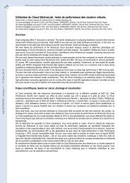
![[tel-00433556, v1] Relation entre Stress Oxydant et Homéostasie ...](https://img.yumpu.com/19233319/1/184x260/tel-00433556-v1-relation-entre-stress-oxydant-et-homeostasie-.jpg?quality=85)