et al.
et al.
et al.

Create successful ePaper yourself
Turn your PDF publications into a flip-book with our unique Google optimized e-Paper software.
REVIEW<br />
776<br />
(remodeled) osteons are surrounded by a cement<br />
line (<strong>al</strong>so of high miner<strong>al</strong> content) (31). In mamm<strong>al</strong>ian<br />
cortic<strong>al</strong> bone, the following intrinsic<br />
toughening mechanisms have been identified:<br />
molecular uncoiling and intermolecular sliding<br />
of collagen, fibrillar sliding of collagen bonds,<br />
and microcracking of the miner<strong>al</strong> matrix (19).<br />
Extrinsic mechanisms are collagen fibril bridging,<br />
uncracked ligament bridging, and crack deflection<br />
and twisting (19). Rarely does a limb bone<br />
snap in two with smooth fracture surfaces; the<br />
crack is often deflected orthogon<strong>al</strong> to the crack<br />
front direction. In the case of (rehydrated) elk<br />
(Cervus elaphus) antler bone (shown in Fig. 2C)<br />
(32), which has the highest toughness of any<br />
bone type by far (33), the hyperminer<strong>al</strong>ized regions<br />
around the primary osteons lead to crack<br />
deflection, and the high amount of collagen<br />
(~60 volume %) adds mechanisms of crack r<strong>et</strong>ardation<br />
and creates crack bridges behind the<br />
crack front. The toughening effect in antlers has<br />
been estimated as: crack deflection, 60%; uncracked<br />
ligament bridges, 35%; and collagen<br />
as well as fibril bridging, 5% (33). A particularly<br />
important feature in bone is that the fracture<br />
toughness increases as the crack propagates, as<br />
shown in the plot. This plot demonstrates the<br />
crack extension resistance curve, or R-curve,<br />
behavior, which is the rate of the tot<strong>al</strong> energy<br />
dissipated as a function of the crack size. This<br />
occurs by the activation of the extrinsic toughening<br />
mechanisms. In this manner, it becomes<br />
gradu<strong>al</strong>ly more difficult to advance the crack. In<br />
human bone, the cracks are deflected and/or<br />
A B<br />
Ab<strong>al</strong>one shell: Nacre<br />
Miner<strong>al</strong> bridges<br />
1 cm<br />
Mesolayers<br />
Deer antler<br />
Subvelv<strong>et</strong>/compact<br />
Subvelv<strong>et</strong>/c<br />
Compact<br />
Transition zone<br />
Cancellous<br />
0.1 mm<br />
300 m<br />
Primary osteons<br />
Collagen fibrils<br />
Human<br />
cortic<strong>al</strong> bone<br />
Chitin fibril n<strong>et</strong>work<br />
C D<br />
Toughness, J (kJm -2 )<br />
100<br />
10<br />
1<br />
0.1<br />
0.01<br />
0<br />
2 m<br />
500 00 nm 50 nm<br />
Miner<strong>al</strong> cryst<strong>al</strong>lites<br />
Transverse<br />
500 nm<br />
500 nm<br />
Elk antler<br />
Human<br />
cortic<strong>al</strong> bone<br />
In-plane longitudin<strong>al</strong><br />
ASTM v<strong>al</strong>id ASTM inv<strong>al</strong>id<br />
0.2 0.4 0.6<br />
Crack extension, a (mm)<br />
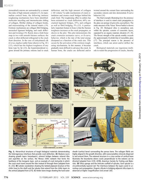
Fig. 2. Hierarchic<strong>al</strong> structures of tough biologic<strong>al</strong> materi<strong>al</strong>s demonstrating<br />
the h<strong>et</strong>erogeneous interfaces that provide crack deflection. (A) Ab<strong>al</strong>one nacre<br />
showing growth layers (mesolayers), miner<strong>al</strong> bridges b<strong>et</strong>ween miner<strong>al</strong> tiles<br />
and asperities on the surface, the fibrous chitin n<strong>et</strong>work that forms the<br />
backbone of the inorganic layer, and an example of crack tortuosity in which<br />
the crack must travel around the tiles instead of through them [adapted from<br />
(4, 21)]. (B) Lobster exoskel<strong>et</strong>on showing the twisted plywood structure of the<br />
chitin (next to the shell) and the tubules that extend from the chitin layers to<br />
the anim<strong>al</strong> [adapted from (27)]. (C) Antler bone image showing the hard outer<br />
˜1 nm<br />
Lobster<br />
twisted around the cement lines surrounding the<br />
secondary osteons and <strong>al</strong>so demonstrate R-curve<br />
behavior (34).<br />
The fin<strong>al</strong> example illustrating how the presence<br />
of interfaces is used to r<strong>et</strong>ard crack propagation is<br />
the glass sea sponge (Euplectella aspergillum). The<br />
entire structure of the Venus’ flower bask<strong>et</strong> is shown<br />
in Fig. 2D. Biologic<strong>al</strong> silica is amorphous and,<br />
within the spicules, consists of concentric layers,<br />
separated by an organic materi<strong>al</strong>, silicatein (35, 36).<br />
The flexure strength of the spicule notably exceeds<br />
(by approximately fivefold) that of monolithic glass<br />
(37). The princip<strong>al</strong> reason is the presence of<br />
interfaces, which can arrest and/or deflect the<br />
crack.<br />
Biologic<strong>al</strong> materi<strong>al</strong>s use ingenious m<strong>et</strong>hods<br />
to r<strong>et</strong>ard the progression of cracks, thereby<br />
50 nm 200 nm<br />
˜3 nm<br />
10 m<br />
1 m<br />
200 m<br />
Spicules<br />
20 mm<br />
15 FEBRUARY 2013 VOL 339 SCIENCE www.sciencemag.org<br />
˜20 nm<br />
Deep sea<br />
sponge<br />
˜10 m<br />
Skel<strong>et</strong>on<br />
sheath (cortic<strong>al</strong> bone) surrounding the porous bone. The collagen fibrils are<br />
highly <strong>al</strong>igned in the growth direction, with nanocryst<strong>al</strong>line miner<strong>al</strong>s dispersed<br />
in and around them. The osteon<strong>al</strong> structure in a cross section of cortic<strong>al</strong> bone<br />
illustrates the boundaries where cracks perpendicular to the osteons can be<br />
directed [adapted from (33)]. ASTM, American Soci<strong>et</strong>y for Testing and Materi<strong>al</strong>s.<br />
(D) Silica sponge and the intricate scaffold of spicules. Each spicule is a<br />
circumferenti<strong>al</strong>ly layered rod: The interfaces b<strong>et</strong>ween the layers assist in arresting<br />
crack propagation. Organic silicate in bridging adjacent silica layers is<br />
observed at higher magnification (red arrow) (36).<br />
on February 14, 2013<br />
www.sciencemag.org<br />
Downloaded from