Panel Disdussion
Panel Disdussion
Panel Disdussion
You also want an ePaper? Increase the reach of your titles
YUMPU automatically turns print PDFs into web optimized ePapers that Google loves.
3rd International Congress of of Nuclear Medicine & & 15th 15th Iranian Annual Annual Congress of<br />
Congress of Nuclear Medicine<br />
Shahid Beheshti Shahid Beheshti University University of Medical of Medical Sciences Sciences 19-21 19-21 May May 2011<br />
Dual isotope cardiac imaging using Tl-Tc cross-contamination<br />
correction by subtraction method<br />
Mohsen Kohanpour 1 , Hossein Rajabi 2 , Mohsen Beheshti 3 , Faraz<br />
Kalantari 2 , Majid Pouladian 1<br />
1 Islamic Azad University<br />
2 Department of Medical Physics, Tarbiat Modares University<br />
3 Paracelsus Medical University<br />
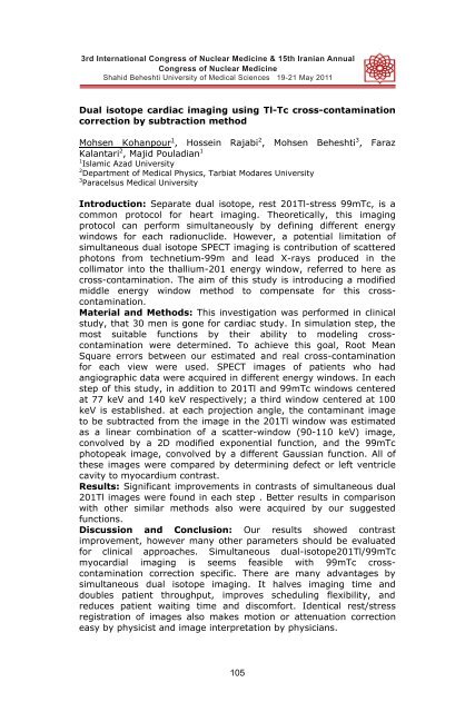
Introduction: Separate dual isotope, rest 201Tl-stress 99mTc, is a<br />
common protocol for heart imaging. Theoretically, this imaging<br />
protocol can perform simultaneously by defining different energy<br />
windows for each radionuclide. However, a potential limitation of<br />
simultaneous dual isotope SPECT imaging is contribution of scattered<br />
photons from technetium-99m and lead X-rays produced in the<br />
collimator into the thallium-201 energy window, referred to here as<br />
cross-contamination. The aim of this study is introducing a modified<br />
middle energy window method to compensate for this crosscontamination.<br />
Material and Methods: This investigation was performed in clinical<br />
study, that 30 men is gone for cardiac study. In simulation step, the<br />
most suitable functions by their ability to modeling crosscontamination<br />
were determined. To achieve this goal, Root Mean<br />
Square errors between our estimated and real cross-contamination<br />
for each view were used. SPECT images of patients who had<br />
angiographic data were acquired in different energy windows. In each<br />
step of this study, in addition to 201Tl and 99mTc windows centered<br />
at 77 keV and 140 keV respectively; a third window centered at 100<br />
keV is established. at each projection angle, the contaminant image<br />
to be subtracted from the image in the 201Tl window was estimated<br />
as a linear combination of a scatter-window (90-110 keV) image,<br />
convolved by a 2D modified exponential function, and the 99mTc<br />
photopeak image, convolved by a different Gaussian function. All of<br />
these images were compared by determining defect or left ventricle<br />
cavity to myocardium contrast.<br />
Results: Significant improvements in contrasts of simultaneous dual<br />
201Tl images were found in each step . Better results in comparison<br />
with other similar methods also were acquired by our suggested<br />
functions.<br />
Discussion and Conclusion: Our results showed contrast<br />
improvement, however many other parameters should be evaluated<br />
for clinical approaches. Simultaneous dual-isotope201Tl/99mTc<br />
myocardial imaging is seems feasible with 99mTc crosscontamination<br />
correction specific. There are many advantages by<br />
simultaneous dual isotope imaging. It halves imaging time and<br />
doubles patient throughput, improves scheduling flexibility, and<br />
reduces patient waiting time and discomfort. Identical rest/stress<br />
registration of images also makes motion or attenuation correction<br />
easy by physicist and image interpretation by physicians.<br />
105


