

the cynipoid genus paramblynotus - American Museum of Natural ...
the cynipoid genus paramblynotus - American Museum of Natural ...
the cynipoid genus paramblynotus - American Museum of Natural ...

Create successful ePaper yourself
Turn your PDF publications into a flip-book with our unique Google optimized e-Paper software.
2007 LIU ET AL.: REVISION OF PARAMBLYNOTUS (HYMENOPTERA) 59<br />
distinct, arising from posterior two-thirds <strong>of</strong><br />
basal vein. Marginal cell 2.3 times as long as<br />
wide. Bulla on Sc+R 1 absent.<br />
Abdominal petiole 0.5 times as long as<br />
wide in lateral view. Relative length <strong>of</strong> T3–7:<br />
1.6:1.0:1.3:3.0:1.4; T3–5 glabrous; T6 with<br />
a row <strong>of</strong> setigerous punctures; T7 entirely<br />
finely punctate with a row <strong>of</strong> setigerous<br />
punctures. T8 completely covered by T7.<br />
All legs densely punctate with pubescence<br />
except metacoxa dorsally glabrous. Metacoxa<br />
prominently expanded anteroventrally<br />
into a lobular process. Metatibia apically<br />
with four small, thin, apically pointed teeth.<br />
1mt/2–5mt 5 0.62.<br />
MALE: Unknown.<br />
Within <strong>the</strong> trisetosus group, P. coxatus<br />
forms a distinct monophyletic clade, <strong>the</strong><br />
trisetosus clade, with P. fuscapiculus, rwandensis,<br />
trisetosus, zairensis, cameroonensis,<br />
kekenboschi, jacksoni, and carinatus. This<br />
clade differs from <strong>the</strong> o<strong>the</strong>r species <strong>of</strong> <strong>the</strong><br />
trisetosus species group in that (1) upper<br />
mesopleuron and speculum glabrous;<br />
(2) posterior margin <strong>of</strong> T7 <strong>of</strong> female metasoma<br />
not emarginate, covering T8 entirely;<br />
and (3) metepisternum with a median nude,<br />
glabrous area. P. coxatus differs from all<br />
o<strong>the</strong>r Paramblynotus species by <strong>the</strong> presence<br />
<strong>of</strong> anteroventral lobular expansion on its<br />
metacoxa and a vertical impression along <strong>the</strong><br />
posterior margin <strong>of</strong> <strong>the</strong> lower mesopleuron.<br />
O<strong>the</strong>rwise, this species is very close to P.<br />
fuscapiculus.<br />
TYPE MATERIAL: HOLOTYPE: R, South<br />
Africa: Natal, 1982-I-10–15, J. Londt coll.<br />
(CNCI).<br />
DISTRIBUTION: South Africa: Natal.<br />
ETYMOLOGY: From Latin, coxa, coxa.<br />
The name coined for its unique coxa feature<br />
<strong>of</strong> anteroventral lobular expansion.<br />
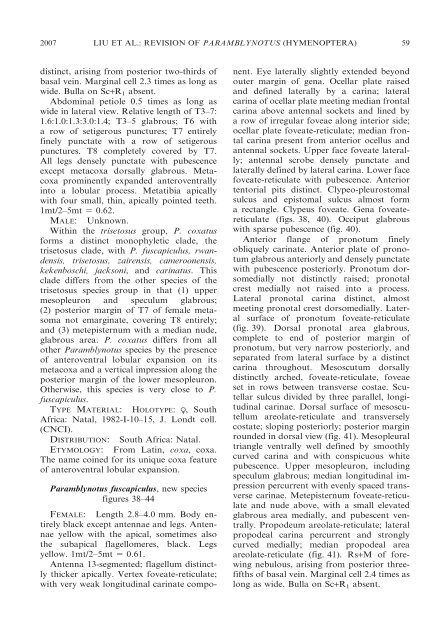
Paramblynotus fuscapiculus, new species<br />
figures 38–44<br />
FEMALE:<br />
Length 2.8–4.0 mm. Body entirely<br />
black except antennae and legs. Antennae<br />
yellow with <strong>the</strong> apical, sometimes also<br />
<strong>the</strong> subapical flagellomeres, black. Legs<br />
yellow. 1mt/2–5mt 5 0.61.<br />
Antenna 13-segmented; flagellum distinctly<br />
thicker apically. Vertex foveate-reticulate;<br />
with very weak longitudinal carinate component.<br />
Eye laterally slightly extended beyond<br />
outer margin <strong>of</strong> gena. Ocellar plate raised<br />
and defined laterally by a carina; lateral<br />
carina <strong>of</strong> ocellar plate meeting median frontal<br />
carina above antennal sockets and lined by<br />
a row <strong>of</strong> irregular foveae along interior side;<br />
ocellar plate foveate-reticulate; median frontal<br />
carina present from anterior ocellus and<br />
antennal sockets. Upper face foveate laterally;<br />
antennal scrobe densely punctate and<br />
laterally defined by lateral carina. Lower face<br />
foveate-reticulate with pubescence. Anterior<br />
tentorial pits distinct. Clypeo-pleurostomal<br />
sulcus and epistomal sulcus almost form<br />
a rectangle. Clypeus foveate. Gena foveatereticulate<br />
(figs. 38, 40). Occiput glabrous<br />
with sparse pubescence (fig. 40).<br />
Anterior flange <strong>of</strong> pronotum finely<br />
obliquely carinate. Anterior plate <strong>of</strong> pronotum<br />
glabrous anteriorly and densely punctate<br />
with pubescence posteriorly. Pronotum dorsomedially<br />
not distinctly raised; pronotal<br />
crest medially not raised into a process.<br />
Lateral pronotal carina distinct, almost<br />
meeting pronotal crest dorsomedially. Lateral<br />
surface <strong>of</strong> pronotum foveate-reticulate<br />
(fig. 39). Dorsal pronotal area glabrous,<br />
complete to end <strong>of</strong> posterior margin <strong>of</strong><br />
pronotum, but very narrow posteriorly, and<br />
separated from lateral surface by a distinct<br />
carina throughout. Mesoscutum dorsally<br />
distinctly arched, foveate-reticulate, foveae<br />
set in rows between transverse costae. Scutellar<br />
sulcus divided by three parallel, longitudinal<br />
carinae. Dorsal surface <strong>of</strong> mesoscutellum<br />
areolate-reticulate and transversely<br />
costate; sloping posteriorly; posterior margin<br />
rounded in dorsal view (fig. 41). Mesopleural<br />
triangle ventrally well defined by smoothly<br />
curved carina and with conspicuous white<br />
pubescence. Upper mesopleuron, including<br />
speculum glabrous; median longitudinal impression<br />
percurrent with evenly spaced transverse<br />
carinae. Metepisternum foveate-reticulate<br />
and nude above, with a small elevated<br />
glabrous area medially, and pubescent ventrally.<br />
Propodeum areolate-reticulate; lateral<br />
propodeal carina percurrent and strongly<br />
curved medially; median propodeal area<br />
areolate-reticulate (fig. 41). Rs+M <strong>of</strong> forewing<br />
nebulous, arising from posterior threefifths<br />
<strong>of</strong> basal vein. Marginal cell 2.4 times as<br />
long as wide. Bulla on Sc+R 1 absent.


