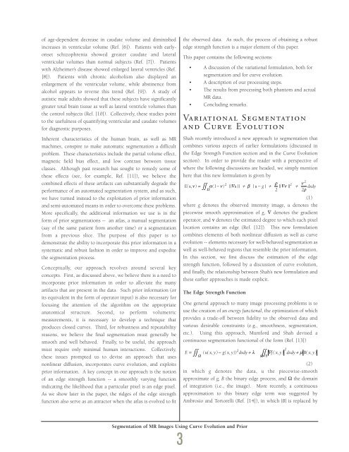
of age-dependent decrease in caudate volume and diminishedincreases in ventricular volume (Ref. [6]). Patients with earlyonsetschizophrenia showed greater caudate and lateralventricular volumes than normal subjects (Ref. [7]). Patientswith Alzheimer’s disease showed enlarged lateral ventricles (Ref.[8]). Patients with chronic alcoholism also displayed anenlargement of the ventricular volume, while abstinence fromalcohol appears to reverse this trend (Ref. [9]). A study ofautistic male adults showed that these subjects have significantlygreater total brain tissue as well as lateral ventricle volumes thanthe control subjects (Ref. [10]). Collectively, these studies pointto the usefulness of quantifying ventricular and caudate volumesfor diagnostic purposes.Inherent characteristics of the human brain, as well as MRmachines, conspire to make automatic segmentation a difficultproblem. These characteristics include the partial volume effect,magnetic field bias effect, and low contrast between tissueclasses. Although past research has sought to remedy some ofthese effects (see, for example, Ref. [11]), we believe thecombined effects of these artifacts can substantially degrade theperformance of an automated segmentation system, and as such,we have turned instead to the exploitation of prior informationand semi-automated means in order to overcome these problems.More specifically, the additional information we use is in theform of prior segmentations -- an atlas, a manual segmentation(say of the same patient from another time) or a segmentationfrom a previous slice. The purpose of this paper is todemonstrate the ability to incorporate this prior information in asystematic and robust fashion in order to improve and expeditethe segmentation process.Conceptually, our approach revolves around several keyconcepts. First, as discussed above, we believe there is a need toincorporate prior information in order to alleviate the manyartifacts that are present in the data. Such prior information (orits equivalent in the form of operator input) is also necessary forfocusing the attention of the algorithm on the appropriateanatomical structure. Second, to perform volumetricmeasurements, it is necessary to develop a technique thatproduces closed curves. Third, for robustness and repeatabilityreasons, we believe the final segmentation must generally besmooth and well behaved. Finally, to be useful, the approachmust require only minimal human interactions. Collectively,these issues prompted us to devise an approach that usesnonlinear diffusion, incorporates curve evolution, and exploitsprior information. A key concept in our approach is the notionof an edge strength function -- a smoothly varying functionindicating the likelihood that a particular pixel is an edge pixel.As we show later in the paper, the ridges of the edge strengthfunction also serve as an attractor when the atlas is evolved to fitthe observed data. As such, the process of obtaining a robustedge strength function is a major element of this paper.This paper contains the following sections:• A discussion of the variational formulation, both forsegmentation and for curve evolution.• A description of our processing steps.• The results from processing both phantom and actualMR data.• Concluding remarks.Variational Segmentationand Curve EvolutionShah recently introduced a new approach to segmentation thatcombines various aspects of earlier formulations (discussed inthe Edge Strength Function section and in the Curve Evolutionsection). In order to provide the reader with a perspective ofwhere the following discussions are headed, we simply mentionhere that this new formulation is given by2ρνE( u, ν) = ∫∫ α( 1 2 2−ν) || ∇ u || + β | u − g | + || ∇ ν || + dxdyΩ 2 2ρ(1)where g denotes the observed intensity image, u denotes thepiecewise smooth approximation of g, ∇ denotes the gradientoperator, and ν denotes the estimated degree to which each pixellocation contains an edge (Ref. [12]). This new formulationcombines elements of both nonlinear diffusion as well as curveevolution -- elements necessary for well-behaved segmentation aswell as well-behaved regions that resemble the prior information.In this section, we first discuss the estimation of the edgestrength function, followed by a discussion of curve evolution,and finally, the relationship between Shah’s new formulation andthese earlier approaches is made explicit.The Edge Strength FunctionOne general approach to many image processing problems is touse the creation of an energy functional, the optimization of whichprovides a trade-off between fidelity to the observed data andvarious desirable constraints (e.g., smoothness, segmentation,etc.). Using this approach, Mumford and Shah devised acontinuous segmentation functional of the form (Ref. [13])2 2E = ∫∫ ( u( x, y ) − g( x, y )) dxdy + λ ∫∫ ∇ f( x, y ) dxdy + µ B( x, y )ΩΩ − B(2)in which g denotes the data, u the piecewise-smoothapproximate of g, B the binary edge process, and Ω the domainof integration (i.e., the image). More recently, a continuousapproximation to this binary edge term was suggested byAmbrosio and Tortorelli (Ref. [14]), in which |B| is replaced bySegmentation of MR Images Using Curve Evolution and Prior3
(3)where ν now represents a continuous edge process, which maybe interpreted as the probability of the presence of an edge atevery pixel. Alternatively, ν represents the “edginess” of eachpixel. For this reason, we refer to ν as the edge strengthfunction. Note that whereas B is an impulse function (it has anamplitude of 1 wherever it is more “cost effective” to incur apenalty of µ instead of incurring the penalty associated with alarge gradient), Eq. (3) is a continuous approximation in thesense that ν is continuous, and Λ ρ → ⎪Β⎪ as ρ → 0 (Ref. [15]).Using the continuous edge approximation of Eq. (3), asegmentation functional of the formE = ∫∫ (( u− g ) 2 + ∇u 2 ( − ) 2 + ( ∇ 2 2µ νλ 1 ν ρ ν + )) dxdyΩ2 ρ(4)was demonstrated in Ref. [15]. There are three terms in thisfunctional -- a data fidelity term, a smoothness term, and an edgepenalty term. Furthermore, the u and ν minimizing E are the“optimal” piecewise smooth estimate of the intensity, and theedge strength function, respectively. Lastly, note that nonlineardiffusion for smoothing the image while retaining edge sharpnessis achieved by suppressing smoothing (i.e., ⎪∇u⎪ wherever ν ishigh). The gradient descent equations (based on the Eulerequations for determining the conditions of optimality) we use tosolve this functional are:∂u∂t∂ν∂tCurve Evolution12 νΛρ = ∫∫ ( ρ∇ ν + ) dxdy2 Ω ρ2 β= −2∇ν⋅∇ u+ ( 1−ν)∇ u−u−gα( 1−ν) ( )Let Γ denote a simple closed curve, and let ν denote the edgestrength function defined earlier. In order to move Γ to where νis high, we look for stationary points ofwhere s denotes the arc length along Γ, and q is a fixed constant.Let C(p,t) : I x [0,∞] → Ω be the evolving family of curves, whereΩ denotes the image domain, I denotes the unit interval, and tdenotes time. We require that C(0,t) = C(1,t) for all values of t,and that the image of C(p,0) in Ω coincides with Γ. Theevolution of curve C is governed by∂C∂t2 ν 2α= ∇ ν − + ( 1−ν) ∇u2ρ ρ∫( 1−ν )q dsΓ= [ q∇ν⋅N−( 1−ν)κ]N22(5)(6)(7)(8)where N is the outward normal, and κ is the curvature definedsuch that it is positive when Γ is a circle.To implement the evolution of Γ, assume that Γ is embedded ina surface f 0 : Ω → R as a level curve. Let f(t, x, y) denote theevolving surface such that f(0, x, y) = f 0 (x, y). In order to let allthe level curves of f 0 evolve simultaneously, considerwhere Γ c = {(x, y)|f(t, x, y) = c}. This functional can betransformed into the image domain via the coarea formula,resulting in the functionalThe gradient descent equation for this functional is given by(9)(10)∂f= − q∇ν⋅∇ f + ( 1−ν) ∇f curv( f ) (11)∂twhere curv(f) is the curvature of the level curves of f given by(12)That is, points along the curve f evolve with a velocity consistingof two components: a component that drives f toward ν, and acomponent proportional to the curvature (Ref. [16]).A New Segmentation FunctionalAs noted at the beginning of this section, Shah introduced22 ρ 2 νE( u, ν) = ∫∫ α( 1−ν)∇ u + β u − g + ∇ ν + dxdyΩ2 2ρ(13)as a common framework that incorporates elements of nonlineardiffusion and curve evolution (Ref. [12]). The associatedgradient descent equations are:∂u∂t∫curv( f ) =∫ −∞∞∫∫( 1−ν ) q ds cdcΓc( 1−ν )qR∇fdxdy2 2fy fxx − 2 fx fy fxy + fx fyy2 2( fx+ fy)3 2βν u ν u curv u u u −= −2∇ ⋅∇ + ( 1− ) ∇ ( ) − ∇gα( 1−ν) | u−g|∂ν 2 ν 2α= ∇ ν − + ( 1−ν) ∇u∂t2ρ ρwith boundary conditions:(14)(15)∂u∂ν= 0;= 0(16)∂n∂Ω∂n∂Ωalong the image boundary. Superficially, this formulationappears very similar to Eq. (4) in the sense that this formulationSegmentation of MR Images Using Curve Evolution and Prior4
- Page 2 and 3: Letter from thePresident and CEO,Vi
- Page 4 and 5: Information TechnologyMilton AdamsE
- Page 6 and 7: BiographyMilton Adams has been at D
- Page 9 and 10: Figure 1 represents a functional de
- Page 11 and 12: Programs. In effect, these controll
- Page 13 and 14: Although the terminal area traffic
- Page 15 and 16: Table 2. ATFM performance evaluatio
- Page 17 and 18: In the experiments, a nominal capac
- Page 19 and 20: [3] Wambsganss, Michael C. “Colla
- Page 21 and 22: Guidance, Navigation, and Control A
- Page 23 and 24: A Control Lyapunov FunctionApproach
- Page 25 and 26: x( 0) ∈ X and w(t) ∈Wfor all t
- Page 27 and 28: (b) Select a quadratic RCLF V i (x)
- Page 29 and 30: at each grid point. In the case w 1
- Page 31 and 32: References[1] Ball, J.A. and A.J. v
- Page 33 and 34: Guidance, Navigation, and Control A
- Page 35 and 36: Relative and Differential GPSData T
- Page 37 and 38: The first term on the right in the
- Page 39 and 40: H R# δρ R,GPS -H A# δρ A,GPSThi
- Page 41 and 42: selection; and (3) shown that the a
- Page 43 and 44: Guidance, Navigation, and Control A
- Page 45: Segmentation of MR ImagesUsing Curv
- Page 49 and 50: Experimental ResultsThe results of
- Page 51 and 52: Table 1. A summary of segmentation
- Page 53 and 54: Guidance, Navigation,and ControlJim
- Page 55 and 56: BiographyGeorge SchmidtGeorge Schmi
- Page 57 and 58: clock and ephemeris errors, as well
- Page 59 and 60: maintained in a rigid structure, wh
- Page 61 and 62: Table 5. “Typical” absolute GPS
- Page 63 and 64: performed, then the target location
- Page 65 and 66: tightly-coupled system, however, ca
- Page 67 and 68: Concluding RemarksRecent progress i
- Page 69 and 70: As real-time systems evolve into th
- Page 71 and 72: Advanced Fault-TolerantComputing fo
- Page 73 and 74: The Viking and Voyager were both in
- Page 75 and 76: Containment Regions (FCRs). There a
- Page 77 and 78: well as reversing the whole process
- Page 79 and 80: As real-time systems evolve into th
- Page 81 and 82: Automated Station-Keepingfor Satell
- Page 83 and 84: Figure 2. Minimum elevation angles
- Page 85 and 86: anomaly M and/or the ascending node
- Page 87 and 88: However, since optimization and rec
- Page 89 and 90: is maintained in the Northern Hemis
- Page 91 and 92: autonomy. It must have the ability
- Page 93 and 94: [31] Neelon, Joseph G., Jr., Paul J
- Page 95 and 96: Draper’s primary goal is to Drape
- Page 97 and 98:
)Rotordynamic Modelingof an Activel
- Page 99 and 100:
Eq. (9) becomes:λ[ R ] { Φ } = [
- Page 101 and 102:
chosen to be 24, for a total of 48
- Page 103 and 104:
InertialInstruments/MechanicalDesig
- Page 105 and 106:
BiographyJeffrey Borenstein is curr
- Page 107 and 108:
process step. Process information i
- Page 109 and 110:
Figure 4. Control chart for boron d
- Page 111 and 112:
References[1] Barbour, N., J. Conne
- Page 113 and 114:
Draper Laboratory continues to engi
- Page 115 and 116:
Validating the Validating Tool:Defi
- Page 117 and 118:
calculates miscellaneous terms, suc
- Page 119 and 120:
Table 1. Suggested specification sh
- Page 121 and 122:
User Accuracy as aFunction of Simul
- Page 123 and 124:
20-min averaging, this clock lockin
- Page 125 and 126:
Table 2. Sample high-level summary
- Page 127 and 128:
AcknowledgmentR.L. Greenspan, J.A.
- Page 129 and 130:
Systems IntegrationRich MartoranaPe
- Page 131 and 132:
BiographyAnthony Kourepenis is an A
- Page 133 and 134:
control is employed to maintain the
- Page 135 and 136:
Table 1. Summary of automotive yaw
- Page 137 and 138:
Resolution (60 Hz) deg/h10000000100
- Page 139 and 140:
References[1] Greiff, P., B. Boxenh
- Page 141 and 142:
Guidance, Navigation, and Control A
- Page 143 and 144:
An Integrated Safety AnalysisMethod
- Page 145 and 146:
Infrastructure ModelsSystemRequirem
- Page 147 and 148:
Figures 6 and 7 illustrate the bloc
- Page 149 and 150:
Notice that each flight track descr
- Page 151 and 152:
Table 7. Safety statistics at 1700-
- Page 153 and 154:
Guidance, Navigation, and Control A
- Page 155 and 156:
An Optimal Guidance Law forPlanetar
- Page 157 and 158:
Note that the states in the three d
- Page 159 and 160:
Crossrange (Kft)10090807060504030Cl
- Page 161 and 162:
The 1997 Charles StarkDraper PrizeT
- Page 163 and 164:
The 1997 Charles StarkDraper Prize1
- Page 165 and 166:
“Draper encourages its personnel
- Page 167 and 168:
Gimballed Vibrating GyroscopeHaving
- Page 169 and 170:
“Draper encourages its personnel
- Page 171 and 172:
Optical Source Isolator withPolariz
- Page 173 and 174:
“Draper encourages its personnel
- Page 175 and 176:
Hunting Suppressor forPolyphase Ele
- Page 177 and 178:
“Draper encourages its personnel
- Page 179 and 180:
Sensor Having an Off-Frequency Driv
- Page 181 and 182:
proof mass from transients and enha
- Page 183 and 184:
1997 Published PapersThe following
- Page 185 and 186:
monitoring of space structures and
- Page 187 and 188:
measured by kinematic degrees of fr
- Page 189 and 190:
i.e., what percent of the earth’s
- Page 191 and 192:
McConley, M. W.; Dahleh, M. A.; Fer
- Page 193 and 194:
unaffordable, or even misguided. Bu
- Page 195 and 196:
The Draper DistinguishedPerformance
- Page 197:
Educational Activitiesat Draper Lab


