- Page 2 and 3:
FOURTH EDITION LANGE Q&A INTERNAL
- Page 4 and 5:
Professional Want to learn more? We
- Page 6 and 7:
iv Contents 9. Muscles and Joints .
- Page 8 and 9:
This page intentionally left blank
- Page 10 and 11:
This page intentionally left blank
- Page 12 and 13:
2 1: Cardiology 5. A 42-year-old ma
- Page 14 and 15:
4 1: Cardiology 14. A patient with
- Page 16 and 17:
6 1: Cardiology (A) (B) (C) (D) (E)
- Page 18 and 19:
8 1: Cardiology 31. A 63-year-old w
- Page 20 and 21:
10 1: Cardiology I aVR V1 V4 II aVL
- Page 22 and 23:
12 1: Cardiology 47. A 25-year-old
- Page 24 and 25:
14 1: Cardiology 53. Figure 1-13 is
- Page 26 and 27:
16 1: Cardiology 59. A 78-year-old
- Page 28 and 29:
18 1: Cardiology 74. A 64-year-old
- Page 30 and 31:
20 1: Cardiology 99. A 65-year-old
- Page 32 and 33:
22 1: Cardiology 115. A 56-year-old
- Page 34 and 35:
24 1: Cardiology Calcium plays a ro
- Page 36 and 37:
26 1: Cardiology and other bradyarr
- Page 38 and 39:
28 1: Cardiology most useful initia
- Page 40 and 41:
30 1: Cardiology 73. (D) In Wolff-P
- Page 42 and 43:
32 1: Cardiology 102. (D) RVMI is c
- Page 44 and 45:
34 1: Cardiology function should be
- Page 46 and 47:
36 2: Skin DIRECTIONS (Questions 6
- Page 48 and 49:
38 2: Skin Figure 2-4. (Reproduced,
- Page 50 and 51:
40 2: Skin Figure 2-7. (Reproduced,
- Page 52 and 53:
42 2: Skin 32. A 32-year-old woman
- Page 54 and 55:
44 2: Skin DIRECTIONS (Questions 43
- Page 56 and 57:
46 2: Skin be difficult to differen
- Page 58 and 59:
48 2: Skin specific antibodies in t
- Page 60 and 61:
50 2: Skin 50. (C) Chloroquine is u
- Page 62 and 63:
52 3: Endocrinology 5. A 17-year-ol
- Page 64 and 65:
54 3: Endocrinology 12. A 56-year-o
- Page 66 and 67:
56 3: Endocrinology 23. A 19-year-o
- Page 68 and 69:
58 3: Endocrinology 34. A 33-year-o
- Page 70 and 71:
60 3: Endocrinology 50. A 25-year-o
- Page 72 and 73:
62 3: Endocrinology 62. A 21-year-o
- Page 74 and 75:
64 3: Endocrinology 73. A 55-year-o
- Page 76 and 77:
66 3: Endocrinology 91. Which of th
- Page 78 and 79:
Answers and Explanations 1. (C) Pri
- Page 80 and 81:
70 3: Endocrinology It is most comm
- Page 82 and 83:
72 3: Endocrinology polygenic in it
- Page 84 and 85:
74 3: Endocrinology 57. (B) Risk of
- Page 86 and 87:
76 3: Endocrinology also be caused
- Page 88 and 89:
78 3: Endocrinology occur only in a
- Page 90 and 91:
80 4: Gastroenterology 5. A 57-year
- Page 92 and 93:
82 4: Gastroenterology 23. A 64-yea
- Page 94 and 95:
84 4: Gastroenterology 29. A 45-yea
- Page 96 and 97:
86 4: Gastroenterology 39. A couple
- Page 98 and 99:
88 4: Gastroenterology 54. A 22-yea
- Page 100 and 101:
90 4: Gastroenterology 66. Which of
- Page 102 and 103:
Answers and Explanations 1. (F) Car
- Page 104 and 105:
94 4: Gastroenterology 18. (B) Desp
- Page 106 and 107:
96 4: Gastroenterology 43. (B) In n
- Page 108 and 109:
98 4: Gastroenterology 67. (A) Anab
- Page 110 and 111:
100 5: Hematology 4. A 62-year-old
- Page 112 and 113:
102 5: Hematology 17. A 23-year-old
- Page 114 and 115:
104 5: Hematology 28. A 63-year-old
- Page 116 and 117:
106 5: Hematology 36. A 4-month-old
- Page 118 and 119:
108 5: Hematology 49. A 19-year-old
- Page 120 and 121:
110 5: Hematology 59. A 28-year-old
- Page 122 and 123:
Answers and Explanations 1. (D) The
- Page 124 and 125:
114 5: Hematology 19. (E) Steroids
- Page 126 and 127:
116 5: Hematology 42. (C) Chorionic
- Page 128 and 129:
118 5: Hematology a chronic disease
- Page 130 and 131:
120 6: Oncology 3. A 42-year-old ma
- Page 132 and 133:
122 6: Oncology 14. You are seeing
- Page 134 and 135:
124 6: Oncology 25. A 63-year-old m
- Page 136 and 137:
126 6: Oncology Questions 38 throug
- Page 138 and 139:
128 6: Oncology Questions 60 throug
- Page 140 and 141:
130 6: Oncology H. pylori is anothe
- Page 142 and 143:
132 6: Oncology 25. (B) Barrett’s
- Page 144 and 145:
134 6: Oncology with other thyroid
- Page 146 and 147:
136 7: Diseases of the Nervous Syst
- Page 148 and 149:
138 7: Diseases of the Nervous Syst
- Page 150 and 151:
140 7: Diseases of the Nervous Syst
- Page 152 and 153:
142 7: Diseases of the Nervous Syst
- Page 154 and 155:
144 7: Diseases of the Nervous Syst
- Page 156 and 157:
146 7: Diseases of the Nervous Syst
- Page 158 and 159:
Answers and Explanations 1. (B) Alz
- Page 160 and 161:
150 7: Diseases of the Nervous Syst
- Page 162 and 163:
152 7: Diseases of the Nervous Syst
- Page 164 and 165:
154 7: Diseases of the Nervous Syst
- Page 166 and 167:
This page intentionally left blank
- Page 168 and 169:
158 8: Kidneys 5. A 74-year-old man
- Page 170 and 171:
160 8: Kidneys 18. A 56-year-old ma
- Page 172 and 173:
162 8: Kidneys 28. A 74-year-old wo
- Page 174 and 175:
164 8: Kidneys 44. A 74-year-old wo
- Page 176 and 177:
166 8: Kidneys 63. A 60-year-old wo
- Page 178 and 179:
Answers and Explanations 1. (D) Ure
- Page 180 and 181:
170 8: Kidneys 21. (E) Intrathoraci
- Page 182 and 183:
172 8: Kidneys 44. (B) Diuretics ar
- Page 184 and 185:
174 8: Kidneys 75. (A) The autosoma
- Page 186 and 187:
176 9: Muscles and Joints 5. A youn
- Page 188 and 189:
178 9: Muscles and Joints 16. A 27-
- Page 190 and 191:
180 9: Muscles and Joints 26. A 77-
- Page 192 and 193:
182 9: Muscles and Joints 35. A 67-
- Page 194 and 195:
184 9: Muscles and Joints 46. A 72-
- Page 196 and 197:
186 9: Muscles and Joints 56. A ver
- Page 198 and 199:
188 9: Muscles and Joints 71. Most
- Page 200 and 201:
190 9: Muscles and Joints 85. A 39-
- Page 202 and 203:
192 9: Muscles and Joints 8. (B) It
- Page 204 and 205:
194 9: Muscles and Joints 29. (B) A
- Page 206 and 207: 196 9: Muscles and Joints 50. (D) T
- Page 208 and 209: 198 9: Muscles and Joints 76. (D) D
- Page 210 and 211: 200 10: Infection 5. A 17-year-old
- Page 212 and 213: 202 10: Infection 14. A 56-year-old
- Page 214 and 215: 204 10: Infection 25. A 4-year-old
- Page 216 and 217: 206 10: Infection 36. A 9-year-old
- Page 218 and 219: 208 10: Infection 48. The dental co
- Page 220 and 221: 210 10: Infection 58. A previously
- Page 222 and 223: 212 10: Infection 72. A 21-year-old
- Page 224 and 225: 214 10: Infection 82. Three individ
- Page 226 and 227: 216 10: Infection 100. A 42-year-ol
- Page 228 and 229: 218 10: Infection relatively benign
- Page 230 and 231: 220 10: Infection 28. (B) Adequate
- Page 232 and 233: 222 10: Infection organisms include
- Page 234 and 235: 224 10: Infection 66. (F) Shigella
- Page 236 and 237: 226 10: Infection 92. (D) Sulfonami
- Page 238 and 239: This page intentionally left blank
- Page 240 and 241: 230 11: Immunology and Allergy 6. A
- Page 242 and 243: 232 11: Immunology and Allergy 15.
- Page 244 and 245: 234 11: Immunology and Allergy 32.
- Page 246 and 247: 236 11: Immunology and Allergy such
- Page 248 and 249: 238 11: Immunology and Allergy 30.
- Page 250 and 251: 240 12: Diseases of the Respiratory
- Page 252 and 253: 242 12: Diseases of the Respiratory
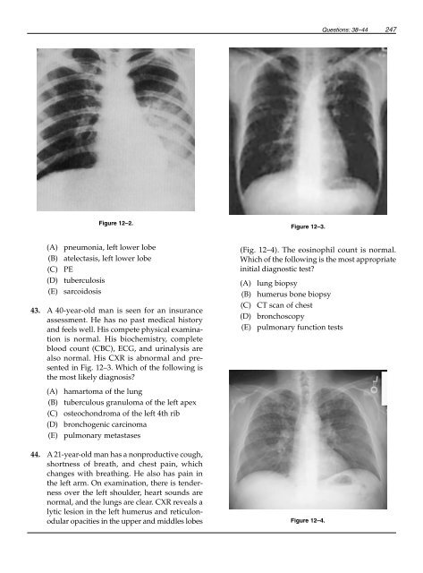
- Page 254 and 255: 244 12: Diseases of the Respiratory
- Page 258 and 259: 248 12: Diseases of the Respiratory
- Page 260 and 261: 250 12: Diseases of the Respiratory
- Page 262 and 263: Answers and Explanations 1. (C) Hyp
- Page 264 and 265: 254 12: Diseases of the Respiratory
- Page 266 and 267: 256 12: Diseases of the Respiratory
- Page 268 and 269: 258 12: Diseases of the Respiratory
- Page 270 and 271: 260 13: Clinical Pharmacology 6. A
- Page 272 and 273: 262 13: Clinical Pharmacology 22. A
- Page 274 and 275: 264 13: Clinical Pharmacology 40. W
- Page 276 and 277: 266 13: Clinical Pharmacology 57. A
- Page 278 and 279: 268 13: Clinical Pharmacology 10. (
- Page 280 and 281: 270 13: Clinical Pharmacology 30. (
- Page 282 and 283: 272 13: Clinical Pharmacology 52. (
- Page 284 and 285: This page intentionally left blank
- Page 286 and 287: 276 14: Comprehensive Review 6. A 3
- Page 288 and 289: 278 14: Comprehensive Review 21. Wh
- Page 290 and 291: 280 14: Comprehensive Review 38. A
- Page 292 and 293: 282 14: Comprehensive Review 53. A
- Page 294 and 295: 284 14: Comprehensive Review 69. A
- Page 296 and 297: 286 14: Comprehensive Review 85. Wh
- Page 298 and 299: 288 14: Comprehensive Review Figure
- Page 300 and 301: 290 14: Comprehensive Review 98. A
- Page 302 and 303: 292 14: Comprehensive Review For ea
- Page 304 and 305: Answers and Explanations 1. (A) Thi
- Page 306 and 307:
296 14: Comprehensive Review enceph
- Page 308 and 309:
298 14: Comprehensive Review antige
- Page 310 and 311:
300 14: Comprehensive Review peak i
- Page 312 and 313:
302 14: Comprehensive Review 76. (D
- Page 314 and 315:
304 14: Comprehensive Review At tim
- Page 316 and 317:
This page intentionally left blank
- Page 318 and 319:
This page intentionally left blank
- Page 320 and 321:
310 Index Antineutrophil cytoplasmi
- Page 322 and 323:
312 Index Dobutamine, 263, 270 Dono
- Page 324 and 325:
314 Index Infective endocarditis (C
- Page 326 and 327:
316 Index Pleuritis, 194 Pneumococc
- Page 328:
318 Index Toxoplasmosis, 148, 226 T