ZMBH J.Bericht 2000 - Zentrum für Molekulare Biologie der ...
ZMBH J.Bericht 2000 - Zentrum für Molekulare Biologie der ...
ZMBH J.Bericht 2000 - Zentrum für Molekulare Biologie der ...

Create successful ePaper yourself
Turn your PDF publications into a flip-book with our unique Google optimized e-Paper software.
Christine Clayton<br />
Molecular Cell Biology of Trypanosomes<br />
Introduction<br />
Salivarian trypanosomes are unicellular parasites that<br />
live in the blood and tissue fluids of mammals. They<br />
cause sleeping sickness in humans and also infect<br />
domestic animals and wildlife, mainly in subSaharan<br />
Africa. The geographical restriction is determined by<br />
the fact that the parasites are usually transmitted from<br />
one mammal to the next by Tsetse flies. They develop<br />
and multiply within the insect midgut and can un<strong>der</strong>go<br />
a sexual cycle before infection of another mammal<br />
via the insect salivary glands. Salivarian trypanosomes<br />
do not multiply inside cells, unlike the closely<br />
related tropical disease parasites Trypanosoma cruzi<br />
and Leishmania. At present about 300 000 people are<br />
infected with the sleeping sickness trypanosomes Trypanosoma<br />
brucei rhodesiense and Trypanosoma gambiense.<br />
The disease is invariably fatal unless treated,<br />
and very few drugs are available, especially to combat<br />
the late stages of the disease when the parasites invade<br />
the brain. Moreover, most of the available drugs are<br />
either too expensive or unacceptably toxic and drug<br />
resistance is developing.<br />
Trypanosomes and Leishmanias belong to the genus<br />
Kinetoplastida, which branched extremely early in<br />
eucaryotic evolution. As a consequence they exhibit<br />
a number of remarkable deviations from standard<br />
eucaryotic paradigms. In the realm of gene expression,<br />
most transcription is polycistronic, all mRNAs are<br />
trans spliced, there is almost no developmental regulation<br />
of transcription, and the mitochondrial RNAs<br />
are extensively edited. Energy and redox metabolism<br />
also exhibit unique features: for example, the glycolytic<br />
enzymes are compartmentalised in an organelle,<br />
54<br />
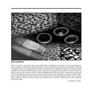
Figure 1: Electron micrographs of trypanosomes. On the left<br />
are normal trypanosomes with many round glycosomes (Gly).<br />
On the right are cells with a 90% reduction in PEXII (Evelyn<br />
Baumgart, Anatomy Dept.).<br />
the glycosome. At the same time, the parasites show<br />
many conserved features. For example, the glycosome<br />
is a specialised type of peroxisome, and trans splicing<br />
is mechanistically related to cis splicing.<br />
Our work concentrates on Trypanosoma brucei brucei,<br />
a close relative of the sleeping sickness trypanosomes<br />
which is not infective for humans. The parasites grow<br />
in laboratory rodents or in vitro and life-cycle related<br />
differentiation can be mimicked in culture. They are<br />
diploid, carrying about 60 Mb of DNA on 11 chromosome<br />
pairs; a sequencing project is in progress. A full<br />
palette of genetic manipulation methods is available,<br />
including gene replacement by homologous recombination<br />
and inducible gene expression. Conditional<br />
knockouts can be made by placing the gene of interest<br />
un<strong>der</strong> control of a tetracycline-inducible promoter,<br />
then deleting the remaining gene copies via homologous<br />
recombination.<br />
In our research on trypanosomes, we try to keep two<br />
aims in view. Firstly, we concentrate on aspects of<br />
the parasites that uniquely distinguish them from their<br />
mammalian hosts, and may therefore represent suitable<br />
targets for specific anti-parasitic chemotherapy.<br />
Secondly, by examining the similarities and differences<br />
between fundamental processes in trypanosomes,<br />
mammals and yeast we can clearly distinguish<br />
those aspects that are conserved throughout eucaryotic<br />
evolution and therefore likely to be essential from<br />
those that are required for specialised lifestyles.<br />
Control of gene expression<br />
Maciej Drodzd, Antonio Gonzales, Claudia Hartmann,<br />
Henriette Irmer, Luis Quilada<br />
Specialised surface molecules are essential for the<br />
survival and virulence of pathogens. In the case of<br />
trypanosomes, the forms that grow in the mammal<br />
(bloodstream forms) wear a uniform coat of variant<br />
surface glycoprotein (VSG), expressed from a single<br />
gene chosen from a repertoire of up to 1000 different<br />
VSG genes. The VSG expressed can be changed either<br />
through recombination or by transcriptional switches.<br />
This enables the parasites to change their surface coat,<br />
thereby evading the immune response. In the Tsetse<br />
midgut, the VSG is replaced by the EP and GPEET<br />
repetitive, acidic proteins. All these surface proteins<br />
are retained on the plasma membrane by a glycosyl<br />
phosphatidylinositol (GPI) lipid anchor.<br />
Normally, protein-coding genes can only be functionally<br />
expressed by RNA polymerase II. This is because<br />
the mRNAs have to be modified at the 5‘-end by<br />
enzymes that are associated exclusively with RNA<br />
polymerase II. The Kinetoplastids are not subject to<br />
this restriction because the 5‘ spliced lea<strong>der</strong> is capped.<br />
The VSG and the EP/GPEET genes are transcribed<br />
by RNA polymerase I as part of polycistronic tran-<br />
scription units. Nearly all other protein-coding genes<br />
are transcribed by RNA polymerase II, but again transcription<br />
is polycistronic. Although the expression of<br />
many genes has to be strongly regulated to enable<br />
survival in the disparate environments of Tsetse and<br />
mammal, there is no evidence for any developmental<br />
control of RNA polymerase II transcription. Instead,<br />
regulation usually requires sequences which are found<br />
in the 3‘-untranslated regions and control RNA degradation<br />
and/or translation. Polymerase I-mediated transcription<br />
of the VSG genes is, exceptionally, subject to<br />
strong developmental regulation, but the EP/GPEET<br />
gene transcription is only weakly regulated.<br />
To compensate for the lack of transcriptional regulation,<br />
the EP/GPEET mRNAs are extremely unstable<br />
and very poorly translated in bloodstream forms,<br />
so that no protein product is detectable. A uridinerich<br />
26 nt sequence in the 3‘-untranslated region of<br />
EP/GPEET mRNA is essential for this control. We<br />
have examined the mechanism of RNA degradation<br />
in detail. Relatively stable RNAs such as the actin<br />
mRNA appear to be degraded by the standard mechanism<br />
seen in eucaryotes: initial destruction of the<br />
poly(A) tail at the 3‘-end is followed by degradation<br />
of the rest of the mRNA. The EP/GPEET mRNAs<br />
are in contrast rapidly degraded in bloodstream forms<br />
without prior degradation of the poly(A) tail. Detailed<br />
characterisation of the EP RNA secondary structure<br />
indicated that the 26mer is in an exposed, extended<br />
conformation, so it should be accessible to RNA-binding<br />
proteins or specific endonucleases. Searches for<br />
specific 26mer-binding proteins by several methods<br />
have however proved fruitless.<br />
Searches of the available genome sequence have<br />
revealed a number of potential trypanosome proteins<br />
that are very similar to yeast enzymes involved in<br />
mRNA degradation and processing of stable RNAs, or<br />
55