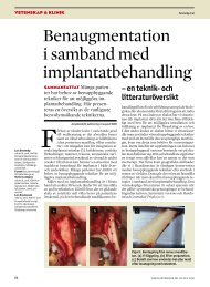
NordISKT TEmA 2009 - Tandläkartidningen
NordISKT TEmA 2009 - Tandläkartidningen
NordISKT TEmA 2009 - Tandläkartidningen

Create successful ePaper yourself
Turn your PDF publications into a flip-book with our unique Google optimized e-Paper software.
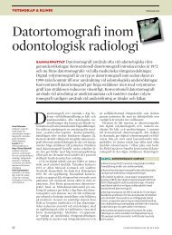
VETENSKAP & KLINIK<br />
TEMA BILDGIVANDE TEKNIKER<br />
derna för att förbättra bildkvaliteten. Den digitala<br />
tekniken har dessutom den fördelen att man<br />
kan göra analyser direkt på datorskärmen med<br />
något av de många programvarupaket som finns<br />
på marknaden. För specialisttandläkare i ortodonti<br />
som använder Björks [68, 69] analysmetod<br />
är tiops [57] ett givet val.<br />
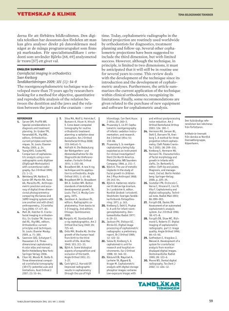
english summary<br />
Craniofacial imaging in orthodontics<br />
Sven Kreiborg<br />
<strong>Tandläkartidningen</strong> <strong>2009</strong>; 101 (1): 54–9<br />
The roentgencephalometric technique was developed<br />
more than 75 years ago by researchers<br />
looking for a method for objective, quantitative<br />
and reproducible analysis of the relation between<br />
the dentition and the jaws and the relation<br />
between the jaws and the cranium – over<br />
time. Today, cephalometric radiographs in the<br />
lateral projection are routinely used worldwide<br />
by orthodontists for diagnostics, treatment<br />
planning and follow-up. Several other cephalometric<br />
projections have been suggested to<br />
include the third dimension, but with limited<br />
success. However, although the technique, in<br />
principle, is limited to two dimensions, it must<br />
be anticipated that it will still be in routine use<br />
for several years to come. This review deals<br />
with the development of the technique since its<br />
introduction and the development of cephalometric<br />
analyses. Furthermore, the article summarizes<br />
the current application of the technique<br />
within clinical orthodontics, recognizing its<br />
limitations. Finally, some recommendations are<br />
given related to the purchase of new equipment<br />
and software for cephalometric analysis.<br />
referenser<br />
1. Sarver DM, Proffit WR.<br />
Special considerations in<br />
diagnosis and treatment<br />
planning. In: Graber TM,<br />
Vanarsdall RL, Vig KWL,<br />
editors. Orthodontics:<br />
current priciples and techniques.<br />
St. Louis: Elsevier<br />
Mosby; 2005. p. 24.<br />
2. Tsang KHS, Cooke MS.<br />
Comparison of cephalometric<br />
analysis using a nonradiographic<br />
sonic digitizer<br />
(DigiGraph Workstation)<br />
with conventional radiography.<br />
Eur J Orthod 1999;<br />
21: 1–13.<br />
3. Weinberg SM, Naidoo S,<br />
Govier DP, Martin RA, Kane<br />
AA, Marazita ML. Anthropometric<br />
precision and accuracy<br />
of digital three-dimensional<br />
photogrammetry:<br />
comparing the Genex and<br />
3dMD imaging systems with<br />
one another and with direct<br />
anthropometry. J Craniofac<br />
Surg 2006; 17: 477–83.<br />
4. Mah JK, Hatcher D. Craniofacial<br />
imaging in orthodontics.<br />
In: Graber TM, Vanarsdall<br />
RL, Vig KWL, editors.<br />
Orthodontics: current<br />
principles and techniques.<br />
St. Louis: Elsevier Mosby;<br />
2005. p. 71–100.<br />
5. Swennen GRJ, Schutyser F,<br />
Hausamen J-E. Threedimensional<br />
cephalometry.<br />
A color atlas and manual.<br />
Berlin Heidelberg New York:<br />
Springer Verlag; 2006.<br />
6. Chan HJ, Woods M, Stella D.<br />
Three dimensional computed<br />
craniofacial tomography<br />
(3D-CT): potential uses and<br />
limitations. Aust Orthod J<br />
2007; 23: 55–64.<br />
7. Silva MA, Wolf U, Heinicke F,<br />
Bumann A, Visser H, Hirsch<br />
E. Cone-beam computed<br />
tomography for routine<br />
orthodontic treatment<br />
planning: a radiation dose<br />
evaluation. Am J Orthod<br />
Dentofacial Orthop 2008;<br />
133: 640.e1–5.<br />
8. Hofrath H. Die Bedeutung<br />
der Röntgenfern- und<br />
Abstandaufnahme für die<br />
Diagnostik der Kieferanomalien.<br />
Fortschr Orthod<br />
1931; 1: 232–58.<br />
9. Broadbent BH. A new X-ray<br />
technique and its application<br />
to orthodontia. Angle<br />
Orthod 1931; 1: 45–66.<br />
10. Broadbent BH Sr, Broadbent<br />
BH Jr, Golden WH. Bolton<br />
standards of dentofacial<br />
developmental growth. St.<br />
Louis: The C.V. Mosby Company,<br />
1975.<br />
11. Jacobson A, Jacobson RL,<br />
editors. Radiographic cephalometry.<br />
From basics to<br />
3-D imaging. 2nd edition.<br />
Chicago: Quintessence;<br />
2006.<br />
12. Margolis HI. Standardized<br />
x-ray cephalographics. Am J<br />
Orthod Oral Surg 1940; 26:<br />
725–40.<br />
13. Ortiz MH, Brodie AG. On the<br />
growth of the human head<br />
from birth to the third<br />
month of life. Anat Rec<br />
1949; 103: 311–33.<br />
14. Björk A. Some biological<br />
aspects of prognathism and<br />
occlusion of the teeth.<br />
Angle Orthod 1951; 21:<br />
3–27.<br />
15. Cartright LJ, Harvold EP.<br />
Improved radiographic<br />
results in cephalometry<br />
through the use of high<br />
kilovoltage. Can Dent Assoc<br />
J 1954; 20: 260–3.<br />
16. Pruzansky S, Lis EF. Cephalometric<br />
roentgenography<br />
of infants: sedation instrumentation,<br />
and research.<br />
Am J Orthod 1954; 51:<br />
159–86.<br />
17. Pruzansky S. Is roentgencephalometry<br />
being fully<br />
exploited as an instrument<br />
for clinical investigation?<br />
Dent Clin North America.<br />
Philadelphia: WB Saunders<br />
Company; 1966. p. 211–7.<br />
18. Björk A. The use of metallic<br />
implants in the study of<br />
facial growth in children.<br />
Am J Phys Anthropol 1968;<br />
29: 243–54.<br />
19. Björk A. Kæbernes relationer<br />
til det øvrige kranium.<br />
In: Lundström A, editor.<br />
Nordisk lärobok i ortodonti.<br />
Stockholm: Sveriges Tandläkarforbunds<br />
Förlagsförening;<br />
1971. p. 163.<br />
20. Kreiborg S, Dahl E, Prydsø<br />
U. A unit for infant roentgencephalometry.<br />
Dentomaxillofac<br />
Radiol 1977;<br />
6: 29–33.<br />
21. Jackson PH, Dickson GC,<br />
Birnie DJ. Digital image<br />
processing of cephalometric<br />
radiographs: a preliminary<br />
report. Br J Orthod 1985;<br />
12: 122–32.<br />
22. Solow B, Kreiborg S. A<br />
cephalometric unit for<br />
research and hospital environments.<br />
Eur J Orthod<br />
1988; 10: 346–52.<br />
23. Näslund EB, Møystad A,<br />
Larheim TA, Øgaard B,<br />
Kruger M. Cephalometric<br />
analysis with digital storage<br />
phosphor images: extreme<br />
low-exposure images with<br />
and without postprocessing<br />
noise reduction. Am J<br />
Orthod Dentofacial Orthop<br />
2003; 124: 190–7.<br />
24. Hermann NV, Jensen BL,<br />
Dahl E, Darvann TA, Kreiborg<br />
S. A method for three<br />
projection infant cephalometry.<br />
Cleft Palate Craniofac<br />
J 2001; 38: 299–316.<br />
25. Kreiborg S, Hermann NV,<br />
Darvann TA. Characteristics<br />
of facial morphology and<br />
growth in infants with<br />
clefts. In: Berkowitz S,<br />
editor. Cleft lip and palate.<br />
Diagnosis, and management.<br />
2nd ed. Berlin Heidelberg:<br />
Springer-Verlag;<br />
2006. p. 225–35.<br />
26. Scutellari PN, Orzincolo C,<br />
Verna C, Vincenzi E, Licci R,<br />
Vita F. Cephalometry and<br />
digital radiography. Technical<br />
note. Radiol Med 1993;<br />
86: 899–903.<br />
27. Forsyth DB, Davies DN.<br />
Assessment of an automated<br />
cephalometric analysis<br />
system. Eur J Orthod 1996;<br />
18: 471–8.<br />
28. Forsyth DB, Shaw WC, Richmond<br />
S, Roberts CT. Digital<br />
imaging of cephalometric<br />
radiographs, part 2: image<br />
quality. Angle Orthod 1996;<br />
66: 43–50.<br />
29. Gotfredsen E, Kragskov J,<br />
Wenzel A. Development of a<br />
system for craniofacial<br />
analysis from monitordisplayed<br />
digital images.<br />
Dentomaxillofac Radiol<br />
1999; 28: 123–6.<br />
30. Moore WS. Dental digital<br />
radiography. Tex Dent J<br />
2002; 11: 404–12.<br />
Den fullständiga referenslistan<br />
kan rekvireras<br />
från författaren.<br />
Artikeln är översatt<br />
från danska av Nordisk<br />
Oversættergruppe,<br />
Köpenhamn.<br />
tandläkartidningen årg 101 nr 1 <strong>2009</strong><br />
59