- Page 1 and 2: NOVINI VO PEDIJATRIJATA GODI{NA REV
- Page 3: Od glavniot redaktor Po~ituvani kol
- Page 6 and 7: Mladenović M, Radlović N, Lekovi
- Page 8 and 9: Milena C. Lo Giudice Although all t
- Page 10 and 11: Milena C. Lo Giudice patient will h
- Page 12 and 13: Milena C. Lo Giudice have on the ch
- Page 14 and 15: Страна 14
- Page 16 and 17: Nedeqko Radlovi] nje. Budući da su
- Page 18 and 19: Nedeqko Radlovi] (maksimalno 15 mg
- Page 20 and 21: Gjeorgjina Kuli-Lito Introduction K
- Page 22 and 23: Gjeoorgjina Kuli-Liito The fever ty
- Page 24 and 25: Gjeorgjina Kuli-Lito stenoses in ch
- Page 26 and 27: Страна 26
- Page 28 and 29: @eqko Ron~evi] Will stethoscope sur
- Page 30 and 31: @eqko Ron~evi] Electronic stethosco
- Page 32 and 33: @eqko Ron~evi] 8. Mangione S, Duffy
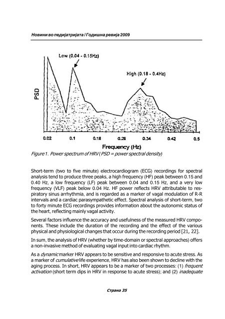
- Page 36 and 37: Nada Pop-Jordanova response (long-t
- Page 38 and 39: Nada Pop-Jordanova In the following
- Page 40 and 41: Nada Pop-Jordanova From Fig. 4 it i
- Page 42 and 43: Nada Pop-Jordanova References: [1]
- Page 44 and 45: Nada Pop-Jordanova [22] Friesen GM,
- Page 46 and 47: Mirjana Ko~ova Ha[imoto tiroiditis
- Page 48 and 49: Mirjana Ko~ova Slika 2. Devoj~e na
- Page 50 and 51: Mirjana Ko~ova c) Ultrazvu~niot pre
- Page 52 and 53: Mirjana Ko~ova smetaat deka kaj ovi
- Page 54 and 55: Mirjana Ko~ova 10. Pituitary pseudo
- Page 56 and 57: Страна 56
- Page 58 and 59: Qup~o Nikolovski Summary: Regardles
- Page 60 and 61: Qup~o Nikolovski Sugeriran antibiot
- Page 62 and 63: Qup~o Nikolovski - Toj mo`e da se k
- Page 64 and 65: Qup~o Nikolovski Sugeriran re`im Pr
- Page 66 and 67: Qup~o Nikolovski decata so akcent n
- Page 68 and 69: Qup~o Nikolovski Antibiotici i dija
- Page 70 and 71: Страна 70
- Page 72 and 73: Sowa Peova Abstrakt Efikasno rano o
- Page 74 and 75: Sowa Peova Ranoto klini~ko prepozna
- Page 76 and 77: Sowa Peova - In vitro analiza na in
- Page 78 and 79: Sowa Peova metodi pred da se pojava
- Page 80 and 81: Sowa Peova Страна 80
- Page 82 and 83: Страна 82
- Page 84 and 85:
Dafina Kuzmanovska bubre`na insufic
- Page 86 and 87:
Dafina Kuzmanovska 1. Abnormalen ra
- Page 88 and 89:
Dafina Kuzmanovska Isto taka, nema
- Page 90 and 91:
Dafina Kuzmanovska 11. Feather AS,
- Page 92 and 93:
Aco Kostovski na holestaza ne se pr
- Page 94 and 95:
Aco Kostovski dukcijata na SREBP -
- Page 96 and 97:
Aco Kostovski niraat sintezata na l
- Page 98 and 99:
Aco Kostovski lema neselektirana st
- Page 100 and 101:
Aco Kostovski 2. Tip 1 ili 2 diabet
- Page 102 and 103:
Aco Kostovski parcijalna lipodistro
- Page 104 and 105:
Aco Kostovski patis od drugi mo`ni
- Page 106 and 107:
Aco Kostovski Lipoproteini so mala
- Page 108 and 109:
Aco Kostovski kontrola na obeznosta
- Page 110 and 111:
Aco Kostovski za razlika od general
- Page 112 and 113:
Aco Kostovski NEALKOHOLNA MASTNA BO
- Page 114 and 115:
Lekovi] Z. SY GILBERT IN CHILDREN:
- Page 116 and 117:
Страна 116
- Page 118 and 119:
Aspazija Sofijanova ABSTRAKT Mnogu
- Page 120 and 121:
Aspazija Sofijanova en~iwa. Zatoa d
- Page 122 and 123:
Aspazija Sofijanova Odgovor na Krat
- Page 124 and 125:
Aspazija Sofijanova cira so seriozn
- Page 126 and 127:
Aspazija Sofijanova sporo. Hepati~n
- Page 128 and 129:
Aspazija Sofijanova asociran so pos
- Page 130 and 131:
Aspazija Sofijanova 16. Meyer PG, O
- Page 132 and 133:
Aspazija Sofijanova 48. Goldman HB.
- Page 134 and 135:
Страна 134
- Page 136 and 137:
Du[ko Fidanovski Abstract With intr
- Page 138 and 139:
Du[ko Fidanovski dilna te`ina od 50
- Page 140 and 141:
Du[ko Fidanovski koncentracii pomal
- Page 142 and 143:
Du[ko Fidanovski Lekuvawe so surfak
- Page 144 and 145:
Du[ko Fidanovski EMRT i golem rizik
- Page 146 and 147:
Du[ko Fidanovski 17. Kotecha S, Wil
- Page 148 and 149:
Du[ko Fidanovski 46. Corbet A, Long
- Page 150 and 151:
Страна 150
- Page 152 and 153:
Filip Duma fluid. Precise diagnose
- Page 154 and 155:
Filip Duma rata i mozo~noto tkivo,
- Page 156 and 157:
Filip Duma Diskusija i rezultati: K
- Page 158 and 159:
Kata Martinova endokrinite `lezdi i
- Page 160 and 161:
Kata Martinova Evaluacija i monitor
- Page 162 and 163:
Kata Martinova SQUID (superconducti
- Page 164 and 165:
Kata Martinova sostojba da odr`uva
- Page 166 and 167:
Kata Martinova 10. Grady RW., Berdo
- Page 168 and 169:
Vladimir ^adikovski The computer th
- Page 170 and 171:
Vladimir ^adikovski 5. Should peopl
- Page 172 and 173:
Nadica Ristoska Bojkovska Istorijat
- Page 174 and 175:
Nadica Ristoska Bojkovska � Virus
- Page 176 and 177:
Nadica Ristoska Bojkovska na CMV in
- Page 178 and 179:
Nadica Ristoska Bojkovska Takrolimu
- Page 180 and 181:
Nadica Ristoska Bojkovska Literatur
- Page 182 and 183:
Velibor Tasi] Hipourikemija e labor
- Page 184 and 185:
Velibor Tasi] Ovoj lek go zgolemuva
- Page 186 and 187:
Velibor Tasi] diabetes mellitus, ma
- Page 188 and 189:
Страна 188
- Page 190 and 191:
Emilija Vla[ki diagnostic investiga
- Page 192 and 193:
Emilija Vla[ki pnevmociti so difuzn
- Page 194 and 195:
Emilija Vla[ki dijagnosti~koto zna~
- Page 196 and 197:
Emilija Vla[ki na~eni promeni treba
- Page 198 and 199:
Katarina Stavri] na prisustvoto na
- Page 200 and 201:
Katarina Stavri] fagite i eozinofil
- Page 202 and 203:
Katarina Stavri] telite i decata do
- Page 204 and 205:
Страна 204
- Page 206 and 207:
Katerina Obo~ki What is the Practal
- Page 208 and 209:
Katerina Obo~ki Tret fakt: izlo`eno
- Page 210 and 211:
Katerina Obo~ki - izbegnuvawe na ek
- Page 212:
Katerina Obo~ki Zaklu~ok: Sekoga[ t