

Self-Assembly of Synthetic and Biological Polymeric Systems of ...
Self-Assembly of Synthetic and Biological Polymeric Systems of ...
Self-Assembly of Synthetic and Biological Polymeric Systems of ...

Create successful ePaper yourself
Turn your PDF publications into a flip-book with our unique Google optimized e-Paper software.
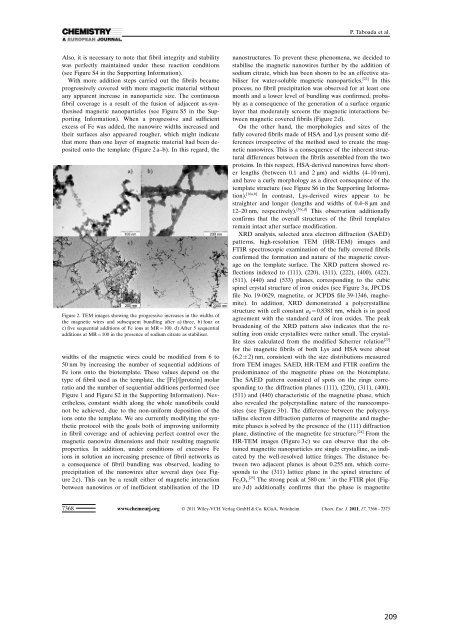
Also, it is necessary to note that fibril integrity <strong>and</strong> stability<br />
was perfectly maintained under these reaction conditions<br />
(see Figure S4 in the Supporting Information).<br />
With more addition steps carried out the fibrils became<br />
progressively covered with more magnetic material without<br />
any apparent increase in nanoparticle size. The continuous<br />
fibril coverage is a result <strong>of</strong> the fusion <strong>of</strong> adjacent as-synthesised<br />
magnetic nanoparticles (see Figure S5 in the Supporting<br />
Information). When a progressive <strong>and</strong> sufficient<br />
excess <strong>of</strong> Fe was added, the nanowire widths increased <strong>and</strong><br />
their surfaces also appeared rougher, which might indicate<br />
that more than one layer <strong>of</strong> magnetic material had been deposited<br />
onto the template (Figure 2 a–b). In this regard, the<br />
Figure 2. TEM images showing the progressive increases in the widths <strong>of</strong><br />
the magnetic wires <strong>and</strong> subsequent bundling after a) three, b) four or<br />
c) five sequential additions <strong>of</strong> Fe ions at MR = 100. d) After 5 sequential<br />
additions at MR = 100 in the presence <strong>of</strong> sodium citrate as stabiliser.<br />
widths <strong>of</strong> the magnetic wires could be modified from 6 to<br />
50 nm by increasing the number <strong>of</strong> sequential additions <strong>of</strong><br />
Fe ions onto the biotemplate. These values depend on the<br />
type <strong>of</strong> fibril used as the template, the [Fe]/ACHTUNGTRENUNG[protein] molar<br />
ratio <strong>and</strong> the number <strong>of</strong> sequential additions performed (see<br />
Figure 1 <strong>and</strong> Figure S2 in the Supporting Information). Nevertheless,<br />
constant width along the whole nan<strong>of</strong>ibrils could<br />
not be achieved, due to the non-uniform deposition <strong>of</strong> the<br />
ions onto the template. We are currently modifying the synthetic<br />
protocol with the goals both <strong>of</strong> improving uniformity<br />
in fibril coverage <strong>and</strong> <strong>of</strong> achieving perfect control over the<br />
magnetic nanowire dimensions <strong>and</strong> their resulting magnetic<br />
properties. In addition, under conditions <strong>of</strong> excessive Fe<br />
ions in solution an increasing presence <strong>of</strong> fibril networks as<br />
a consequence <strong>of</strong> fibril bundling was observed, leading to<br />
precipitation <strong>of</strong> the nanowires after several days (see Figure<br />
2 c). This can be a result either <strong>of</strong> magnetic interaction<br />
between nanowires or <strong>of</strong> inefficient stabilisation <strong>of</strong> the 1D<br />
7368<br />
P. Taboada et al.<br />
nanostructures. To prevent these phenomena, we decided to<br />
stabilise the magnetic nanowires further by the addition <strong>of</strong><br />
sodium citrate, which has been shown to be an effective stabiliser<br />
for water-soluble magnetic nanoparticles. [22] In this<br />
process, no fibril precipitation was observed for at least one<br />
month <strong>and</strong> a lower level <strong>of</strong> bundling was confirmed, probably<br />
as a consequence <strong>of</strong> the generation <strong>of</strong> a surface organic<br />
layer that moderately screens the magnetic interactions between<br />
magnetic covered fibrils (Figure 2 d).<br />
On the other h<strong>and</strong>, the morphologies <strong>and</strong> sizes <strong>of</strong> the<br />
fully covered fibrils made <strong>of</strong> HSA <strong>and</strong> Lys present some differences<br />
irrespective <strong>of</strong> the method used to create the magnetic<br />
nanowires. This is a consequence <strong>of</strong> the inherent structural<br />
differences between the fibrils assembled from the two<br />
proteins. In this respect, HSA-derived nanowires have shorter<br />
lengths (between 0.1 <strong>and</strong> 2 mm) <strong>and</strong> widths (4–10 nm),<br />
<strong>and</strong> have a curly morphology as a direct consequence <strong>of</strong> the<br />
template structure (see Figure S6 in the Supporting Information).<br />
[16a,b] In contrast, Lys-derived wires appear to be<br />
straighter <strong>and</strong> longer (lengths <strong>and</strong> widths <strong>of</strong> 0.4–8 mm <strong>and</strong><br />
12–20 nm, respectively). [16c,d] This observation additionally<br />
confirms that the overall structures <strong>of</strong> the fibril templates<br />
remain intact after surface modification.<br />
XRD analysis, selected area electron diffraction (SAED)<br />
patterns, high-resolution TEM (HR-TEM) images <strong>and</strong><br />
FTIR spectroscopic examination <strong>of</strong> the fully covered fibrils<br />
confirmed the formation <strong>and</strong> nature <strong>of</strong> the magnetic coverage<br />
on the template surface. The XRD pattern showed reflections<br />
indexed to (111), (220), (311), (222), (400), (422),<br />
(511), (440) <strong>and</strong> (533) planes, corresponding to the cubic<br />
spinel crystal structure <strong>of</strong> iron oxides (see Figure 3 a, JPCDS<br />
file No. 19-0629, magnetite, or JCPDS file 39-1346, maghemite).<br />
In addition, XRD demonstrated a polycrystalline<br />
structure with cell constant a 0 = 0.8381 nm, which is in good<br />
agreement with the st<strong>and</strong>ard card <strong>of</strong> iron oxides. The peak<br />
broadening <strong>of</strong> the XRD pattern also indicates that the resulting<br />
iron oxide crystallites were rather small. The crystallite<br />
sizes calculated from the modified Scherrer relation [23]<br />
for the magnetic fibrils <strong>of</strong> both Lys <strong>and</strong> HSA were about<br />
(6.2 2) nm, consistent with the size distributions measured<br />
from TEM images. SAED, HR-TEM <strong>and</strong> FTIR confirm the<br />
predominance <strong>of</strong> the magnetite phase on the biotemplate.<br />
The SAED pattern consisted <strong>of</strong> spots on the rings corresponding<br />
to the diffraction planes (111), (220), (311), (400),<br />
(511) <strong>and</strong> (440) characteristic <strong>of</strong> the magnetite phase, which<br />
also revealed the polycrystalline nature <strong>of</strong> the nanocomposites<br />
(see Figure 3 b). The difference between the polycrystalline<br />
electron diffraction patterns <strong>of</strong> magnetite <strong>and</strong> maghemite<br />
phases is solved by the presence <strong>of</strong> the (111) diffraction<br />
plane, distinctive <strong>of</strong> the magnetite fcc structure. [24] From the<br />
HR-TEM images (Figure 3 c) we can observe that the obtained<br />
magnetite nanoparticles are single crystalline, as indicated<br />
by the well-resolved lattice fringes. The distance between<br />
two adjacent planes is about 0.255 nm, which corresponds<br />
to the (311) lattice plane in the spinel structure <strong>of</strong><br />
Fe 3O 4. [25] The strong peak at 580 cm 1 in the FTIR plot (Figure<br />
3 d) additionally confirms that the phase is magnetite<br />
www.chemeurj.org 2011 Wiley-VCH Verlag GmbH & Co. KGaA, Weinheim Chem. Eur. J. 2011, 17, 7366 – 7373


