1st Joint ESMAC-GCMAS Meeting - Análise de Marcha
1st Joint ESMAC-GCMAS Meeting - Análise de Marcha
1st Joint ESMAC-GCMAS Meeting - Análise de Marcha
Create successful ePaper yourself
Turn your PDF publications into a flip-book with our unique Google optimized e-Paper software.
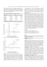
Table 1. Subject <strong>de</strong>mographics<br />
Group Age pre N<br />
(post #1)<br />
Follow-up time<br />
(post #1)<br />
- 185 -<br />
N<br />
(post #2)<br />
Follow-up time<br />
(post #2)<br />
S1 6.8 (2.3) 28 1.1 (0.3) 7 3.6 (0.6)<br />
S2 5.5 (1.3) 69 1.2 (0.7) 20 3.5 (0.5)<br />
Key: mean (standard <strong>de</strong>viation), all times in years.<br />
Discussion<br />
This study showed that response of subjects to L1-S1 and L1-S2 SDR surgery was equivalent for<br />
a specific set of kinematic and spasticity outcome measures. There has been significant<br />
controversy over whether S2 rootlets should be inclu<strong>de</strong>d in SDR surgery. Supporting S2<br />
inclusion was Lang’s study showing that sparing S2 left residual plantarflexor spasticity.<br />
Opposing S2 inclusion was Molenaers’ study showing that including S2 promoted crouch gait.<br />
This study appears to contradict both of those prior studies. The data analyzed here shows<br />
equivalent outcomes, between S1 and S2 surgeries, for both plantarflexor spasticity and selected<br />
kinematic variables. The explanation for this seems to lie in clinical principles un<strong>de</strong>rlying the<br />
surgical technique as applied here. For the majority of subjects, rootlets <strong>de</strong>monstrating<br />
pathological electrophysiology were sectioned while rootlets with appropriate response were<br />
spared (n.b. some subjects did have S2 rootlets spared for a variety of reasons not directly related<br />
to electrophysiological response). Clearly the question of including vs. sparing S2 rootlets<br />
remains unresolved. Further analysis into long(er) term outcomes, including foot-related<br />
outcomes, is warranted.<br />
Maximum Dorsiflexion [<strong>de</strong>g]<br />
Knee Flexion @ Initial Contact [<strong>de</strong>g]<br />
15<br />
10<br />
5<br />
0<br />
-5<br />
45<br />
40<br />
35<br />
30<br />
25<br />
20<br />
15<br />
10<br />
5<br />
0<br />
typical<br />
pre pst #1 pst #2<br />
typical<br />
pre pst #1 pst #2<br />
Minimum Hip Flexion [<strong>de</strong>g]<br />
Minimum Knee Flexion [<strong>de</strong>g]<br />
10<br />
5<br />
0<br />
-5<br />
20<br />
15<br />
10<br />
5<br />
0<br />
typical<br />
pre pst #1 pst #2<br />
typical<br />
References<br />
1.Lang FF et al., Neurosurgery, 34:847-853<br />
2.Molenaers G, et al., 13 th <strong>ESMAC</strong>, Warsaw, 2004.<br />
3. Schwartz et al. 14 th <strong>ESMAC</strong>, Barcelona, 2005<br />
pre pst #1 pst #2<br />
Pelvic Tilt [<strong>de</strong>g]<br />
25<br />
20<br />
15<br />
10<br />
5<br />
0<br />
pre pst #1 pst #2<br />
S1<br />
S2<br />
typical<br />
Figure 1. Mean and 95%<br />
confi<strong>de</strong>nce interval for<br />
kinematic outcome measures are<br />
shown. No significant<br />
interactions were found. The<br />
trends that appear (minimum hip<br />
flexion and knee flexion and<br />
maximum dorsiflexion) favor<br />
the S2 group. Disconcertingly,<br />
mean pelvic tilt showed a<br />
<strong>de</strong>teriorating trend in both<br />
groups and both intervals.


