1st Joint ESMAC-GCMAS Meeting - Análise de Marcha
1st Joint ESMAC-GCMAS Meeting - Análise de Marcha
1st Joint ESMAC-GCMAS Meeting - Análise de Marcha
You also want an ePaper? Increase the reach of your titles
YUMPU automatically turns print PDFs into web optimized ePapers that Google loves.
O-40<br />
SAGITTAL PLANE LOWER EXTREMITY KINEMATICS IN CHILDREN WITH<br />
HEREDITARY SPASTIC PARAPLEGIA<br />
Nichols, Molly, MPT 1 , Dorociak, Robin, BS 1 , and Aiona, Michael, MD 1<br />
1 Shriners Hospitals for Children, Portland, Oregon, United States<br />
Summary/conclusions<br />
Children with Hereditary Spastic Paraplegia (HSP) <strong>de</strong>monstrate one of four sagittal plane gait<br />
patterns distinct from children with spastic diplegic cerebral palsy as revealed by computerized<br />
gait analysis of the lower extremities. Further analysis with additional subjects and upper<br />
extremity kinematics will substantiate these patterns.<br />
Introduction<br />
Hereditary Spastic Paraplegia refers to a group of more than 30 inherited neurologic disor<strong>de</strong>rs<br />
in which the predominant characteristic is lower extremity spasticity resulting in an atypical<br />
gait pattern [1]. Since the first clinical <strong>de</strong>scriptions of HSP by Strümpell in 1880, few studies<br />
have been published <strong>de</strong>scribing the gait patterns of HSP [2] and to date minimal kinematic<br />
information exists for the pediatric population.<br />
Statement of clinical significance<br />
Defining sagittal plane gait patterns specific to children with HSP will allow the clinician to<br />
differentiate between this population and children with spastic diplegic cerebral palsy, better<br />
directing diagnosis and treatment. The use of computerized gait analysis for children with HSP<br />
reveals patterns that cannot be distinguished by visual gait inspection.<br />
Methods<br />
A retrospective chart review was performed for 18 patients (10 female, 8 male; mean age 12.7<br />
yrs, range 3.9 to 18.7 yrs) with HSP who un<strong>de</strong>rwent routine clinical gait analysis. The<br />
diagnosis of HSP was confirmed by family history (14 patients) or clinically confirmed by a<br />
neurologist (4 patients). Patients inclu<strong>de</strong>d were in<strong>de</strong>pen<strong>de</strong>nt ambulators without orthoses or<br />
prior surgical intervention. Computerized gait analysis was performed using a VICON motion<br />
system. Patient gait trials were processed using Vicon Clinical Manager. One representative<br />
trial for each of the right and left si<strong>de</strong>s was chosen for analysis. Because it was noted that the<br />
patients had symmetric patterns, a representative si<strong>de</strong> was chosen for illustration. Utilizing the<br />
gait classification system <strong>de</strong>scribed by Rodda [3], six clinicians experienced in gait analysis<br />
attempted to classify the HSP patients into the diplegic gait patterns. As 11/18 HSP patients did<br />
not fit into any of these categories, the purpose of this study was to <strong>de</strong>termine whether patterns<br />
specific to HSP could be differentiated.<br />
Results<br />
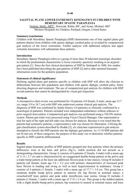
Sagittal plane kinematic profiles of HSP patients grouped into four patterns where the primary<br />
differences were at the knee and pelvis (fig.1). Ankle position did not present as a<br />
distinguishing characteristic. Group A consists of 6 patients (2 female, 4 male) with a mean age<br />
of 11.4 ± 2.7 yrs and is characterized by an exaggerated double-bump pattern at the pelvis and<br />
a triple-bump pattern at the knee (an additional flexion peak in late stance). Group B inclu<strong>de</strong>s 4<br />
patients (all female, mean age 5.4 ± 2.2 yrs) with primary characteristics of increased peak<br />
knee flexion in loading and swing as well as knee hyperextension in midstance. Group C<br />
consists of 4 patients (1 female, 3 male) with a mean age of 17.7 ± 1.0 yrs. This group has a<br />
mo<strong>de</strong>rate double bump pelvic pattern in anterior tilt, hip flexion in terminal stance, a<br />
crouched/stiff knee pattern and peak ankle dorsiflexion near norms. Group D inclu<strong>de</strong>s 4<br />
patients (3 female, 1 male) with a mean age of 17.0 ± 2.4 yrs. This group is the mil<strong>de</strong>st pattern<br />
with a slight double-bump pelvic pattern, hip extension to neutral in terminal stance and knee<br />
- 140 -


