

1st Joint ESMAC-GCMAS Meeting - Análise de Marcha
1st Joint ESMAC-GCMAS Meeting - Análise de Marcha
1st Joint ESMAC-GCMAS Meeting - Análise de Marcha

Create successful ePaper yourself
Turn your PDF publications into a flip-book with our unique Google optimized e-Paper software.
O-26<br />
CLINICALLY SIGNIFICANT SHOULDER JOINT KINEMATICS DESCRIPTION<br />
Morrow, Duane A, MS, Lin, Hui-Ting, MS, Kaufman, Kenton R, PhD, An, Kai-Nan, PhD<br />
Department of Orthopedic Research, Mayo Clinic, Rochester, MN, USA<br />
Summary/conclusions<br />
While there are numerous methods available for the calculation of shoul<strong>de</strong>r joint kinematics,<br />
the ability to present these in clinically significant terms has remained a challenge. Through<br />
realignment of coordinate axes on the humerus, shoul<strong>de</strong>r kinematics can be calculated in terms<br />
that are familiar in clinical settings.<br />
Introduction<br />
The ability to <strong>de</strong>scribe calculated shoul<strong>de</strong>r joint kinematics in clinically relevant terms has, to<br />
date, been difficult. This difficulty has likely been compoun<strong>de</strong>d by the plethora of methods<br />
used to <strong>de</strong>compose the rotation of the shoul<strong>de</strong>r joint. Rotations captured using motion analysis<br />
techniques are analysed most wi<strong>de</strong>ly using either Euler angle or screw axis <strong>de</strong>composition.<br />
While those who use Euler angles in their analyses may choose to follow the rotation sequence<br />
initially proposed by An [1] and recommen<strong>de</strong>d by the ISB [2], This method, however, offers<br />
shoul<strong>de</strong>r motion results in terms of the angle and amount of humeral elevation in lieu of<br />
outright measures of flexion/extension or ab/adduction. Additionally, in the clinical setting,<br />
neural, muscular, or skeletal disor<strong>de</strong>rs may cause individuals to take up postures and<br />
compensations that may make recommen<strong>de</strong>d coordinate assignments or <strong>de</strong>compositions<br />
provi<strong>de</strong> unwanted results. Our laboratory has previously advocated the use of screw-axes for<br />
the analysis of shoul<strong>de</strong>r motion to avoid some of the pitfalls of gimbal lock that may be<br />
associated with Euler angle <strong>de</strong>composition [3]. However, the problem still remains that<br />
shoul<strong>de</strong>r flexion/extension and ab/adduction are measures of the humerus about axes of the<br />
trunk. Internal/external rotation of the humerus can change the alignment of the humeral<br />
sagittal and coronal axes, confounding the ability to examine the motion of the humerus with<br />
respect to the trunk (or scapula, for glenohumeral motion). The current study outlines a method<br />
for presenting shoul<strong>de</strong>r kinematics in terms of clinical rotations.<br />
Statement of clinical significance<br />
A simple correction algorithm may be applied to<br />
the <strong>de</strong>composition of shoul<strong>de</strong>r joint kinematics<br />
to allow for clinically meaningful interpretation<br />
of shoul<strong>de</strong>r joint motions.<br />
Methods<br />
This study was conducted as part of a larger<br />
study for the creation of a database of normal<br />
kinematics during a set of motions used in<br />
clinical upper extremity assessments of the<br />
neck, scapulothoracic, glenohumeral, elbow,<br />
and wrist joints, as well as the thoracohumeral<br />
pseudo-joint. Subjects were asked to perform<br />
select motions (full shoul<strong>de</strong>r flexion, abduction,<br />
touching their head, touching their opposite<br />
shoul<strong>de</strong>r, etc.) in a comfortable motion. For the purposes of this discussion, only motion of the<br />
humerus with respect to the thorax during shoul<strong>de</strong>r flexion will be consi<strong>de</strong>red, due to space<br />
constraints. The motion of retro-reflective markers was tracked at 60 Hz using a 10-camera<br />
Motion Analysis EVa RealTime system (Motion Analysis Corp., Santa Clara, CA, USA).<br />
Three markers, on the xiphoid process, sternal notch, and C7, were used to <strong>de</strong>fine the trunk<br />
- 104 -<br />
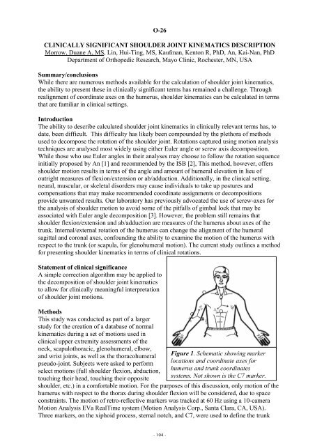
Figure 1. Schematic showing marker<br />
locations and coordinate axes for<br />
humerus and trunk coordinates<br />
systems. Not shown is the C7 marker.


