2008 - Communication Across the Curriculum (CAC)
2008 - Communication Across the Curriculum (CAC)
2008 - Communication Across the Curriculum (CAC)

You also want an ePaper? Increase the reach of your titles
YUMPU automatically turns print PDFs into web optimized ePapers that Google loves.
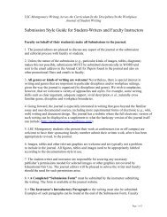
Figure 2. This picture shows <strong>the</strong> difference<br />
between a normal heart (left) and one with<br />
left hypertrophy (right).<br />
R e s e a r c h P a p e r<br />
muscle mass, but <strong>the</strong> blood<br />
pumping remains <strong>the</strong> same<br />
(Montoya 233). Besides <strong>the</strong><br />
lymph nodes and heart, <strong>the</strong><br />
parasite can infect <strong>the</strong> liver,<br />
spleen, and bone marrow,<br />
causing megalias and<br />
anemia. The parasite can<br />
also invade <strong>the</strong> lungs and<br />
sometimes it can produce<br />
meningoencephalitis which<br />
http://images.google.com/<br />
is a condition that is marked<br />
by <strong>the</strong> brain and meninges becoming inflamed (Restrepo et al<br />
607).<br />
Most of <strong>the</strong> patients survive <strong>the</strong> acute phase, enter <strong>the</strong><br />
asymptomatic or latent phase, and <strong>the</strong>n <strong>the</strong> infection becomes<br />
chronic. It’s known that in <strong>the</strong> chronic phase 30% of <strong>the</strong> patients<br />
have cardiac damage. The principal clinic manifestation is<br />
myocarditis, which can be asymptomatic for a long period<br />
of time. Progressively, congestive cardiac insufficiency is<br />
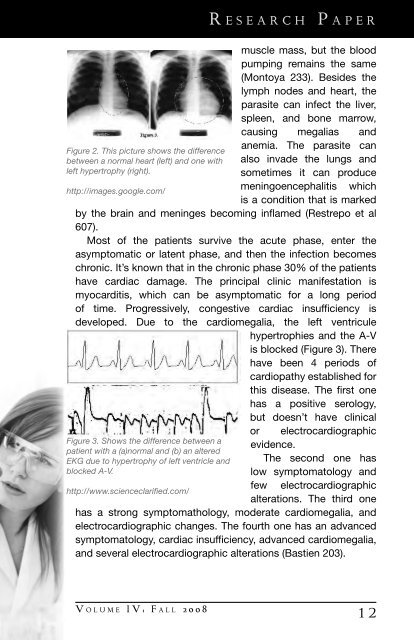
developed. Due to <strong>the</strong> cardiomegalia, <strong>the</strong> left ventricule<br />
hypertrophies and <strong>the</strong> A-V<br />
is blocked (Figure 3). There<br />
have been 4 periods of<br />
cardiopathy established for<br />
this disease. The first one<br />
has a positive serology,<br />
but doesn’t have clinical<br />
or electrocardiographic<br />
Figure 3. Shows <strong>the</strong> difference between a evidence.<br />
patient with a (a)normal and (b) an altered<br />
EKG due to hypertrophy of left ventricle and The second one has<br />
blocked A-V.<br />
low symptomatology and<br />
few electrocardiographic<br />
http://www.scienceclarified.com/<br />
alterations. The third one<br />
has a strong symptomathology, moderate cardiomegalia, and<br />
electrocardiographic changes. The fourth one has an advanced<br />
symptomatology, cardiac insufficiency, advanced cardiomegalia,<br />
and several electrocardiographic alterations (Bastien 203).<br />
V o l u m e I V : F a l l 2 0 0 8<br />
1 2