Pharmaceutical Technology: Controlled Drug Release, Volume 2
Pharmaceutical Technology: Controlled Drug Release, Volume 2
Pharmaceutical Technology: Controlled Drug Release, Volume 2

You also want an ePaper? Increase the reach of your titles
YUMPU automatically turns print PDFs into web optimized ePapers that Google loves.
CH. 9] POLYACRYLATE (EUDRAGIT RETARD) MICROSPHERES 109<br />
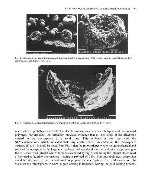
Fig. 4—Scanning electron micrograph of nifedipine-loaded microspheres (31% w/w) at various magnifications. For<br />
experimental conditions, see Fig. 1.<br />
Fig. 5—Scanning electron micrograph of a fractured nifedipine-loaded microsphere (31% w/w).<br />
microspheres, probably as a result of molecular interactions between nifedipine and the Eudragit<br />
polymers. Nevertheless, this inflection provided evidence that at least some of the nifedipine<br />
existed in the microsphere in a solid state. This evidence is consistent with the<br />
SEM examinations, which indicated that drug crystals were embedded on the microsphere<br />
surfaces (Fig. 4). It could be noted from Fig. 4 that the microspheres where not quitespherical and<br />
some of them, especially the large microspheres, collapsed and lost their spherical shape owing to<br />
the existence of an internal void volume as evidenced by Fig. 5, exhibiting the internal structure of<br />
a fractured nifedipine microsphere having a payload of 31%. This morphological depression<br />
could be attributed to the method used to prepare the microspheres for SEM evaluation. To<br />
visualize the microsphere, in SEM, a gold coating is imparted. During the gold coating process,