

Soybean and Bees

Create successful ePaper yourself
Turn your PDF publications into a flip-book with our unique Google optimized e-Paper software.
from genetic male sterile plants (msl,msl) in soybeans. Comparisons were made between<br />
the average numbers of pollen grains <strong>and</strong> the average numbers of coenocytic microspores<br />
with respect to environment where plants were grown, <strong>and</strong> to stamen position in<br />
the flower. Pollen production from fertile plants varied from 374 to 760 pollen grains per<br />
anther, among genetic lines <strong>and</strong> environments.<br />
Photo: Decio Luiz Gazzoni<br />
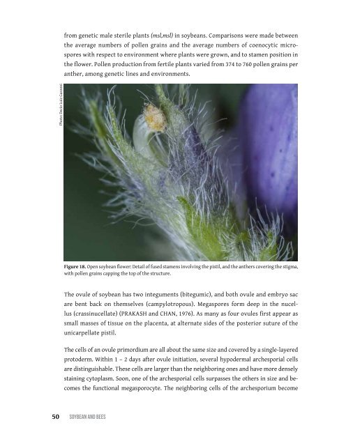
Figure 18. Open soybean flower: Detail of fused stamens involving the pistil, <strong>and</strong> the anthers covering the stigma,<br />
with pollen grains capping the top of the structure.<br />
The ovule of soybean has two integuments (bitegumic), <strong>and</strong> both ovule <strong>and</strong> embryo sac<br />
are bent back on themselves (campylotropous). Megaspores form deep in the nucellus<br />
(crassinucellate) (Prakash <strong>and</strong> Chan, 1976). As many as four ovules first appear as<br />
small masses of tissue on the placenta, at alternate sides of the posterior suture of the<br />
unicarpellate pistil.<br />
The cells of an ovule primordium are all about the same size <strong>and</strong> covered by a single-layered<br />
protoderm. Within 1 – 2 days after ovule initiation, several hypodermal archesporial cells<br />
are distinguishable. These cells are larger than the neighboring ones <strong>and</strong> have more densely<br />
staining cytoplasm. Soon, one of the archesporial cells surpasses the others in size <strong>and</strong> becomes<br />
the functional megasporocyte. The neighboring cells of the archesporium become<br />
50 SoybeAn <strong>and</strong> bees