

Anorectal Manometry in 3D NEW! - Swiss-knife.org
Anorectal Manometry in 3D NEW! - Swiss-knife.org
Anorectal Manometry in 3D NEW! - Swiss-knife.org

You also want an ePaper? Increase the reach of your titles
YUMPU automatically turns print PDFs into web optimized ePapers that Google loves.
erative complications. Overall morbidity was 2.5%. With a median follow up of 14 months, 1 Clavien<br />
grade I (hemoptysis), 2 grade II (1 pneumonia and 1 ombilical seroma) and 2 grade III (1 bowel<br />
anastomosis leak and 1 umbilical <strong>in</strong>cisional hernia) complications occurred. All patients were recommend<strong>in</strong>g<br />
this approach and extremely satisfied with cosmetic results after LESS procedure.<br />
Conclusion: LESS visceral surgery is feasible for advanced laparoscopic surgeons but rema<strong>in</strong> difficult<br />
<strong>in</strong> relation to its specificities. Technical developments are especially needed to improve feasibility and<br />
thus safety of advanced visceral LESS procedures. It offers improved cosmesis, and may offer decreased<br />
pa<strong>in</strong> and shorter complete recovery. Complications are consistent with the published data<br />
for laparoscopic visceral surgery. Whether LESS surgery represents a progress <strong>in</strong> m<strong>in</strong>imally <strong>in</strong>vasive<br />
surgery rema<strong>in</strong> to be demonstrated formally <strong>in</strong> formal randomized trials.<br />
33.5<br />
Transanale Tumorabtragung mittels S<strong>in</strong>gle-Port-Video-Rektoskopie – e<strong>in</strong>e neue Anwendungsmöglichkeit<br />
für den “SILS“-Port<br />
B. Boldog, M. Weber (Schaffhausen)<br />
Objective: Transanale Tumorabtragungen bei hoch gelegenen Pathologien im Rektosigmoid s<strong>in</strong>d e<strong>in</strong>e<br />
chirurgisch technische Herausforderung. Die transanale endoskopische Mikrochirurgie (TME) ist zudem<br />
mit e<strong>in</strong>em hohen Geräteaufwand und e<strong>in</strong>er erheblichen Sph<strong>in</strong>kterdehnung verbunden. Transabdom<strong>in</strong>elle<br />
Verfahren s<strong>in</strong>d mit der Morbidität der tiefen Rektumchirurgie assoziert.<br />
Methods and Results: Anamnese und kl<strong>in</strong>ischer Befund: E<strong>in</strong> 67 Jahre alter Patient wird wegen rezidivierenden<br />
unteren gastro<strong>in</strong>test<strong>in</strong>alen Blutungen zugewiesen. Kolonoskopisch zeigt sich e<strong>in</strong> grosses<br />
breitbasiges Adenom (Histologie) 20 cm ab ano mit der Indikation zur chirurgischen transanalen<br />
Tumorexzision Methode: Patient <strong>in</strong> SSL <strong>in</strong> Intubationsnarkose. Unter anoskopischer Kontrolle platzieren<br />
e<strong>in</strong>es 20 french Foleykatheter oral von Tumor im Sigma. Füllen des Ballons unter Sicht mit 12 ml NaCl.<br />
Transanales E<strong>in</strong>setzen des „SILS“ Port-s (Covidien). Herstellen e<strong>in</strong>es Pneumo-Rectums mit CO 2 (10<br />
mmHg). Nach oral, neben dem Ballon <strong>in</strong>s Sigma durchtretendes CO 2 wird fortlaufend durch den offenen<br />
Foleykatheter nach transanal dekomprimiert. Unter Kamera-Kontrolle (5mm Optik) exakte Lokalisation<br />
des 20 mm grossen Tumors. Mittels „laparoskopischer“ 5 mm Fasszange und 10 mm Endo-GIA<br />
(Covidien) kann der Tumor tangential an der Basis im Gesunden abgetragen werden.<br />
Conclusion: SILS und NOTES ergeben unerwartete neue „sp<strong>in</strong>n-off“ Möglichkeiten <strong>in</strong> anderen Bereichen.<br />
Der Fallbericht zeigt e<strong>in</strong>e neue attraktive Anwendungsmöglichkeit des SILS-Ports. Die S<strong>in</strong>gle-<br />
Port-Video-Rektoskopie nach Boldog ist e<strong>in</strong>e neue schonende Operationstechnik.<br />
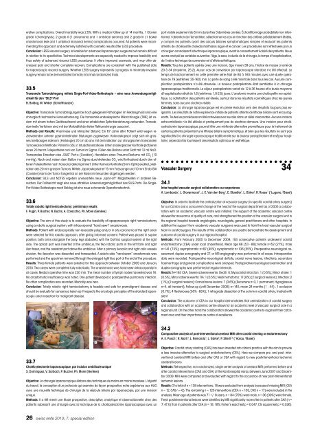
33.6<br />
Totally robotic right hemicolectomy: prelim<strong>in</strong>ary results<br />
F. Pug<strong>in</strong>, P. Bucher, N. Buchs, A. Carecchio, Ph. Morel (Genève)<br />
Objective: The aim of this study is to evaluate the feasibility of lapaparoscopic right hemicolectomy<br />
us<strong>in</strong>g a robotic surgical system, with <strong>in</strong>tracorporeal “hand sewn” anastomosis.<br />
Methods: Patient with endoscopically non-resecable polyp and/or <strong>in</strong> situ carc<strong>in</strong>oma of the right colon<br />
were selected for this robotic approach, after giv<strong>in</strong>g <strong>in</strong>formed consent. Patient was placed <strong>in</strong> sup<strong>in</strong>e<br />
position, both arms alongside the body, legs abducted, with the DaV<strong>in</strong>ci surgical system at the right<br />
side. The optical port was <strong>in</strong>serted at the umbilicus, the two robotic ports <strong>in</strong> the left flank and right<br />
iliac fossa, and the assistant port above the umbilicus. After a primary ileocolic and right colic vessels<br />
division, the ileocolon was dissected and transsected. A side-to-side “hand-sewn” anastomosis was<br />
performed and the specimen removed through the enlarged right iliac port at the end of the procedure.<br />
Results: Three female patients were selected for this approach between October 2009 and January<br />
2010. Two cases were completed fully robotically. The anastomosis was hand-sewn <strong>in</strong>tracorporally <strong>in</strong><br />
all cases. Median operative time was 230 m<strong>in</strong>. The mean number of lymph nodes harvested was 16.<br />
No anastomotic <strong>in</strong>sufficiency was noted. One patient developed a postoperative pulmonary <strong>in</strong>fection.<br />
No other complication was recorded. Mortality was zero.<br />
Conclusion: Totally robotic right hemicolectomy is feasible and safe for premalignant disease and<br />
should be evaluate for cancerous lesion as it respects the oncologic pr<strong>in</strong>ciples of the standard laparoscopic<br />
colon resection for malignant disease.<br />
33.7<br />
Cholécystectomie laparoscopique, par <strong>in</strong>cision ombilicale unique<br />
S. Dom<strong>in</strong>guez, V. Sarbach, P. Bucher, Ph. Morel (Genève)<br />
Objective: La chirurgie laparoscopique élabore des techniques de mo<strong>in</strong>s en mo<strong>in</strong>s <strong>in</strong>vasives. L’objectif<br />
du travail, la conception d‘un protocole qui exam<strong>in</strong>e de façon prospective notre expérience aux HUG<br />
avec une nouvelle technique de chirurgie de la vésicule biliaire par laparoscopie, par une <strong>in</strong>cision<br />
unique.<br />
Methods: Il a été mené une étude prospective, descriptive, analytique et observationnelle chez des<br />
patients subissant une chirurgie avec la technique de la cholécystectomie laparoscopique avec un<br />
26 swiss <strong>knife</strong> 2010; 7: special edition<br />
port visible seulement de 5 mm durant les 3 dernières années. Échantillonnage probabiliste non <strong>in</strong>tentionnel,<br />
l‘obtention de l‘échantillon, sélectionner les cas en fonction des critères préalablement établis,<br />
y compris les patients ayant des calculs biliaires symptomatiques simples et excluant les patients<br />
atte<strong>in</strong>ts de cholécystite choledocholithiasis aiguë et le cancer. Les procédures sont effectuées par un<br />
chirurgien connaissant la technique laparoscopique, avant le consentement éclairé des patients. Nous<br />
avons analysé les variables suivantes: l‘âge, le sexe, la durée de la chirurgie, le temps d‘hospitalisation,<br />
de l‘<strong>in</strong>dice technique de conversion et d‘effets esthétiques.<br />
Results: Tous les patients opérés avec une <strong>in</strong>cision, âge moyen 39 ans, l‘<strong>in</strong>dice de masse a varié de<br />
20 à 34 (moyenne, 25.2). Aucun cas de conversion par laparoscopie standard n’a été effectué. Le<br />
temps de fonctionnement en cette première série était de 80 à 160 m<strong>in</strong>utes avec une durée opératoire<br />
de 79 (extrêmes: 35-160) m<strong>in</strong>. La perte de sang a été m<strong>in</strong>imale dans tous les cas. Aucune complication<br />
postopératoire n’a été observée. La douleur postopératoire était semblable à la chirurgie<br />
laparoscopique traditionnelle. Le séjour postopératoire variait de 12 à 36 heures et la durée moyenne<br />
d‘hospitalisation était de 1,6 (extrêmes: 1,0-2,5) jours. L‘anatomie montre une cholécystite non spécifique.<br />
La satisfaction des patients est élevée, surtout dans les résultats cosmétiques chez les jeunes<br />
femmes, sans aucune cicatrice visible.<br />
Conclusion: La chirurgie laparoscopique est en ple<strong>in</strong>e évolution vers des résultats toujours plus exigeants.<br />
Les résultats de notre expérience <strong>in</strong>itiale de patients atte<strong>in</strong>ts de lithiase biliaire sont encourageants.<br />
Toutes les procédures ont été achevées avec succès dans un délai raisonnable. Aucune <strong>in</strong>cision<br />
extra-ombilicale n’a été utilisée et pratiquement pas de cicatrice demeure. Une <strong>in</strong>cision pour cholécystectomie<br />
laparoscopique, pourrait être une méthode alternative prometteuse pour le traitement de<br />
certa<strong>in</strong>s patients présentant une lithiase biliaire symptomatique, et bien que les résultats ne sont pas<br />
significatifs à la chirurgie laparoscopique traditionnelle sur la douleur postopératoire et le séjour hospitalier,<br />
cependant ils fournissent des résultats optimaux en esthétique.<br />
Vascular Surgery 34<br />
34.1<br />
Inter-hospital vascular surgical collaboration: our experience<br />
A. Lombardo 1 , L. Giovannacci 1 , J. C. Van den Berg 1 , C. Staedler 1 , L. Gürke 2 , R. Rosso 1 ( 1 Lugano, 2 Basel)<br />
Objective: In order to facilitate the centralization of vascular surgery (<strong>in</strong> specific carotid artery surgery)<br />
for our Canton and a concurrent change of the head of the surgical department as of 2005 a collaboration<br />
with an academic vascular centre was <strong>in</strong>itiated. The support of the academic vascular centre<br />
allowed for assurance of quality of care, and strengthened the position of the vascular surgical unit <strong>in</strong><br />
the regional hospital towards angiologists, neurologists, general practitioners and other hospitals. In<br />
addition the support from academic vascular surgeons was used to tra<strong>in</strong> the local vascular surgical<br />
team <strong>in</strong> carotid surgery. The results of this collaboration are used to demonstrate the development and<br />
outcome of carotid surgery <strong>in</strong> our regional hospital.<br />
Methods: From February 2005 to December 2009, 193 consecutive patients underwent carotid<br />
endarterectomy (CEA) under local anaesthesia. Mean age 68 (53 - 88); female n=52 (27%), male<br />
n=141 (73%); asymptomatic n=87 (45%); symptomatic n=106 (55%). Preoperative neurological assessment,<br />
duplex sonography and CT- or MR-angiography was performed <strong>in</strong> all cases. Intraoperative<br />
data were recorded. Postoperative neurological deficits, cranial nerve lesions, <strong>in</strong>fections, secondary<br />
haemorrhage and general complications were analysed. Postoperative neurological exam<strong>in</strong>ation and<br />
duplex sonography was performed at regular <strong>in</strong>tervals.<br />
Results: N=193 CEA, Severe adverse events: Death: 0; Myocardial <strong>in</strong>farction: 1 (0.5%); M<strong>in</strong>or stroke: 1<br />
(0.5%), M<strong>in</strong>or adverse events: TIA: 1 (0.5%); Neck hematoma: 17 (8%) (2 surgical revision); Infection: 2<br />
(1%) (2 surgical revision); Cranial nerve lesions: 7 (3.6%) (Recurrens n=3, 1 permanent, Hypoglossus<br />
n=4, all transient), Follow-up (until December 2008) n=140, mean 24 months (1 - 44) , 1 occlusion<br />
(0.7%); 4 Restenoses (50% - 65%); 1 retrograde dissection of the common carotid artery, treated with<br />
stent<br />
Conclusion: The outcome of CEA <strong>in</strong> our hospital demonstrates that centralization of carotid surgery<br />
and collaboration with an academic centre allows for an academic level of vascular surgical care <strong>in</strong> a<br />
regional unit. On the other hand the collaboration allowed the academic centre to augment their catchment<br />
area and their importance as centre of excellence.<br />
34.2<br />
Comparative analysis of post-<strong>in</strong>terventional cerebral MRI after carotid stent<strong>in</strong>g or endarterectomy<br />
A. E. Pasch 1 , R. Marti 1 , L. Remonda 1 , L. Gürke 2 , P. Stierli 1,2 ( 1 Aarau, 2 Basel)<br />
Objective: Carotid artery stent<strong>in</strong>g (CAS) has been <strong>in</strong>vented <strong>in</strong>to cl<strong>in</strong>ical practice with the aim to provide<br />
a less <strong>in</strong>vasive alternative to surgical endarterectomy (CEA). Here we compare pre- and post- <strong>in</strong>terventional<br />
cerebral MRI before and after CAS or CEA with regard to new post<strong>in</strong>terventional ischemic<br />
cerebral lesions.<br />
Methods: Retrospective, non randomized, s<strong>in</strong>gle center analysis of cerebral MRI performed before and<br />
after carotid <strong>in</strong>terventions (CAS and CEA) at the Kantonsspital Aarau between June 2007 and December<br />
2009. MRI were compared and evaluated with regard to the occurence of new post-<strong>in</strong>terventional<br />
ischemic lesions.<br />
Results: Of a total of n = 138 <strong>in</strong>terventions, 18 were excluded from analysis because of miss<strong>in</strong>g MRI (CEA<br />
n = 12, CAS n = 6). The rema<strong>in</strong><strong>in</strong>g n = 120 <strong>in</strong>terventions (CEA n = 103, CAS n = 17) were <strong>in</strong>cluded <strong>in</strong> the<br />
analysis. Mean age of patients was 71 +/- 8 years, n = 84 (70%) were male, n = 36 (30%) were female.<br />
Fresh post<strong>in</strong>terventional lesions were identified by MRI significantly more often <strong>in</strong> patients after CAS (n =<br />
7; 41%) than <strong>in</strong> patients after CEA (n = 18; 18%; Fisher’s exact test p = 0.047; Chi square test p = 0.026).


