

Anorectal Manometry in 3D NEW! - Swiss-knife.org
Anorectal Manometry in 3D NEW! - Swiss-knife.org
Anorectal Manometry in 3D NEW! - Swiss-knife.org

Create successful ePaper yourself
Turn your PDF publications into a flip-book with our unique Google optimized e-Paper software.
the anorectal part of the <strong>in</strong>test<strong>in</strong>e. All these lesions are accessible by perform<strong>in</strong>g a digital rectal exam<br />
and a bedside anorectoscopy +/- biopsy with a rigid anorectoscop. These two exam<strong>in</strong>ations are simple<br />
and fast, <strong>in</strong>expensive and safe and offer to obta<strong>in</strong> the right diagnosis <strong>in</strong> half of the patients with<br />
lower G-I-bleed<strong>in</strong>g. Additional time-consum<strong>in</strong>g and expensive <strong>in</strong>vestigations, as CT-scan, gastroscopy,<br />
angiography and others may be avoided.<br />
43.2<br />
Sacral nerve stimulation for faecal <strong>in</strong>cont<strong>in</strong>ence: loss of function considerably leads to <strong>in</strong>crease of<br />
costs<br />
D. Kisner, D. Hahnloser, D. D<strong>in</strong>do (Zurich)<br />
Objective: Sacral nerve stimulation (SNS) was repeatedly shown to be successful <strong>in</strong> the treatment of<br />
faecal <strong>in</strong>cont<strong>in</strong>ence and to be cost-effective. However, <strong>in</strong> some patients, the electrode and the implantable<br />
pulse generator (IPG) have to be removed. The aim of this study was to analyse the reasons for<br />
device removal. Moreover, the f<strong>in</strong>ancial burden of failed SNS was calculated.<br />
Methods: Consecutive patients with faecal <strong>in</strong>cont<strong>in</strong>ence, who underwent percutaneous nerve evaluation<br />
at the Department of Surgery of the University Hospital Zurich between 2001 and 2009, were evaluated.<br />
Data were prospectively recorded. For cost analyses, only direct costs have been considered.<br />
Results: One hundred and three patients (80 females, 23 males) underwent percutaneous nerve evaluation.<br />
Seventy-n<strong>in</strong>e patients (77%) with successful test-stimulation underwent permanent implantation<br />
of the IPG. After a median follow-up of 42 months (range 2-102), 69 of the 79 patients (87%) had<br />
a steady reduction <strong>in</strong> <strong>in</strong>cont<strong>in</strong>ence symptoms (improvement of >50% compared to basel<strong>in</strong>e). Dur<strong>in</strong>g<br />
follow-up, 22 IPG had to be removed (28%); <strong>in</strong> 10 patients, the whole device (electrode and IPG) was<br />
taken out (29%). Twelve patients underwent re-implantation of the IPG (17%). Reasons for the explantation<br />
of the IPG were <strong>in</strong>fection (n=2), loss of cl<strong>in</strong>ical effect (n=10) and malfunction of the IPG (n=8).<br />
Only 2 IPG were replaced due to loss of battery function (planned). In our cohort of 103 patients, the<br />
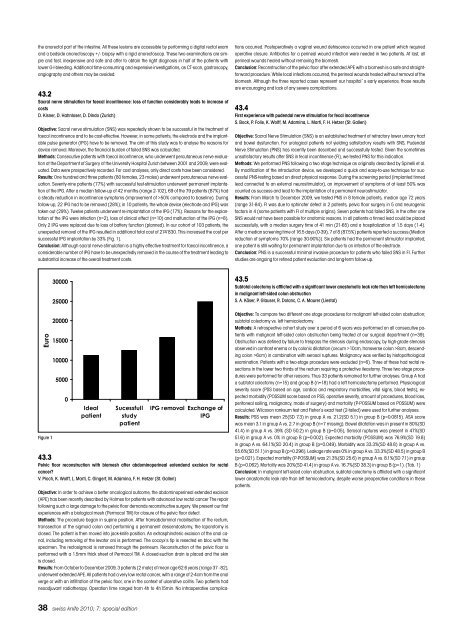
unexpected removal of the IPG resulted <strong>in</strong> additional total cost of 274’830. This <strong>in</strong>creased the cost per<br />
successful IPG implantation by 33% (Fig. 1).<br />
Conclusion: Although sacral nerve stimulation is a highly effective treatment for faecal <strong>in</strong>cont<strong>in</strong>ence, a<br />
considerable number of IPG have to be unexpectedly removed <strong>in</strong> the course of the treatment lead<strong>in</strong>g to<br />
substantial <strong>in</strong>crease of the overall treatment costs.<br />
�����<br />
Figure 1<br />
�����<br />
�����<br />
�����<br />
�����<br />
�����<br />
����<br />
�<br />
�����<br />
�������<br />
����������<br />
�����<br />
�������<br />
38 swiss <strong>knife</strong> 2010; 7: special edition<br />
����������� �����������<br />
���<br />
43.3<br />
Pelvic floor reconstruction with biomesh after abdom<strong>in</strong>oper<strong>in</strong>eal extendend excision for rectal<br />
cancer?<br />
V. Pioch, K. Wolff, L. Marti, C. G<strong>in</strong>gert, M. Adam<strong>in</strong>a, F. H. Hetzer (St. Gallen)<br />
Objective: In order to achieve a better oncological outcome, the abdom<strong>in</strong>oper<strong>in</strong>eal extended excision<br />
(APE) has been recently described by Holmes for patients with advanced low rectal cancer The repair<br />
follow<strong>in</strong>g such a large damage to the pelvic floor demands reconstructive surgery. We present our first<br />
experiences with a biological mesh (Permacol TM) for closure of the pelvic floor defect.<br />
Methods: The procedure began <strong>in</strong> sup<strong>in</strong>e position. After transabdom<strong>in</strong>al mobilisation of the rectum,<br />
transsection of the sigmoid colon and perform<strong>in</strong>g a permanent descendostomy, the laparotomy is<br />
closed. The patient is then moved <strong>in</strong>to jack-<strong>knife</strong> position. An extrasph<strong>in</strong>cteric excision of the anal canal,<br />
<strong>in</strong>clud<strong>in</strong>g remov<strong>in</strong>g of the levator ani is performed. The coccyx’s tip is resected en bloc with the<br />
specimen. The rectosigmoid is removed through the per<strong>in</strong>eum. Reconstruction of the pelvic floor is<br />
performed with a 1.5mm thick sheet of Permacol TM. A closed-suction dra<strong>in</strong> is placed and the sk<strong>in</strong><br />
is closed.<br />
Results: From October to December 2009, 3 patients (2 male) of mean age 62.6 years (range 37 - 82),<br />
underwent extended APE. All patients had a very low rectal cancer, with a range of 2-4cm from the anal<br />
verge or with an <strong>in</strong>filtration of the pelvic floor, one <strong>in</strong> the context of ulcerative colitis. Two patients had<br />
neoadjuvant radiotherapy. Operation time ranged from 4h to 4h15m<strong>in</strong>. No <strong>in</strong>traoperative complica-<br />
tions occurred. Postoperatively a vag<strong>in</strong>al wound dehiscence occurred <strong>in</strong> one patient which required<br />
operative closure. Antibiotics for a per<strong>in</strong>eal wound <strong>in</strong>fection were needed <strong>in</strong> two patients. At last, all<br />
per<strong>in</strong>eal wounds healed without remov<strong>in</strong>g the biomesh.<br />
Conclusion: Reconstruction of the pelvic floor after extended APE with a biomesh is a safe and straightforward<br />
procedure. While local <strong>in</strong>fections occurred, the per<strong>in</strong>eal wounds healed without removal of the<br />
biomesh. Although the three reported cases represent our hospital`s early experience, those results<br />
are encourag<strong>in</strong>g and lack of any severe complications.<br />
43.4<br />
First experience with pudendal nerve stimulation for fecal <strong>in</strong>cont<strong>in</strong>ence<br />
S. Bock, P. Folie, K. Wolff, M. Adam<strong>in</strong>a, L. Marti, F. H. Hetzer (St. Gallen)<br />
Objective: Sacral Nerve Stimulation (SNS) is an established treatment of refractory lower ur<strong>in</strong>ary tract<br />
and bowel dysfunction. For urological patients not yield<strong>in</strong>g satisfactory results with SNS, Pudendal<br />
Nerve Stimulation (PNS) has recently been described and successfully tested. Given the sometimes<br />
unsatisfactory results after SNS <strong>in</strong> fecal <strong>in</strong>cont<strong>in</strong>ence (FI), we tested PNS for this <strong>in</strong>dication.<br />
Methods: We performed PNS follow<strong>in</strong>g a two stage technique as orig<strong>in</strong>ally described by Sp<strong>in</strong>elli et al.<br />
By modification of the <strong>in</strong>troduction device, we developed a quick and easy-to-use technique for successful<br />
PNS-test<strong>in</strong>g based on direct physical response. Dur<strong>in</strong>g the screen<strong>in</strong>g period (implanted t<strong>in</strong>ned<br />
lead connected to an external neurostimulator), an improvement of symptoms of at least 50% was<br />
counted as success and lead to the implantation of a permanent neurostimulator.<br />
Results: From March to December 2009, we tested PNS <strong>in</strong> 8 female patients, median age 72 years<br />
(range 31-84). FI was due to sph<strong>in</strong>cter defect <strong>in</strong> 2 patients, pelvic floor surgery <strong>in</strong> 5 and neurogenic<br />
factors <strong>in</strong> 4 (some patients with FI of multiple orig<strong>in</strong>s). Seven patients had failed SNS, <strong>in</strong> the other one<br />
SNS would not have been possible for anatomic reasons. In all patients a t<strong>in</strong>ned lead could be placed<br />
successfully, with a median surgery time of 41 m<strong>in</strong> (21-65) and a hospitalization of 1.5 days (1-4).<br />
After a median screen<strong>in</strong>g time of 16.5 days (0-39), 7 of 8 (87.5%) patients reported a success (Median<br />
reduction of symptoms 70% (range 30-90%)). Six patients had the permanent stimulator implanted;<br />
one patient is still wait<strong>in</strong>g for permanent implantation due to an <strong>in</strong>fection of the electrode.<br />
Conclusion: PNS is a successful m<strong>in</strong>imal <strong>in</strong>vasive procedure for patients who failed SNS <strong>in</strong> FI. Further<br />
studies are ongo<strong>in</strong>g for ref<strong>in</strong>ed patient evaluation and long-term follow-up.<br />
43.5<br />
Subtotal colectomy is afflicted with a significant lower anastomotic leak rate than left hemicolectomy<br />
<strong>in</strong> malignant left-sided colon obstruction<br />
S. A. Käser, P. Glauser, R. Dolanc, C. A. Maurer (Liestal)<br />
Objective: To compare two different one stage procedures for malignant left-sided colon obstruction;<br />
subtotal colectomy vs. left hemicolectomy.<br />
Methods: A retrospective cohort study over a period of 6 years was performed on all consecutive patients<br />
with malignant left-sided colon obstruction be<strong>in</strong>g treated at our surgical department (n=39).<br />
Obstruction was def<strong>in</strong>ed by failure to trespass the stenosis dur<strong>in</strong>g endoscopy, by high-grade stenosis<br />
observed <strong>in</strong> contrast enema or by colonic dilatation (cecum >10cm, transverse colon >8cm, descend<strong>in</strong>g<br />
colon >6cm) <strong>in</strong> comb<strong>in</strong>ation with serosal ruptures. Malignancy was verified by histopathological<br />
exam<strong>in</strong>ation. Patients with a two-stage procedure were excluded (n=6). Three of these had rectal resections<br />
<strong>in</strong> the lower two thirds of the rectum requir<strong>in</strong>g a protective ileostomy. Three two stage procedures<br />
were performed for other reasons. Thus 33 patients rema<strong>in</strong>ed for further analyses. Group A had<br />
a subtotal colectomy (n=15) and group B (n=18) had a left hemicolectomy performed. Physiological<br />
severity score (PSS based on age, cardiac and respiratory morbidities, vital signs, blood tests), expected<br />
morbidity (POSSUM score based on PSS, operative severity, amount of procedures, blood loss,<br />
peritoneal soil<strong>in</strong>g, malignancy, mode of surgery) and mortality (P-POSSUM based on POSSUM) were<br />
calculated. Wilcoxon ranksum test and Fisher’s exact test (2-tailed) were used for further analyses.<br />
Results: PSS was mean 25(SD 7.3) <strong>in</strong> group A vs. 21.2(SD 5.1) <strong>in</strong> group B (p=0.0815). ASA score<br />
was mean 3.1 <strong>in</strong> group A vs. 2.7 <strong>in</strong> group B (n=7 miss<strong>in</strong>g). Bowel dilatation was <strong>in</strong> present <strong>in</strong> 80%(SD<br />
41.4) <strong>in</strong> group A vs. 39% (SD 50.2) <strong>in</strong> group B (p=0.05), Serosal ruptures was present <strong>in</strong> 47%(SD<br />
51.6) <strong>in</strong> group A vs. 0% <strong>in</strong> group B (p=0.002). Expected morbidity (POSSUM) was 76.9%(SD 19.8)<br />
<strong>in</strong> group A vs. 64.1%(SD 20.4) <strong>in</strong> group B (p=0.049). Morbidity was 33.3%(SD 48.8) <strong>in</strong> group A vs.<br />
55.6%(SD 51.1) <strong>in</strong> group B (p=0.296). Leakage rate was 0% <strong>in</strong> group A vs. 33.3%(SD 48.5) <strong>in</strong> group B<br />
(p=0.021). Expected mortality (P-POSSUM) was 21.3%(SD 25.6) <strong>in</strong> group A vs. 8.1%(SD 7.1) <strong>in</strong> group<br />
B (p=0.062). Mortality was 20%(SD 41.4) <strong>in</strong> group A vs. 16.7%(SD 38.3) <strong>in</strong> group B (p=1). (Tab. 1)<br />
Conclusion: In malignant left-sided colon obstruction, subtotal colectomy is afflicted with a significant<br />
lower anastomotic leak rate than left hemicolectomy, despite worse preoperative conditions <strong>in</strong> these<br />
patients.


