

Anorectal Manometry in 3D NEW! - Swiss-knife.org
Anorectal Manometry in 3D NEW! - Swiss-knife.org
Anorectal Manometry in 3D NEW! - Swiss-knife.org

Create successful ePaper yourself
Turn your PDF publications into a flip-book with our unique Google optimized e-Paper software.
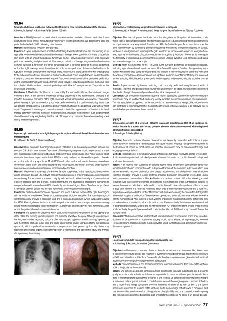
99.64<br />
Traumatic abdom<strong>in</strong>al wall hernia follow<strong>in</strong>g blunt trauma: a case report and review of the literature<br />
V. Pioch 1 , M. Tur<strong>in</strong>a 2 , H.-P. Simmen 2 ( 1 St. Gallen, 2 Zürich)<br />
Objective: A TAWH (traumatic abdom<strong>in</strong>al wall hernia) is def<strong>in</strong>ed as defect of the abdom<strong>in</strong>al wall musculature<br />
and fascia with or without primary sk<strong>in</strong> <strong>in</strong>jury, result<strong>in</strong>g <strong>in</strong> a hernia. We describe the case of a<br />
patient admitted with TAWH follow<strong>in</strong>g blunt trauma.<br />
Methods: Retrospective review of a s<strong>in</strong>gle case.<br />
Results: A 51-year old patient was admitted after fall<strong>in</strong>g down 9 meters from a silo and land<strong>in</strong>g on his<br />
right side. He immediately felt pa<strong>in</strong> and tenderness <strong>in</strong> the right lower quadrant. Cl<strong>in</strong>ically, a superficial<br />
sk<strong>in</strong> lesion with an underly<strong>in</strong>g palpable tumor was found. Follow<strong>in</strong>g primary survey, a CT- scan was<br />
performed reveal<strong>in</strong>g multiple craniofacial fractures, a contusion of the right lung as well as free <strong>in</strong>traabdom<strong>in</strong>al<br />
fluid and a herniation of a small bowel loop with a fist-sized defect of the entire abdom<strong>in</strong>al<br />
wall <strong>in</strong> the right lower quadrant. Immediate laparotomy was performed. Intraoperatively a traumatic<br />
rupture of the meso of the term<strong>in</strong>al ileum was found. Adjacent, the lateral abdom<strong>in</strong>al wall was ruptured<br />
to the subcutaneous tissue. Resection of the harmed Ileum of 20cm length followed by direct anastomosis<br />
and closure of the meso defect ensued. Then, cont<strong>in</strong>uous closure of the peritoneal perforation<br />
on the lateral abdom<strong>in</strong>al wall was performed us<strong>in</strong>g vicryl-0. Follow<strong>in</strong>g exploration of the hernia from<br />
the outside, débridement and closure layer-by-layer with Maxon-0 was performed. The postoperative<br />
course was uneventful.<br />
Conclusion: A TAWH after blunt trauma is a rare entity. The reported <strong>in</strong>cidence of acute hernia ranges<br />
from 02,%-3,6%. In our case the TAWH was already diagnosed <strong>in</strong> the trauma room. Mahajna et al.<br />
reported the case of herniation of the right colon with vessel strangulation, which wasn`t seen <strong>in</strong> the<br />
primary survey. A right hemicolectomy had to be performed on the 2nd posttraumatic day. In our case<br />
we decided <strong>in</strong>traoperatively to perform a primary reconstruction of the abdom<strong>in</strong>al wall without mesh<br />
repair. The potential advantage of a mesh implantation lies <strong>in</strong> the augmentation of the abdom<strong>in</strong>al wall,<br />
thereby potentially lower<strong>in</strong>g the risk of <strong>in</strong>cisional hernia. However, the benefits of such augmentation<br />
should be cautiously weighed aga<strong>in</strong>st the risk of foreign body contam<strong>in</strong>ation when resect<strong>in</strong>g bowel<br />
dur<strong>in</strong>g the same operation.<br />
99.65<br />
Laparoscopic treatment of rare right diaphragmatic rupture with small bowell herniation after blunt<br />
thoracic trauma<br />
H. Hoffmann, D. Oertli, O. Heizmann (Basel)<br />
Objective: Blunt traumatic diaphragmatic rupture (BTDR) is a life-threaten<strong>in</strong>g condition with an <strong>in</strong>cidence<br />
of 0,8-1,6% <strong>in</strong> blunt trauma. The closure of the diaphragm-rupture should be performed immediatly.<br />
The diagnosis is often delayed because of absent typical symptoms or other major <strong>in</strong>juries, which<br />
dom<strong>in</strong>ate the cl<strong>in</strong>ical aspect. An isolated BTDR is a rarity and can be followed by a period of weeks<br />
or months without any symptoms. Most BTDR are located on the left side <strong>in</strong> the musculotend<strong>in</strong>eal<br />
<strong>in</strong>tersection. Right BTDR are rarely described and less frequent. Herniation of colon, small bowell or<br />
liver may occure and can result <strong>in</strong> ileus, necrosis and perforation.<br />
Methods: We present a rare case of a 68 year female, hospitalized <strong>in</strong> the neurological departement<br />
due to park<strong>in</strong>son disease. She felt with her right hemithorax onto a chair. Initially subjective symptoms<br />
were miss<strong>in</strong>g. The exam<strong>in</strong>ation showed a slightly reduced breath without any signs of pneumothorax,<br />
a decent pressure pa<strong>in</strong> and a bruise. 4 days after trauma she developed a progrediend pulmonal decompensation<br />
with a saturation of 89%, attendantly she showed signs of ileus. The chest x-rays offered<br />
a herniation of small bowell <strong>in</strong>to the right hemithorax with consecutive ileus-signs.<br />
Results: We performed a laparoscopic approach and found a 4x5cm rupture of the right diaphragma<br />
with herniation of small bowell. The small bowell appeared vital after reposition. The transdiaphragmatical<br />
thoracoscopy showed a collapsed lung and a dislocated ripfracture, which supposably caused<br />
the BTDR. After irrigation of the thoracic cavity we performed a direct laparoscopic tensionfree runn<strong>in</strong>gsuture<br />
with non-absorbable tie (0/0 Ethibond). A dra<strong>in</strong> was positioned <strong>in</strong> the right hemithorax. Afterwards<br />
the patient showed an uneventful course.<br />
Conclusion: The delay of the diagnosis was 4 days, which shows the variety of the cl<strong>in</strong>ical appearance<br />
of the BTDR. The miss<strong>in</strong>g typical symptoms can mask the severity of the <strong>in</strong>jury. Although large prospective<br />
long-term-studies regard<strong>in</strong>g outcome after laparoscopic approach are still miss<strong>in</strong>g, laparoscopy<br />
was the method of choice <strong>in</strong> our case and could be performed savely. Compared to the thoracoscopic<br />
approach, which is prefered by some authors, we performed the laparoscopy. It mostly allows easy<br />
reposition of herniated <strong>org</strong>ans, sufficient <strong>in</strong>spection of the thoracic and abdom<strong>in</strong>al cavity and immediate<br />
laparotomy if necessary.<br />
99.66<br />
Introduction of contemporary surgery for varicosis ve<strong>in</strong>s <strong>in</strong> mongolia<br />
C. Twerenbold 1 , A. Rotzer 2 , P. Nussbaumer 3 , <strong>Swiss</strong> Surgical Team ( 1 W<strong>in</strong>terthur, 2 Glarus, 3 Lachen)<br />
Objective: After the collapse of the Soviet Union the Mongolian health system fell <strong>in</strong>to a deep crisis<br />
with a lack of consumable supplies and technical equipment. Educational and tra<strong>in</strong><strong>in</strong>g opportunities<br />
for physicians became very limited. Founded <strong>in</strong> 1998, the <strong>Swiss</strong> Surgical Team aims to improve the<br />
local health system by conduct<strong>in</strong>g periodic educational missions to Mongolian hospitals. In Europe,<br />
saphenous ve<strong>in</strong> ligation and stripp<strong>in</strong>g is the gold standard for varicose ve<strong>in</strong> surgery. In Mongolia however,<br />
treatment still consists of local phlebectomies through long <strong>in</strong>cisions. We aimed to <strong>in</strong>vestigate<br />
the feasibility of <strong>in</strong>troduc<strong>in</strong>g a contemporary procedure utiliz<strong>in</strong>g available local resources and us<strong>in</strong>g<br />
varicose ve<strong>in</strong> surgery as an example.<br />
Methods: From the 22nd May to the 14th June 2009 our team performed 50 surgical procedures,<br />
<strong>in</strong>clud<strong>in</strong>g eight <strong>in</strong>terventions for varicose ve<strong>in</strong>s at the General Hospital of Darchan. Preoperatively every<br />
patient was exam<strong>in</strong>ated us<strong>in</strong>g a trendelenburg test <strong>in</strong> order to identify <strong>in</strong>sufficient perforans ve<strong>in</strong>s and<br />
the valvular competence. After saphenous ve<strong>in</strong> ligation a sterilized monofilament fish<strong>in</strong>g l<strong>in</strong>e was used<br />
for ve<strong>in</strong> stripp<strong>in</strong>g. M<strong>in</strong>iphlebectomy was performed us<strong>in</strong>g stab <strong>in</strong>cisions and a locally available crochet<br />
hook.<br />
Results: Saphenous ve<strong>in</strong> ligation and stripp<strong>in</strong>g could be easily performed us<strong>in</strong>g the described local<br />
materials. The <strong>in</strong>tra- and postoperative course was uneventful <strong>in</strong> all cases. Our experience confirmed<br />
that the local surgical community could easily learn the new procedure.<br />
Conclusion: Our Mongolian experience suggests that <strong>in</strong> develop<strong>in</strong>g countries simple contemporary<br />
procedures may be readily <strong>in</strong>troduced us<strong>in</strong>g locally available <strong>in</strong>expensive materials and <strong>in</strong>struments.<br />
This trial establishes an approach for the <strong>in</strong>troduction of new contemporary surgical techniques which<br />
can contribute to the improvement of the local health system, otherwise unlikely to be achieved due to<br />
prohibitively expensive conventional materials.<br />
99.67<br />
Arthroscopic elevation of a reversed Hill-Sachs lesion and simultaneous ORIF of an ispilateral acromion<br />
fracture <strong>in</strong> a patient with locked posterior shoulder dislocation comb<strong>in</strong>ed with a displaced<br />
acromion fracture: a case report<br />
P. Gruen<strong>in</strong>ger, C. Meier (Zürich)<br />
Objective: Traumatic posterior shoulder dislocations are frequently associated with anterior impression<br />
fractures of the humeral head (reversed Hill-Sachs lesion). Whereas non-operative treatment is<br />
the treatment of choice for most cases, an operative <strong>in</strong>tervention may be considered for large and<br />
engag<strong>in</strong>g osseous defects.<br />
Methods: Presentation of a m<strong>in</strong>imally-<strong>in</strong>vasive arthroscopic technique to elevate a large reversed Hill-<br />
Sachs lesion <strong>in</strong> a patient with a locked posterior shoulder dislocation <strong>in</strong> comb<strong>in</strong>ation with a displaced<br />
fracture of the acromion.<br />
Results: A 34-year old man susta<strong>in</strong>ed an isolated trauma to his left shoulder consist<strong>in</strong>g of a posterior<br />
shoulder dislocation <strong>in</strong> comb<strong>in</strong>ation with a displaced fracture of the acromion. A CT scan which was<br />
performed due to recurrent dislocation after closed reduction and immobilisation <strong>in</strong> <strong>in</strong>ternal rotation<br />
(Gilchrist bandage) showed a locked posterior shoulder dislocation with a large reversed Hill-Sachs<br />
lesion, a reversed double chondral Bankart lesion and an <strong>in</strong>tact rotator cuff. In the follow<strong>in</strong>g, closed<br />
reduction was successfully performed and reta<strong>in</strong>ed <strong>in</strong> a handshake brace. Arthroscopic surgery to<br />
elevate the osseous defect was performed <strong>in</strong> comb<strong>in</strong>ation with plate osteosynthesis of the acromion<br />
5 days after trauma. The reversed Hill-Sachs lesion was arthroscopically visualized and a tibial ACL<br />
target device was placed <strong>in</strong> the centre of the lesion with the K-wire enter<strong>in</strong>g the bone <strong>in</strong> the distal greater<br />
tuberosity through a short <strong>in</strong>cision. The cortex was opened and a cannula was <strong>in</strong>serted over the K-wire<br />
<strong>in</strong>to the humeral head. After removal of the K-wire the impression was elevated and the defect filled with<br />
cancellous bone harvested from the anterior iliac crest. Postoperatively, the shoulder was immobilized<br />
<strong>in</strong> a handshake brace for 3 weeks and no <strong>in</strong>ternal rotation >0° was allowed for 6 weeks. Three months<br />
postoperatively the patient presented with a stable shoulder, full ROM and bony consolidation of the<br />
humeral head.<br />
Conclusion: While non-operative treatment with immobilisation <strong>in</strong> a handshake brace after closed reduction<br />
may be successful <strong>in</strong> most cases, surgery should be considered for large engag<strong>in</strong>g reversed<br />
Hill-Sachs lesions. Osseous defects may be elevated us<strong>in</strong>g our technique via a m<strong>in</strong>imally-<strong>in</strong>vasive arthroscopic<br />
approach.<br />
99.69<br />
Leiomyosarcome de la ve<strong>in</strong>e petite saphène: un diagnostic rare<br />
M. L. Matthey, E. Pezzetta, O. Mart<strong>in</strong>et (Montreux)<br />
Objective: Les léiomyosarcomes vasculaires sont des tumeurs rares et le plus souvent localisées dans<br />
la ve<strong>in</strong>e cave <strong>in</strong>férieure; peu de cas touchant le système ve<strong>in</strong>eux périphérique des membres <strong>in</strong>férieurs<br />
ont été rapportés dans la littérature. Dans cette situation les symptômes sont généralement tardifs et<br />
aspécifiques avec un pronostic globalement défavorable.<br />
Methods: nous présentons un cas de léiomyosarcome touchant un branche de la ve<strong>in</strong>e petite saphène<br />
traité chirurgicalement avec succès.<br />
Results: une patiente de 88 ans connue pour une <strong>in</strong>suffisance ve<strong>in</strong>euse superficielle, qui a présenté<br />
quelques mois après le traitement d’une varicophlébite du membre <strong>in</strong>férieur gauche des douleurs<br />
dans le mollet ipsilatéral évoquant la présence d’une récidive. La persistance des symptômes malgré<br />
le traitement anticoagulant <strong>in</strong>stauré a conduit à une réévalutation angiologique. L examen échodoppler<br />
a montré une image compatible avec un thrombus <strong>in</strong>tralum<strong>in</strong>al de 4cm au se<strong>in</strong> d’une varice<br />
accessoire provenant de la ve<strong>in</strong>e petite saphène. Cette même image est retrouvée 5 mois plus tard<br />
lors d’un contrôle, une <strong>in</strong>tervention chirurgicale est a<strong>in</strong>si planifiée avec une crossectomie et stripp<strong>in</strong>g<br />
des ve<strong>in</strong>es petites saphènes bilatérales avec phlébectomies étagées. Au cours d’un paquet pseudo-<br />
swiss <strong>knife</strong> 2010; 7: special edition 77


