

Anorectal Manometry in 3D NEW! - Swiss-knife.org
Anorectal Manometry in 3D NEW! - Swiss-knife.org
Anorectal Manometry in 3D NEW! - Swiss-knife.org

Create successful ePaper yourself
Turn your PDF publications into a flip-book with our unique Google optimized e-Paper software.
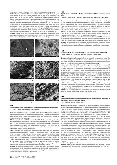
scope, the SEM processes <strong>in</strong>cluded dehydration, critical po<strong>in</strong>t dry<strong>in</strong>g and plat<strong>in</strong>um sputter<strong>in</strong>g.<br />
Results: At day 1 post Tx, the allograft showed overall signs of destruction (Fig. A): alveoli were <strong>in</strong>jured<br />
with thicken<strong>in</strong>g of the alveolar wall conta<strong>in</strong><strong>in</strong>g multiple perforations (s<strong>in</strong>gle arrow). The capillary lumen<br />
between alveoli vanished. There was a widen<strong>in</strong>g of the perivascular space around vessels (double arrow)<br />
with <strong>in</strong>vasion of <strong>in</strong>flammatory cells and erythrocytes. Furthermore, bronchioles and alveoli were<br />
covered with a thick layer of mucus. In contrast, the non-transplanted lung showed a normal architecture<br />
and alveoli and vessels rema<strong>in</strong>ed <strong>in</strong> close contact to each other (Fig. B, Fig. E). At day 5 post<br />
Tx, the architecture of the alveoli was completely lost with <strong>in</strong>vasion of multiple cells <strong>in</strong>to the alveolar<br />
and perivascular space. Leukocytes and macrophages were found numerously <strong>in</strong> the perivascular<br />
tissue, with accumulation of ma<strong>in</strong>ly erythrocytes <strong>in</strong> the alveolar space (Fig. D). The architecture of the<br />
non transplanted lung on day 5 was normal. Interest<strong>in</strong>gly, leukocytes percolated the basal membrane<br />
of the bronchioles (Fig. F) with concomitant macrophage <strong>in</strong>vasion <strong>in</strong>to term<strong>in</strong>al bronchioles (Fig. C).<br />
Conclusion: The SEM detects patho-morphological changes of acute rejection at an early state. The <strong>in</strong>terstitial<br />
space, small vessels, alveoli and bronchioles of the allograft were severely altered by <strong>in</strong>vasion<br />
of immune cells, with a significant <strong>in</strong>volvement also of the non-transplanted lung.<br />
50 swiss <strong>knife</strong> 2010; 7: special edition<br />
�<br />
������������������ �<br />
� �<br />
�<br />
55.6<br />
Survival of trimodal therapy of malignant pleural mesothelioma with extrapleural pneumonectomy<br />
R. Fahrner, G. L. Carboni, A. Ochsenbe<strong>in</strong>, R. A. Schmid (Berne)<br />
Objective: S<strong>in</strong>ce multimodal therapy of malignant pleural mesothelioma (MPM) was <strong>in</strong>troduced survival<br />
was improved particulary <strong>in</strong> subpopulations with epithelial histology, R0-resection and absence of<br />
broad chestwall <strong>in</strong>vasion. This study aimed to evaluate the longterm outcomes after trimodal therapy<br />
<strong>in</strong>clud<strong>in</strong>g extrapleural pneumonectomy (EPP).<br />
Methods: Between October 1st 2000 to December 31st 2005 41 patients with histologically verified<br />
MPM were treated <strong>in</strong> our <strong>in</strong>stitution. After confirmation of the diagnosis with surgical biopsy (epithelial<br />
type n=17, mixed type n=4) and negative lymph node status <strong>in</strong> mediast<strong>in</strong>oscopy 21 patients (51%)<br />
underwent trimodal therapy with a median follow-up of 655 days (63-2567 days) and the other 20<br />
patients underwent palliative treatment. Postoperative complications, 1-, 2- and 5-year survial rates<br />
and recurrence rates were analyzed retrospectively.<br />
Results: Patients with trimodal therapy had a median age of 64 years (40-75, 18 men). Neoadjuvant<br />
chemotherapy consisted ma<strong>in</strong>ly <strong>in</strong> a comb<strong>in</strong>ation of plat<strong>in</strong>um based agents (n=19), gemcitab<strong>in</strong>e<br />
(n=15) or pemetrexed (n=4). EPP was done as standard surgical procedure, time from diagnosis and<br />
EPP was 117 days. Complications such as <strong>in</strong>fections (n=11), arrhythmia (n=4), bleed<strong>in</strong>g (n=2), patch<br />
avulsion (n=1) but no bronchus stump <strong>in</strong>sufficiency occurred. 19 patients received adjuvant radiotherapy.<br />
1-year-survival rate was 71% <strong>in</strong> the trimodal treatment group versus 21% <strong>in</strong> the palliative group<br />
(p=0.005). Survival rates <strong>in</strong> the trimodal treatment group were 28% after two years and 10% after five<br />
years. There were no statistical significant differences seen regard<strong>in</strong>g sex, age, tumor stage or cell type.<br />
Tumor recurrence was detected after one and two years respectively 40% and 82%.<br />
Conclusion: At diagnosis the majority of patients has already a advanced stage disease and palliative<br />
approaches rema<strong>in</strong> the only option. In the subpopulation of patients which underwent trimodal therapy<br />
survival was better than <strong>in</strong> the palliative treatment group but still long term survival is worse than <strong>in</strong><br />
bronchogenic carc<strong>in</strong>oma, consider<strong>in</strong>g the high morbidity of EPP.<br />
�<br />
�<br />
�<br />
55.7<br />
Atypical expression and distribution of embryonic stem cell marker, OCT4, <strong>in</strong> human lung adenocarc<strong>in</strong>oma<br />
G. Karoubi 1 , L. Cortes-Dericks 1 , M. Gugger 1 , D. Galetta 2 , L. Spaggiari 2 , R. A. Schmid 1 ( 1 Berne, 2 Milan)<br />
Objective: Lung cancer is one of the lead<strong>in</strong>g causes of cancer-related deaths <strong>in</strong> the world. Although<br />
the orig<strong>in</strong> still rema<strong>in</strong>s to be resolved, a prevail<strong>in</strong>g hypothesis implies the <strong>in</strong>volvement of cancer stem<br />
cells (CSCs) responsible for tumor <strong>in</strong>itiation, ma<strong>in</strong>tenance and progression. OCT4, a major regulator<br />
of embryonic stem cell self-renewal, encodes two ma<strong>in</strong> spliced variants designated as OCT4A and<br />
OCT4B, and has recently been shown to have a dual role; as a potential adult stem cell marker, and as<br />
a cancer stem cell marker <strong>in</strong> germl<strong>in</strong>e and somatic tumors.<br />
Methods: In this study we sought to <strong>in</strong>vestigate the expression and <strong>in</strong>tracellular distribution of OCT4A<br />
and OCT4B isoforms us<strong>in</strong>g flow cytometry, Western blot and quantitative RT-PCR analyses <strong>in</strong> normal<br />
and lung adenocarc<strong>in</strong>oma cell l<strong>in</strong>es, primary cultures and tissue biopsies.<br />
Results: We demonstrate for the first time, the presence of rare OCT4A+ and OCT4B+ cells <strong>in</strong> normal<br />
lung. Notably, we observed higher levels of expression and atypical cytoplasmic distribution of OCT4A<br />
and not OCT4B, <strong>in</strong> the malignant sett<strong>in</strong>g, strongly <strong>in</strong>dicat<strong>in</strong>g an oncogenic role <strong>in</strong> lung adenocarc<strong>in</strong>oma.<br />
Conclusion: We postulate that OCT4A+ cells represent a putative cancer stem cell population. Identification<br />
of these cells and the biological processes vital for their subsistence, will guide the development<br />
of diagnostic and therapeutic cl<strong>in</strong>ical approaches with the goal of elim<strong>in</strong>at<strong>in</strong>g lung adenocarc<strong>in</strong>oma.<br />
55.8<br />
Functional results of a new screwless fixation device for rib fractures <strong>in</strong> patients with flail chest<br />
T. Strauss, H. Hoffmann, J. Bremerich, M. Siegemund, M. Tamm, D. Lard<strong>in</strong>ois (Basel)<br />
Objective: Selected patients with severe antero-lateral flail chest and impaired respiratory function who<br />
should recover quickly have an <strong>in</strong>dication for operative stabilisation of the ribs. This is a prospective<br />
study of chest wall <strong>in</strong>tegrity and pulmonary function <strong>in</strong> these patients who underwent chest wall stabilisation<br />
with a new screwless fixation device (STRATOSTM, MedXpert, Germany).<br />
Methods: Between May 2008 and December 2009 14 patients (10m, 4w) with a mean age of 57<br />
years (43-74) were operated for traumatic flail chest. The mean number of affected ribs was 8 (3-12),<br />
the sternum was fractured <strong>in</strong> 4 cases. Titanium rib clamps were placed and fixed to the stable parts of<br />
the most affected ribs and connected by titanium plates. Cl<strong>in</strong>ical outcome, pulmonary test<strong>in</strong>g and dynamic<br />
assessment of the chest wall mobility by c<strong>in</strong>e MRI were performed 6 months follow<strong>in</strong>g surgery.<br />
Results: 9 patients (64%) had various comb<strong>in</strong>ations of <strong>in</strong>juries of the thorax, head, abdomen and extremities.<br />
9 patients (64%) underwent unilateral, 5 patients (35%) bilateral stabilisation with a medium<br />
delay of 5,4 days (1-14) from admission. 30 day mortality was 0%. Immediate postoperative extubation<br />
was feasible <strong>in</strong> 5 patients (35%). No material dislocation was observed dur<strong>in</strong>g follow up. Two<br />
patients were reoperated due to <strong>in</strong>fection and hematoma. After operation patients rema<strong>in</strong>ed <strong>in</strong> hospital<br />
for 24.3 days <strong>in</strong> the mean (8-71), patients with monotrauma only 11.5 days. At 6 months prelim<strong>in</strong>ary<br />
data show no restriction <strong>in</strong> the pulmonary function test<strong>in</strong>g and c<strong>in</strong>eMRI shows symmetric movement<br />
of the chest wall. Time to return to normal daily activity was 44 days after discharge. Shoulder function<br />
was impaired after 6 months <strong>in</strong> 4 patients.<br />
Conclusion: Our results suggest that antero-lateral flail chest <strong>in</strong>juries accompanied by respiratory <strong>in</strong>sufficiency<br />
can be effectively stabilised by screwless titanium fixation device.<br />
55.9<br />
Primary pedunculated muscle flap coverage of bronchial and tracheal defects as an alternative to<br />
direct closure or bronchotracheal sleeve resection<br />
P. Dorn, M. Kuhn, M. Odermatt, M. Furrer (Chur)<br />
Objective: Bronchial airways after lung resections are usually closed either by manual or mechanical<br />
suture. To prevent bronchopleural fistula (BPF) additional re<strong>in</strong>forcement of the bronchial stump by<br />
a pedunculated muscle flap (PMF) is often recommended. In very central tumours sleeve resection<br />
with anastomosis is generally preferred to direct closure with the aim of m<strong>in</strong>imiz<strong>in</strong>g airway stenosis<br />
or avoid<strong>in</strong>g <strong>in</strong>complete resection. As a further alternative we describe a technique where only a PMF<br />
is used to cover an open and unsutured central airway defect by means of three high-risk patients.<br />
Methods: CASE 1: A 40 year-old man underwent extended right pneumonectomy because of central<br />
non small cell lung cancer (NSCLC) with severe poststenotic pneumonia. The tracheal defect at the<br />
bifurcation was covered by a PMF without preced<strong>in</strong>g bronchial suture closure. There was no evidence<br />
of air leakage <strong>in</strong> <strong>in</strong>traoperative test<strong>in</strong>g and repetitive bronchoscopy. No complications were seen <strong>in</strong><br />
the postoperative course. CASE 2: A 45 year-old woman underwent extended right pneumonectomy.<br />
After impressive tumour regression due to neoadjuvant chemotherapy the resection marg<strong>in</strong> of the right<br />
bronchus was close to the car<strong>in</strong>a. A PMF was used for coverage of the airway defect. Intraoperative<br />
test<strong>in</strong>g <strong>in</strong>clud<strong>in</strong>g bronchoscopy showed no evidence of air leakage or contralateral prolapse of the<br />
muscle flap. The postoperative course was complicated by ARDS of the rema<strong>in</strong><strong>in</strong>g left lung necessitat<strong>in</strong>g<br />
cont<strong>in</strong>uous positive airway pressure (CPAP). Delayed re-thoracotomy with muscle flap refixation<br />
had to be performed because of secondary air leakage. CASE 3: A 72 year-old man with NSCLC and<br />
neoadjuvant chemotherapy underwent right bilobectomy with extended resection of the right <strong>in</strong>termediate<br />
bronchus up to the ma<strong>in</strong> bronchus. The defect of the ma<strong>in</strong> bronchus was covered us<strong>in</strong>g a<br />
PMF. Intraoperative bronchoscopy and air leakage test<strong>in</strong>g were normal. The postoperative course was<br />
uneventfull.<br />
Results: See above (Methods and Results).<br />
Conclusion: Primary coverage of a central bronchial or tracheal defect us<strong>in</strong>g only a PMF is feasible.<br />
Predom<strong>in</strong>antly <strong>in</strong> high risk patients direct suture at risk or extensive sleeve resection may be avoided.


