

Anorectal Manometry in 3D NEW! - Swiss-knife.org
Anorectal Manometry in 3D NEW! - Swiss-knife.org
Anorectal Manometry in 3D NEW! - Swiss-knife.org

You also want an ePaper? Increase the reach of your titles
YUMPU automatically turns print PDFs into web optimized ePapers that Google loves.
99.75<br />
Isolated dorso-medial dislocation of the first ray at the medial cuneonavicular jo<strong>in</strong>t: a case report<br />
E. Testa, M. Camurati, J. Müller, D. Haeni, M. Arigoni, C. Candrian (Lugano)<br />
Objective: The isolated dislocation of the first ray at the cuneonavicular jo<strong>in</strong>t of the foot is a very rare<br />
<strong>in</strong>jury of the foot. So far, only 10 cases have been reported. Probably the low <strong>in</strong>cidence of these lesion is<br />
due to a very rigid plantar anatomical structure: The medial cuneiform bone is united with the navicular<br />
bone, the first and second metatarsal bone trough a thick and strong plantar ligament, re<strong>in</strong>forced by<br />
the tibialis posterior and anterior tendon as well as the <strong>in</strong>sertion of the peroneus longus tendon. An<br />
additional element of stability is furnished by the naviculocuneiform ligaments.<br />
Methods: We report about a patient with an isolated dislocation of the first ray at the cuneonavicular<br />
jo<strong>in</strong>t of the foot dislocation of the first cuneiform bone without fracture’s associated, its treatment,<br />
follow-up and a review of the literature.<br />
Results: A 39 year old man, known to be affected by ontogenesis imperfecta, after a bike accident with<br />
axial trauma on the first metatarsus, revealed a remarkably swollen and deformed foot with a bump<br />
over the medial border, distal to the medial malleolus. Conventional X-ray’s displayed a dorso-medial<br />
dislocation of the first ray at the cuneonavicular jo<strong>in</strong>t of the foot, with a loss of congruity at both correspond<strong>in</strong>g<br />
jo<strong>in</strong>ts, without any fracture’s. Because of <strong>in</strong>stability after successful closed reduction, percutaneous<br />
temorary artrodeses with K-wires between the medial cuneiform bone and os navicolare and<br />
<strong>in</strong>termediate cuneiform bone was performed. Patient’s foot was immobilised and partial weight bear<strong>in</strong>g<br />
manta<strong>in</strong>ed for 6 weeks, than K-wires removed and gradual weight bear<strong>in</strong>g allowed. After 6 month<br />
the patient was asymptomatic, with a normal range of motion. Radiologically no loss of reduction<br />
was seen, a slight <strong>in</strong>creased space between the medial and <strong>in</strong>termediate cuneiform bone was visible.<br />
Conclusion: Dislocations of the cuneiforme have been treated <strong>in</strong> a variety of methods, rang<strong>in</strong>g from<br />
open or closed reduction, with or without fixation. Some author’s have postulated also immediate def<strong>in</strong>itive<br />
artrodeses to avoid arthritis of the <strong>in</strong>volved jo<strong>in</strong>ts. Given the rarity and variability of these <strong>in</strong>juries,<br />
a common approach is very difficult to establish, although immediate anatomic reduction and retention,<br />
possibly m<strong>in</strong>i-<strong>in</strong>vasive with K-wire’s is essential to ensure a good functional result and to avoid a<br />
degenerative evolution of the lesion.<br />
99.76<br />
Fournier-Gangrän: Vers<strong>org</strong>ung der Testikel bei komplettem Verlust der Skrotalhaut gemäss „Surgery<br />
<strong>in</strong> Africa“<br />
M. Nägeli, A. Bagot, H. Knönagel, E. Grossen, T. Delko, O. Schöb (Schlieren)<br />
Objective: Die Fournier-Gangrän ist e<strong>in</strong>e seltene Sonderform der nekrotisierenden Fasziitis der Perigenitalregion.<br />
Die Mortalität kann trotz adäquater Antibiotikatherapie und radikaler chirurgischer Therapie<br />
bis zu 50% betragen.<br />
Methods: Fallbericht: Vorstellung e<strong>in</strong>es 59jährigen Patienten mit bekanntem Alkoholkonsum mit phlegmonöser<br />
Entzündung des Skrotums bis <strong>in</strong> die rechte Leiste reichend mit ödematös geschwollener und<br />
pränekrotischer Skrotalhaut nach Manipulation e<strong>in</strong>es Furunkels. Sonographisch ke<strong>in</strong> Abszess nachweisbar,<br />
lokal 5mm grosser Hautdefekt an Peniswurzel zum Skrotumübergang. Es wird e<strong>in</strong> skrotales<br />
Débridement mit Resektion der ganzen Skrotalhaut vrogenommen, so dass beide Testikel frei liegen.<br />
Zusätzlich antibiotische Therapie. In 48h Abstand weitere Débridements nach gluteal rechts und pararektal<br />
rechts. Da die Wunde durch Stuhlgang regelmässig verschmutzt wurde, laparoskopische Anlage<br />
e<strong>in</strong>er Entlastungsileostomie. E<strong>in</strong>e Woche nach Initialoperation bei sauberen Wundverhältnissen erfolgt<br />
die Verlagerung beider Hoden <strong>in</strong> subkutane Taschen suprasymphysär beidseits, so dass e<strong>in</strong> Vakuumverband<br />
über das gesamte Per<strong>in</strong>eum und die beiden Skrotalfächer angelegt werden kann.<br />
Results: Nach mehreren Vakuumverband-Wechsel und progredienten Sekundärnähten kam es zu<br />
e<strong>in</strong>er guten Wundheilung. Die hormonelle Hodenfunktion wurde mittels Testosteronbestimmung dokumentiert<br />
und zeigt normale Werte, zudem sonographisch normale Perfusion. Der Patient fühlt sich<br />
durch die Hoden, die suprasymphysär verlagert wurden, nicht gestört. ->Fotodokumentation<br />
Conclusion: Die Fourniergangrän hat e<strong>in</strong> hohes Sterblichkeitsrisiko und erfordert e<strong>in</strong>e aggressive,<br />
frühzeitige chirurgische Therapie. Um auf e<strong>in</strong>e Orchiektomie verzichten zu können (bei e<strong>in</strong>er 21% Orchiektomierate<br />
gemäss Literatur) wurden die Hoden des Patienten, auf die Methode aus e<strong>in</strong>em Afrikabuch<br />
zurückgreifend, subkutan suprasymphysär verlagert.<br />
99.77<br />
Abdom<strong>in</strong>ales CRPS – e<strong>in</strong>e Fallvorstellung<br />
S. Schmidt, R. Schreiber, R. Humm, B. Muff (Bülach)<br />
Objective: Das CRPS (complex regional pa<strong>in</strong> syndrom, früher auch M. Sudeck, Algodystrophie) ist<br />
e<strong>in</strong> nicht seltenes, differenziertes Krankheitsbild, das vor allem an den Extremitäten nach äusserer<br />
E<strong>in</strong>wirkung (Trauma, Entzündung, Operation) auftritt. Häufigste Symptome: Ödeme, Durchblutungsstörungen,<br />
trophische Hautstörungen, Schmerzen, Funktionse<strong>in</strong>schränkung. Es besteht e<strong>in</strong>e<br />
starke Tendenz zur Chronifizierung. Die Pathogenese ist noch nicht abschliessend geklärt.<br />
Methods: 59jähriger Patient, Landwirt, seit mehr als 12 Monaten arbeitsunfähig wegen seit 08/2008<br />
bestehendem Schmerzsyndrom. Als Auslöser wird e<strong>in</strong> Verhebetrauma angegeben. Symptomatik:<br />
Massive, teils brennende, teils dumpfe Schmerzen <strong>in</strong> Abdomen und Ingu<strong>in</strong>algegend. Hautkolorit des<br />
Abdomens wechselnd blau, teils grünlich. Störendes Kältegefühl im Bauch, Leisten, teils bis <strong>in</strong> Füsse.<br />
Subjektives Durchblutungsproblem der Genitalregion. Erektile Dysfunktion. Ungewollter Gewichtsverlust<br />
von 35 kg. Koord<strong>in</strong>ationsstörungen. Erschöpfungszustand. PA: St. n. TEPP bds. 2002, St. n. diagnostischer<br />
Laparoskopie wg. Abdom<strong>in</strong>albeschwerden 2004, St. n. Achillessehnenoperation 1995.<br />
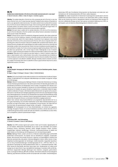
Kl<strong>in</strong>ik: Zyanose der Bauchdecke, teils Striae rubrae (siehe Foto). Hauttemperatur am unteren leicht<br />
niedriger als am oberen Stamm. Hypo- Asensibilität der Genitalregion. Druckdolenz LWS und ISG bds.,<br />
LWS-Flexion e<strong>in</strong>geschränkt. Bisherige Abklärungen (Sono Abdomen, urologische, gastro-enterologische,<br />
neurologische Abklärung, MRI LWS und Becken, <strong>in</strong>ternistische stationäre & rheumatologische<br />
Abklärung, psychiatrisches Gutachten) konnten die Symptomatik nicht erklären. Verdachtsdiagnose:<br />
Abdom<strong>in</strong>ales CRPS oder Chronifiziertes Schmerzsyndrom der Bauchdecken und Leisten bds. nach<br />
Verhebetrauma. ND: Harnblasendysfunktion, BPH Grad I, reaktive Depression.<br />
Conclusion: Bei unklarer Symptomatik mit teils kl<strong>in</strong>isch-morphologischem Korrelat jedoch ohne klar<br />
objektivierbare somatische Genese ist als Ursache an e<strong>in</strong> abdom<strong>in</strong>ales CRPS zu denken. Bisherige<br />
Daten aus der Literatur dazu s<strong>in</strong>d rar, therapeutische Optionen auf empirischen Daten beruhend. Es<br />
wird e<strong>in</strong>e multifaktorielle Ätiologie vermutet. Nach genauer somatischer Abklärung ist e<strong>in</strong> CRPS, obwohl<br />
atypisch, auch am Abdomen differentialdiagnostisch <strong>in</strong> Betracht zu ziehen.<br />
Fotodokumentation (Aufnahme im Liegen und stehend) zur Fallvorstellung: Abdom<strong>in</strong>ales CRPS<br />
swiss <strong>knife</strong> 2010; 7: special edition 79


