

Anorectal Manometry in 3D NEW! - Swiss-knife.org
Anorectal Manometry in 3D NEW! - Swiss-knife.org
Anorectal Manometry in 3D NEW! - Swiss-knife.org

You also want an ePaper? Increase the reach of your titles
YUMPU automatically turns print PDFs into web optimized ePapers that Google loves.
99.55<br />
Complexes de von Meyenburg(hamartomes biliaires): découverte fortuite lors d‘une cholécystectomie<br />
par laparoscopie<br />
D. Douma 1 , K. Djebaili 1 , C. Becciol<strong>in</strong>i 1 , A. Safret 2 , J.-C. Renggli 1 , L. Ducali 1 ( 1 La Chaux-de-Fonds, 2 Neuchâtel)<br />
Objective: Nous rapportons une observation des complexes de von Meyenburg(hamartomes biliaires)<br />
chez une jeune patiente.<br />
Methods: Il s‘agit d‘une patiente de 25 ans, en bonne santé habituelle,qui se plaignait depuis<br />
huit mois de douleurs abdom<strong>in</strong>ales hautes réccurentes associées à un sydrome de reflux gastro-oesophagien,sans<br />
trouble de transit. Pas de notion perte de poids. Cl<strong>in</strong>iquement,elle était<br />
apyrétique,l‘abdomen était sensible dans l‘hypocondre droit et le signe de Murphy était positif. Le bilan<br />
biologique était normal. L‘échographie abdom<strong>in</strong>al mettait en évidence une lithiase vésiculaire sans<br />
signe de cholécystite et sans dilatation des voies biliaires. La gastroscopie réalisée ne montrait aucune<br />
pathologie. Une cholécystectomie par laparoscopie était programmée. Lors de l‘<strong>in</strong>tervention,nous<br />
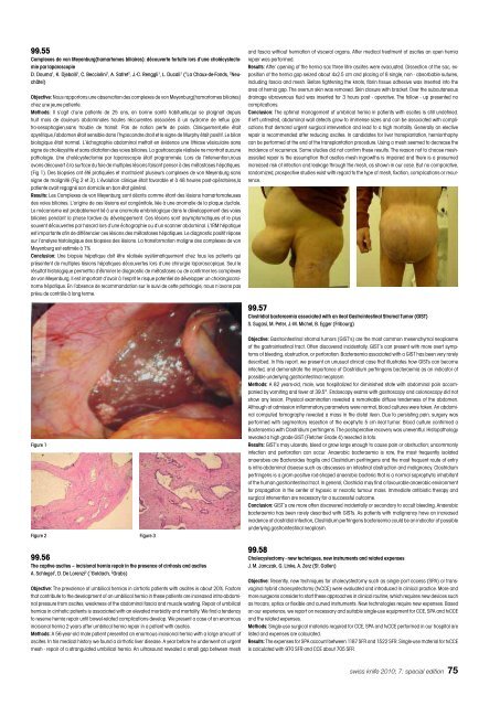
avons découvert à la surface du foie de multiples lésions faisant penser à des métastases hépatiques.<br />
(Fig 1). Des biopsies ont été pratiquées et montraient plusieurs complexes de von Meyenburg sans<br />
signe de malignité (Fig 2 et 3). L‘évolution cl<strong>in</strong>ique était favorable et à 48 heures post-opératoires,la<br />
patiente avait regagné son domicile en bon état général.<br />
Results: Les Complexes de von Meyenburg sont décrits comme étant des lésions hamartomateuses<br />
des voies biliaires. L‘orig<strong>in</strong>e de ces lésions est congénitale, liée à une anomalie de la plaque ductale.<br />
Le mécanisme est probablement lié à une anomalie embriologique dans le développement des voies<br />
biliaires pendant la phase tardive du développement. Ces lésions sont asymptomatiques et le plus<br />
souvent découvertes par hasard lors d‘une échographie ou d‘un scanner abdom<strong>in</strong>al. L‘IRM hépatique<br />
est importante af<strong>in</strong> de différencier ces lésions des métastases hépatiques. Le diagnostic positif répose<br />
sur l‘analyse histologique des biopsies des lésions. La transformation maligne des complexes de von<br />
Meyenburg est estimée à 7%<br />
Conclusion: Une biopsie hépatique doit être réalisée systématiquement chez tous les patients qui<br />
présentent de multiples lésions hépatiques découvertes lors d‘une chirurgie laparoscopique. Seul le<br />
résultat histologique permettra d‘élim<strong>in</strong>er le diagnostic de métastases ou de confirmer les complexes<br />
de von Meyenburg. Il est important d‘avoir à l‘esprit le risque potentiel de développer un cholangicarc<strong>in</strong>ome<br />
hépatique. En l‘absence de recommandation sur le suivi de cette pathologie, nous n‘avons pas<br />
prévu de contrôle à long terme.<br />
Figure 1<br />
Figure 2 Figure 3<br />
99.56<br />
The captive ascites – <strong>in</strong>cisional hernia repair <strong>in</strong> the presence of cirrhosis and ascites<br />
A. Schlegel 1 , D. De Lorenzi 2 ( 1 Goldach, 2 Grabs)<br />
Objective: The prevalence of umbilical hernias <strong>in</strong> cirrhotic patients with ascites is about 20%. Factors<br />
that contribute to the development of an umbilical hernia <strong>in</strong> these patients are <strong>in</strong>creased <strong>in</strong>tra-abdom<strong>in</strong>al<br />
pressure from ascites, weakness of the abdom<strong>in</strong>al fascia and muscle wast<strong>in</strong>g. Repair of umbilical<br />
hernias <strong>in</strong> cirrhotic patients is associated with an elevated morbidity and mortality. We f<strong>in</strong>d a tendency<br />
to reserve hernia repair until bowel-related complications develop. We present a case of an enormous<br />
<strong>in</strong>cisional hernia 2 years after umbilical hernia repair <strong>in</strong> a patient with ascites.<br />
Methods: A 56-year-old male patient presented an enormous <strong>in</strong>cisional hernia with a large amount of<br />
ascites. In his medical history we found a cirrhotic liver disease. A year before he underwent an urgent<br />
mesh - repair of a strangulated umbilical hernia. An ultrasound revealed a small gap between mesh<br />
and fascia without herniation of visceral <strong>org</strong>ans. After medical treatment of ascites an open hernia<br />
repair was performed.<br />
Results: After open<strong>in</strong>g of the hernia sac three litre ascites were evacuated. Dissection of the sac, exposition<br />
of the hernia gap seized about 4x2.5 cm and plac<strong>in</strong>g of 8 s<strong>in</strong>gle, non - absorbable sutures,<br />
<strong>in</strong>clud<strong>in</strong>g fascia and mesh. Before tighten<strong>in</strong>g the knots, fibr<strong>in</strong> tissue adhesive was <strong>in</strong>serted <strong>in</strong>to the<br />
area of hernia gap. The overrun sk<strong>in</strong> was removed. Sk<strong>in</strong> closure with bracket. Over the subcutaneous<br />
dra<strong>in</strong>age vibravenous fluid was <strong>in</strong>serted for 3 hours post - operative. The follow - up presented no<br />
complications.<br />
Conclusion: The optimal management of umbilical hernia <strong>in</strong> patients with ascites is still undef<strong>in</strong>ed.<br />
If left untreated, abdom<strong>in</strong>al wall defects grow to immense sizes and can be associated with complications<br />
that demand urgent surgical <strong>in</strong>tervention and lead to a high mortality. Generally an elective<br />
repair is recommended after reduc<strong>in</strong>g ascites. In candidates for liver transplantation, herniorrhaphy<br />
can be performed at the end of the transplantation procedure. Us<strong>in</strong>g a mesh seemed to decrease the<br />
<strong>in</strong>cidence of recurrence. Some studies did not confirm these results. The reason not to choose meshassisted<br />
repair is the assumption that ascites mesh <strong>in</strong>growths is impaired and there is a presumed<br />
<strong>in</strong>creased risk of <strong>in</strong>fection and leakage through the mesh, as shown <strong>in</strong> our case. But no comparative,<br />
randomized, prospective studies exist with regard to the type of mesh, fixation, complications or recurrence.<br />
99.57<br />
Clostridial bacteraemia associated with an ileal Gastro<strong>in</strong>test<strong>in</strong>al Stromal Tumor (GIST)<br />
S. Sugasi, M. Peter, J.-M. Michel, B. Egger (Fribourg)<br />
Objective: Gastro<strong>in</strong>test<strong>in</strong>al stromal tumors (GIST‘s) are the most common mesenchymal neoplasms<br />
of the gastro<strong>in</strong>test<strong>in</strong>al tract. Often discovered <strong>in</strong>cidentally, GIST‘s can present with more overt symptoms<br />
of bleed<strong>in</strong>g, obstruction, or perforation. Bacteraemia associated with a GIST has been very rarely<br />
described. In this report, we present an unusual cl<strong>in</strong>ical case that illustrates how GISTs can become<br />
<strong>in</strong>fected, and demonstrate the importance of Clostridium perfr<strong>in</strong>gens bacteraemia as an <strong>in</strong>dicator of<br />
possible underly<strong>in</strong>g gastro<strong>in</strong>test<strong>in</strong>al neoplasm.<br />
Methods: A 82 years-old, male, was hospitalized for dim<strong>in</strong>ished state with abdom<strong>in</strong>al pa<strong>in</strong> accompanied<br />
by vomit<strong>in</strong>g and fever at 39.5°. Endoscopy exams with gastroscopy and colonoscopy did not<br />
show any lesion. Physical exam<strong>in</strong>ation revealed a remarkable diffuse tenderness of the abdomen.<br />
Although at admission <strong>in</strong>flammatory parameters were normal, blood cultures were taken. An abdom<strong>in</strong>al<br />
computed tomography revealed a mass <strong>in</strong> the distal ileon. Due to persist<strong>in</strong>g pa<strong>in</strong>, surgery was<br />
performed with segmentary resection of the exophytic 5 cm ileal tumor. Blood culture confirmed a<br />
Bacteraemia with Clostridium perfr<strong>in</strong>gens. The postoperative recovery was uneventful. Histopathology<br />
revealed a high grade GIST (Fletcher Grade 4) resected <strong>in</strong> toto.<br />
Results: GIST‘s may ulcerate, bleed or grow large enough to cause pa<strong>in</strong> or obstruction; uncommonly<br />
<strong>in</strong>fection and perforation can occur. Anaerobic bacteraemia is rare, the most frequently isolated<br />
anaerobes are Bacteroides fragilis and Clostridium perfr<strong>in</strong>gens and the most frequent route of entry<br />
is <strong>in</strong>tra-abdom<strong>in</strong>al disease such as abscesses on <strong>in</strong>test<strong>in</strong>al obstruction and malignancy. Clostridium<br />
perfr<strong>in</strong>gens is a gram-positive rod-shaped anaerobic bacteria that is a normal saprophytic <strong>in</strong>habitant<br />
of the human gastro<strong>in</strong>test<strong>in</strong>al tract. In general, Clostridia may f<strong>in</strong>d a favourable anaerobic environment<br />
for propagation <strong>in</strong> the center of hypoxic or necrotic tumour mass. Immediate antibiotic therapy and<br />
surgical <strong>in</strong>tervention are necessary for a successful outcome.<br />
Conclusion: GIST‘s are more often discovered <strong>in</strong>cidentally or secondary to occult bleed<strong>in</strong>g. Anaerobic<br />
bacteraemia has been rarely described with GISTs. As patients with malignancy have an <strong>in</strong>creased<br />
<strong>in</strong>cidence of clostridial <strong>in</strong>fection, Clostridium perfr<strong>in</strong>gens bacteraemia could be an <strong>in</strong>dicator of possible<br />
underly<strong>in</strong>g gastro<strong>in</strong>test<strong>in</strong>al neoplasm.<br />
99.58<br />
Cholecystectomy - new techniques, new <strong>in</strong>struments and related expenses<br />
J. M. Janczak, G. L<strong>in</strong>ke, A. Zerz (St. Gallen)<br />
Objective: Recently, new techniques for cholecystectomy such as s<strong>in</strong>gle port access (SPA) or transvag<strong>in</strong>al<br />
hybrid cholecystectomy (tvCCE) were evaluated and <strong>in</strong>troduced <strong>in</strong> cl<strong>in</strong>ical practice. More and<br />
more surgeons consider to start these approaches <strong>in</strong> cl<strong>in</strong>ical rout<strong>in</strong>e, which requires new devices such<br />
as trocars, optics or flexible and curved <strong>in</strong>struments. New technologies require new expenses. Based<br />
on our experience, we report on necessary and suitable s<strong>in</strong>gle-use equipment for CCE, SPA and tvCCE<br />
and the related expenses.<br />
Methods: S<strong>in</strong>gle-use surgical materials required for CCE, SPA and tvCCE performed <strong>in</strong> our hospital are<br />
listed and expenses are calculated.<br />
Results: The expenses for SPA account between 1187 SFR and 1522 SFR. S<strong>in</strong>gle-use material for tvCCE<br />
is calculated with 970 SFR and CCE about 705 SFR.<br />
swiss <strong>knife</strong> 2010; 7: special edition 75


