

Microbiology Research - Academic Journals
Microbiology Research - Academic Journals
Microbiology Research - Academic Journals

You also want an ePaper? Increase the reach of your titles
YUMPU automatically turns print PDFs into web optimized ePapers that Google loves.
5160 Afr. J. Microbiol. Res.<br />
1.4<br />
1.2<br />
1<br />
0.8<br />
0.6<br />
0.4<br />
0.2<br />
0<br />
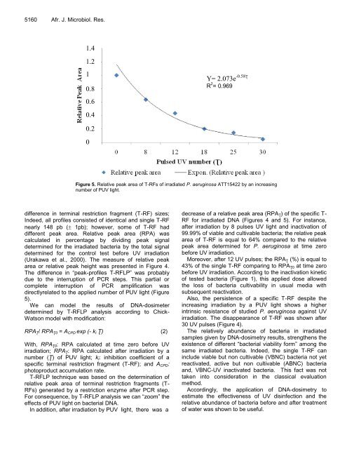
Figure 5. Relative peak area of T-RFs of irradiated P. aeruginosa ATT15422 by an increasing<br />
number of PUV light.<br />
difference in terminal restriction fragment (T-RF) sizes;<br />
Indeed, all profiles consisted of identical and single T-RF<br />
nearly 148 pb (� 1pb); however, some of T-RF had<br />
different peak area. Relative peak area (RPA) was<br />
calculated in percentage by dividing peak signal<br />
determined for the irradiated bacteria by the total signal<br />
determined for the control test before UV irradiation<br />
(Urakawa et al., 2000). The measure of relative peak<br />
area or relative peak height was presented in Figure 4.<br />
The difference in “peak-profiles T-RFLP” was probably<br />
due to the interruption of PCR steps. This partial or<br />
complete interruption of PCR amplification was<br />
directlyrelated to the applied number of PUV light (Figure<br />
5).<br />
We can model the results of DNA-dosimeter<br />
determined by T-RFLP analysis according to Chick-<br />
Watson model with modification:<br />
RPAƮ/ RPAƮ0 = ACPD exp (- ki Ʈ) (2)<br />
With, RPAƮ0: RPA calculated at time zero before UV<br />
irradiation; RPAƮ: RPA calculated after irradiation by a<br />
number (Ʈ) of PUV light; ki: inhibition coefficient of a<br />
specific terminal restriction fragment (T-RF); and ACPD:<br />
photoproduct accumulation rate.<br />
T-RFLP technique was based on the determination of<br />
relative peak area of terminal restriction fragments (T-<br />
RFs) generated by a restriction enzyme after PCR step.<br />
For consequence, by T-RFLP analysis we can “zoom” the<br />
effects of PUV light on bacterial DNA.<br />
In addition, after irradiation by PUV light, there was a<br />
Y= 2.073e -0.58Ʈ<br />
R 2 = 0.969<br />
decrease of a relative peak area (RPAƮ) of the specific T-<br />
RF for irradiated DNA (Figures 4 and 5). For instance,<br />
after irradiation by 8 pulses UV light and inactivation of<br />
99.99% of viable and cultivable bacteria; the relative peak<br />
area of T-RF is equal to 64% compared to the relative<br />
peak area determined for P. aeruginosa at time zero<br />
before UV irradiation.<br />
Moreover, after 12 UV pulses; the RPAƮ (%) is equal to<br />
43% of the single T-RF comparing to RPAƮ0 at time zero<br />
before UV irradiation. According to the inactivation kinetic<br />
of tested bacteria (Figure 1), this applied dose allowed<br />
the loss of bacteria cultivability in usual media with<br />
subsequent reactivation.<br />
Also, the persistence of a specific T-RF despite the<br />
increasing irradiation by a PUV light shows a higher<br />
intrinsic resistance of studied P. aeruginosa against UV<br />
irradiation. The disappearance of T-RF was shown after<br />
30 UV pulses (Figure 4).<br />
The relatively abundance of bacteria in irradiated<br />
samples given by DNA-dosimetry results, strengthens the<br />
existence of different “bacterial viability form” among the<br />
same irradiated bacteria. Indeed, the single T-RF can<br />
include viable but non cultivable (VBNC) bacteria not yet<br />
reactivated, active but non cultivable (ABNC) bacteria<br />
and, VBNC-UV inactivated bacteria. This fact was not<br />
taken into consideration in the classical evaluation<br />
method.<br />
Accordingly, the application of DNA-dosimetry to<br />
estimate the effectiveness of UV disinfection and the<br />
relative abundance of bacteria before and after treatment<br />
of water was shown to be useful.


