- Page 1 and 2: U. S. ARMY MEDICAL DEPARTMENT CENTE
- Page 3 and 4: TABLE OF CONTENTS Lesson Paragraphs
- Page 5 and 6: 9 THE HUMAN CARDIOVASCULAR AND LYMP
- Page 7 and 8: MD0006 v LIST OF ILLUSTRATIONS Figu
- Page 9 and 10: MD0006 vii LIST OF TABLES Table Pag
- Page 11 and 12: Material Furnished: In addition to
- Page 13 and 14: LESSON ASSIGNMENT LESSON 1 Introduc
- Page 15 and 16: terminology. Accountants have debit
- Page 17 and 18: . Head and Neck. The brain, eyes, e
- Page 19 and 20: Figure 1-2. Anatomical position and
- Page 21 and 22: 1-11. NAMES a. Names are chosen to
- Page 23 and 24: d. Mitochondria (Plural). Mitochond
- Page 25 and 26: 5. What is a cell? 6. What is a tis
- Page 27 and 28: 15. In figure 1-6, three points are
- Page 29 and 30: 18. In figure 1-8, parts of a "typi
- Page 31 and 32: 14. a. Midsagittal or median plane.
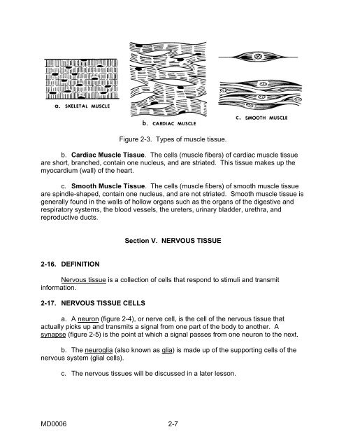
- Page 33 and 34: 2-1. DEFINITION LESSON 2 TISSUES OF
- Page 35 and 36: d. Functions. According to its loca
- Page 37: . Types of Bone Tissues. There are
- Page 41 and 42: 8. Characteristic cells of fibrous
- Page 43 and 44: SOLUTIONS TO EXERCISES, LESSON 2 1.
- Page 45 and 46: LESSON ASSIGNMENT LESSON 3 The Huma
- Page 47 and 48: 3-2. COVERINGS OF THE HUMAN BODY Th
- Page 49 and 50: 3-6. GLANDS The types of glands inc
- Page 51 and 52: 3-13. BURSA a. A bursa (figure 3-3)
- Page 53 and 54: EXERCISES, LESSON 3 REQUIREMENT. Th
- Page 55 and 56: 16. The term serous refers to a . S
- Page 57 and 58: 13. Sebaceous glands produce an oil
- Page 59 and 60: MD0006 4-2 4-13. Describe the gener
- Page 61 and 62: Section II. BONE AS AN INDIVIDUAL O
- Page 63 and 64: 4-5. DEVELOPMENT OF AN INDIVIDUAL B
- Page 65 and 66: Example: The frontal bone. (The fro
- Page 67 and 68: d. Capsule. The "typical" synovial
- Page 69 and 70: (2) In the plane joint, the contact
- Page 71 and 72: Figure 4-3B. Posterior view of the
- Page 73 and 74: (d) The sacrum, which is a bony fus
- Page 75 and 76: Figure 4-6. The human skull (front
- Page 77 and 78: PART UPPER MEMBER LOWER MEMBER GIRD
- Page 79 and 80: Figure 4-8. The human scapula and c
- Page 81 and 82: Figure 4-10. The human hand. Figure
- Page 83 and 84: Figure 4-13. The human foot. MD0006
- Page 85 and 86: 7. In the early fetus, bones are pr
- Page 87 and 88: 17. Name and define the two major s
- Page 89 and 90:
SOLUTIONS TO EXERCISES, LESSON 4 1.
- Page 91 and 92:
19. The regions of the vertebral co
- Page 93 and 94:
5-1. MUSCLE TISSUES LESSON 5 THE HU
- Page 95 and 96:
Figure 5-1. Skeletal and facial mus
- Page 97 and 98:
a. Trunk Musculature. The trunk mus
- Page 99 and 100:
. Sesamoid bones, such as the patel
- Page 101 and 102:
EXERCISES, LESSON 5 REQUIREMENT. Th
- Page 103 and 104:
SOLUTIONS TO EXERCISES, LESSON 5 1.
- Page 105 and 106:
6-1. GENERAL LESSON 6 THE HUMAN DIG
- Page 107 and 108:
a. Teeth. Figure 6-2. Anatomy of th
- Page 109 and 110:
6-4. PHARYNX The pharynx (pronounce
- Page 111 and 112:
common bile duct. The common bile d
- Page 113 and 114:
EXERCISES, LESSON 6 REQUIREMENT. Th
- Page 115 and 116:
14. The texture of the pancreas is
- Page 117 and 118:
10. The process of digestion is fac
- Page 119 and 120:
7-1. INTRODUCTION LESSON 7 THE HUMA
- Page 121 and 122:
SUBDIVISION FUNCTION (1) SUPRALARYN
- Page 123 and 124:
(2) Oropharynx. The portion of the
- Page 125 and 126:
Figure 7-4. Infralaryngeal structur
- Page 127 and 128:
7-8. DIAPHRAGMATIC (ABDOMINAL) BREA
- Page 129 and 130:
9. The functions of the supralaryng
- Page 131 and 132:
SOLUTIONS TO EXERCISES, LESSON 7 1.
- Page 133 and 134:
LESSON ASSIGNMENT LESSON 8 The Huma
- Page 135 and 136:
8-3. THE KIDNEY a. General. (1) The
- Page 137 and 138:
the cortex layer, it once again bec
- Page 139 and 140:
eginning of an embryo (the process
- Page 141 and 142:
(2) Wall structure. The inner linin
- Page 143 and 144:
Figure 8-5. The human male genital
- Page 145 and 146:
(1) Surrounding the urethra is a ce
- Page 147 and 148:
8. The first coiled portion of the
- Page 149 and 150:
21. The external genitalia of the h
- Page 151 and 152:
SOLUTIONS TO EXERCISES, LESSON 8 1.
- Page 153 and 154:
19. The inner lining of the uterus
- Page 155 and 156:
LESSON ASSIGNMENT LESSON 9 The Huma
- Page 157 and 158:
LESSON 9 THE HUMAN CARDIOVASCULAR A
- Page 159 and 160:
(1) Red blood cells (erythrocytes).
- Page 161 and 162:
(1) The arteries carry blood away f
- Page 163 and 164:
(3) Relationship of wall thickness
- Page 165 and 166:
ecome closed for whatever reason, t
- Page 167 and 168:
Figure 9-5. Main arteries of the hu
- Page 169 and 170:
Figure 9-6. Main veins of the human
- Page 171 and 172:
a. Lymphatic Capillaries. Lymphatic
- Page 173 and 174:
5. The most common types of white b
- Page 175 and 176:
18. In the case of collateral circu
- Page 177 and 178:
SOLUTIONS TO EXERCISES, LESSON 9 1.
- Page 179 and 180:
15. The coronary arteries supply "n
- Page 181 and 182:
LESSON ASSIGNMENT LESSON 10 The Hum
- Page 183 and 184:
. The Endocrine System. In the huma
- Page 185 and 186:
e. Of the many hormones produced by
- Page 187 and 188:
hormones are involved in the mobili
- Page 189 and 190:
5. The pituitary body is a small -s
- Page 191 and 192:
SOLUTIONS TO EXERCISES, LESSON 10 1
- Page 193 and 194:
13. During the first half of the me
- Page 195 and 196:
MD0006 11-2 11-12. Define periphera
- Page 197 and 198:
11-5. NEURON PROCESSES Figure 11-1.
- Page 199 and 200:
a. The Synapse. A synapse (figure 1
- Page 201 and 202:
system (ANS). The CNS is made up of
- Page 203 and 204:
Figure 11-5B. Human brain (bottom v
- Page 205 and 206:
d. Ventricles. Within the brain, th
- Page 207 and 208:
Figure 11-7. A schematic diagram of
- Page 209 and 210:
(1) Cranial nerves. The 12 pairs of
- Page 211 and 212:
(4) Visceral motor neurons of the A
- Page 213 and 214:
organ. The cell bodies of the secon
- Page 215 and 216:
(2) Motor pathways. A motor pathway
- Page 217 and 218:
11-26. GENERAL Section IX. THE SPEC
- Page 219 and 220:
c. Internal Structures of the Eyeba
- Page 221 and 222:
(5) Iris. Another structure formed
- Page 223 and 224:
11-30. GENERAL Section X. THE SPECI
- Page 225 and 226:
11-33. THE INTERNAL EAR a. Labyrint
- Page 227 and 228:
Figure 11-14. Diagram of the scalae
- Page 229 and 230:
11-36. SEMICIRCULAR DUCTS (FIGURE 1
- Page 231 and 232:
f. Cerebellum. The cerebellum has b
- Page 233 and 234:
7. Each item below refers to the th
- Page 235 and 236:
18. Groups of related functions are
- Page 237 and 238:
30. If it carries information from
- Page 239 and 240:
46. Name examples of general senses
- Page 241 and 242:
61. The orbit is the cavity in the
- Page 243 and 244:
73. The central column of the cochl
- Page 245 and 246:
SOLUTIONS TO EXERCISES, LESSON 11 1
- Page 247 and 248:
19. The ventricles of the brain are
- Page 249 and 250:
34. In the ANS, the number of neuro
- Page 251 and 252:
52. The outermost layer of the eyeb
- Page 253 and 254:
ossicles are: malleus, incus, and s
- Page 255:
COMMENT SHEET SUBCOURSE MD0006 Basi