NewTom VG User Manual rev 4.0 - Image Works
NewTom VG User Manual rev 4.0 - Image Works
NewTom VG User Manual rev 4.0 - Image Works
You also want an ePaper? Increase the reach of your titles
YUMPU automatically turns print PDFs into web optimized ePapers that Google loves.
Getting started…<br />
4 Getting started…<br />
This chapter provides a brief introduction for the <strong>NewTom</strong> <strong>VG</strong>i system, its power on and off routines and<br />
control devices.<br />
4.1 An introduction to the system<br />
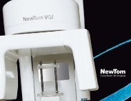
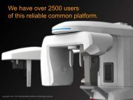
The device <strong>NewTom</strong> <strong>VG</strong>i is a panoramic 3D system, dedicated to dento-maxillo-facial area imaging. It<br />
performs the so called "cone-beam" technology.<br />
<strong>NewTom</strong> <strong>VG</strong>i is intended for diagnosis of the dento-maxillo-facial complex.<br />
It’s designed for:<br />
<br />
<br />
<br />
imaging of temporo mandibular joints;<br />
imaging of mandibula/jaw for surgical planing;<br />
imaging of nasal/sinus and maxillofacial complex;<br />
A patient is made to lean on the head support and centered by means of 2 laser modules and "scout view"<br />
images.<br />
The scanning system performs a completely rotation around the patient's head. Radiological images are<br />
acquired, that are then automatically processed by the system. The result is the slices set that forms the<br />
reconstructed volume. At the end of this process the axial slices set composes the Volumetric Data. Through<br />
these data it is possible to display coronal and sagittal views of the reconstructed volume in real time.<br />
After defining a Region Of Interest (ROI), from the volumetric data the user can start the creation of a study.<br />
The ROI can be inclined from the volumetric data both to obtain perpendicular images and to correct<br />
positioning errors.<br />
Working on the study data it is possible to create panoramic, transaxial and 3D images. You can also work<br />
on these kinds of images measuring distances, angles, putting comment etc.<br />
Finally new images can be saved inside the study.<br />
The study images can be used to compile a report, that can be then printed and/or saved on a physical<br />
support.<br />
To study in deep these themes please refer to the Software <strong>Manual</strong>.<br />
4.2 Working principle<br />
According to the cone-beam technology, the source detector system performs a single rotation around the<br />
patient's head, simultaneously acquiring every necessary data for the volumetric reconstruction. Data<br />
acquired each scan step are the digital images corresponding to the radiographic projection. The raw data<br />
set so collected is used in the volumetric tomographic reconstruction process.<br />
This technology brings some advantages:<br />
• direct reconstruction of any set of the scanned object points without passing through axial reconstruction<br />
and data re-formatting;<br />
• total scan time related to the acquisition electronics, rather than to the x-ray tube power and the<br />
mechanics, usually shorter;<br />
• under same conditions of total scan time, less requirements in regards to the source/tube assembly<br />
power and scan mechanics, with constructive and maintenance advantages.<br />
Rev. 1.0 - 02/15/2009 Page 20 of 72


