Dirac Fermions in Graphene and Graphiteâa view from angle ...
Dirac Fermions in Graphene and Graphiteâa view from angle ...
Dirac Fermions in Graphene and Graphiteâa view from angle ...

Create successful ePaper yourself
Turn your PDF publications into a flip-book with our unique Google optimized e-Paper software.
100 nm<br />
500 nm<br />
a<br />
50 nm<br />
b<br />
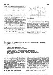
Figure 3.2. SEM images recorded on the surface of a sample grown <strong>in</strong> the same conditions as sample<br />
A. (a) Typical region of the surface, show<strong>in</strong>g a pattern with a length scale on the order of hundreds<br />
of nanometers, <strong>and</strong> a f<strong>in</strong>er pattern with a length scale on the order of 10 nm. (b) Region marked<br />
by a cluster of unidentified structures characterized by six-sided geometry, reflect<strong>in</strong>g the underly<strong>in</strong>g<br />
hexagonal lattice.<br />
produces the complex (6 √ 3×6 √ 3)R30 pattern shown <strong>in</strong> panel (c). Notably, a slight <strong>in</strong>crease of the anneal<strong>in</strong>g<br />
temperature gives rise to the more well developed (6 √ 3 × 6 √ 3)R30 pattern shown <strong>in</strong> panel (d). Panels (e)<br />
<strong>and</strong> (f) show LEED patterns obta<strong>in</strong>ed at a lower electron energy, under the same conditions as panels (c) <strong>and</strong><br />
(d), respectively. Graphite diffraction spots clearly observed only <strong>in</strong> panel (f) <strong>in</strong>dicate that a th<strong>in</strong> graphene<br />
overlayer is formed <strong>in</strong> the last step.<br />
3.2 Characterization of epitaxial graphene<br />
3.2.1 Scann<strong>in</strong>g electron microscopy (SEM)<br />
Fig. 3.2 presents SEM images of a graphene sample. The image shown <strong>in</strong> panel (a) exhibits two patterns,<br />
one <strong>in</strong> a large length scale, correspond<strong>in</strong>g to hundreds of nm, <strong>and</strong> another <strong>in</strong> a small length scale, on the<br />
order of 10 nm. The small length scale co<strong>in</strong>cides with a typical terrace size observed by scann<strong>in</strong>g tunnel<strong>in</strong>g<br />
spectroscopy 42 , while the large length scale may correspond to the s<strong>in</strong>gle-crystal gra<strong>in</strong> size. The image shown<br />
<strong>in</strong> panel (b) exhibits a similar pattern <strong>and</strong>, <strong>in</strong> addition, unidentified structures - white specks each enclosed<br />
by a large black region. Spread out widely over the sample surface, the white specks <strong>and</strong> enclos<strong>in</strong>g black<br />
regions show facets reflect<strong>in</strong>g the underly<strong>in</strong>g hexagonal lattice. An <strong>in</strong>vestigation <strong>in</strong>to potential causes for the<br />
formation of such structures is needed.<br />
3.2.2 X-ray photoemission spectroscopy (XPS)<br />
XPS measurements were carried out us<strong>in</strong>g Mg Kα radiation <strong>and</strong> normal emission geometry at 300K.<br />
Fig. 3.3 shows XPS spectra of the C 1s <strong>and</strong> Si 2p core levels <strong>and</strong> their l<strong>in</strong>e shape fits. For all samples, the Si<br />
2p spectrum consists of a s<strong>in</strong>gle peak located at ≈ 101.6 eV, attributed to the SiC bulk. The C 1s spectrum<br />
consists of three components, labeled as X, G, <strong>and</strong> S. In the analysis that follows we will make use of the<br />
latter two, which are assigned to the graphite overlayer (G) <strong>and</strong> the SiC bulk (S). These assignments are<br />
<strong>in</strong> agreement with previous work 43,44 . In particular, the small chemical shift of peak S relative to pure SiC<br />
by about 0.4 eV has been noted before, <strong>and</strong> was attributed to a Fermi level p<strong>in</strong>n<strong>in</strong>g associated with surface<br />
19