Vol 21 No. 1
Vol 21 No. 1
Vol 21 No. 1

Create successful ePaper yourself
Turn your PDF publications into a flip-book with our unique Google optimized e-Paper software.
J Bagh College Dentistry <strong>Vol</strong>. <strong>21</strong>(1), 2009 Prevalence and distribution …..<br />
apicocoronal dimension of the root surface<br />
exposed, this was done according to criteria<br />
suggested by Miller (16) :<br />
1- Small recession: less than 3mm of root surface<br />
exposed.<br />
2- Moderate recession: 3 to 4 mm of root surface<br />
exposed.<br />
3- Advanced recession: more than 4mm of root<br />
surface exposed to the dental environment.<br />
Measurement of root caries was done using root<br />
caries index (RCI)<br />
(R – D) + (R – F) x 100<br />
RCI = ---------------------------------------------<br />
(R – D) + (R + F) + (R – N)<br />
R – N= Recession present (root surface normal or<br />
sound)<br />
R – D= Recession present (with a decayed root<br />
surface)<br />
R – F=Recession present (with a filled root<br />
surface)<br />
Measurement was done according to age<br />
group and gender. The association between root<br />
caries and gingival recession was evaluated using<br />
logistic regression (LR).<br />
RESULTS<br />
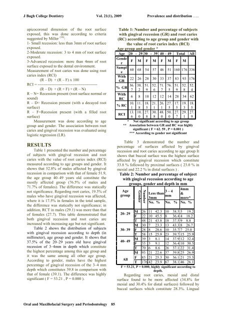
Table 1 presented the number and percentage<br />
of subjects with gingival recession and root<br />
caries with the value of root caries index (RCI)<br />
measured according to age groups and gender. It<br />
shows that 52.8% of males affected by gingival<br />
recession in comparison with that of female 51.9,<br />
the age group 40–49 years old constitute the<br />
mostly affected group (76.5% of males and<br />
71.7% of females). The difference was statically<br />
not significance. Regarding root caries, 19.3% of<br />
males who have gingival recession was affected,<br />
where it is 17.5% in females in the total sample,<br />
the difference was statically not significance; in<br />
addition, RCT in males (29.1) was more than that<br />
of females (27.7). This table demonstrated that<br />
both gingival recession and root caries are<br />
increased with increasing age but not significant.<br />
Table 2 shows the distribution of subjects<br />
with gingival recession according to depth (in<br />
millimeter), age group and gender. It shows that<br />
37.5% of the 20–29 years old have gingival<br />
recession of 3–4mm in depth which constitute<br />
the highest percentage among this age group and<br />
it was the same among all other age group.<br />
According to gender, males have the highest<br />
percentage of gingival recession of the 3–4 mm<br />
depth which constitutes 39.8 in comparison with<br />
that of female (39.1). The difference was highly<br />
significant ( F = 53.<strong>21</strong> , P = 0.000 ).<br />
Table 1: Number and percentage of subjects<br />
with gingival recession (GR) and root caries<br />
(RC) according to age group and gender with<br />
the value of root caries index (RCI)<br />
Age group and gender *<br />
Age 20 - 29 30 - 39 40 – 49 Total All<br />
Gende<br />
F M F M F M F M<br />
r<br />
Sampl<br />
60 68 54 57 46 51 160 176 336<br />
e<br />
With<br />
GR<br />
% GR<br />
With<br />
RC<br />
% RC<br />
RCI<br />
22<br />
36.<br />
7<br />
6<br />
26<br />
38.<br />
2<br />
8<br />
28<br />
51.<br />
9<br />
10<br />
30<br />
52.<br />
6<br />
12<br />
33<br />
71.<br />
7<br />
12<br />
37<br />
76.<br />
5<br />
14<br />
83<br />
51.<br />
9<br />
28<br />
93<br />
52.<br />
8<br />
34<br />
176<br />
52.<br />
4<br />
62<br />
10.<br />
1<br />
11.<br />
8<br />
18.<br />
5<br />
<strong>21</strong>.<br />
1<br />
26.<br />
1<br />
27.<br />
5<br />
17.<br />
5<br />
19.<br />
3<br />
18.<br />
5<br />
13. 10. 27. 30. 48. 50. 27. 29. 27.<br />
6 0 8 8 0 0 7 1 9<br />
* <strong>No</strong>t significant according to age group<br />
** Association between GR and RC was highly<br />
significant ( F = 62. 59 , P = 0.001 )<br />
*** According to gender not significant<br />
Table 3 demonstrated the number and<br />
percentage of surfaces affected by gingival<br />
recession and root caries according to age group It<br />
shows that buccal surface was the highest surface<br />
affected by gingival recession which constitute<br />
33.8 % followed by proximal surfaces ( 23.0 % in<br />
mesial and 22.2 % in distal surfaces ) .<br />
Table 2: Number and percentage of subject<br />
with gingival recession according to age<br />
groups, gender and depth in mm<br />
Age<br />
group<br />
Number<br />
Gender<br />
3 – 4<br />
Less than<br />
mm<br />
3mm<br />
<strong>No</strong>. %<br />
<strong>No</strong>. %<br />
4mm<br />
&<br />
more*<br />
<strong>No</strong>. %<br />
M 26 11 42.3 10 38.5 5 19.2<br />
20- 29<br />
F 22 10 45.5 8 36.4 4 18.2<br />
T 48 <strong>21</strong> 43.8 18 37.5 9 8.8<br />
M 30 7 23.3 13 43.3 8 26.7<br />
30- 39 F 28 8 28.6 10 35.7 7 25.0<br />
T 58 15 25.9 23 39.7 15 25.9<br />
M 39 3 8.1 14 37.9 12 32.4<br />
40- 49<br />
F 33 3 9.1 12 36.4 10 30.3<br />
T 70 6 8.6 26 37.1 22 31.4<br />
M 93 <strong>21</strong> 22.6 37 39.8 25 26.9<br />
F 83 <strong>21</strong> 25.3 30 36.1 <strong>21</strong> 25.3<br />
All<br />
T 176 42 23.9 67 38.1 46 26.1<br />
F = 53.<strong>21</strong>, P = 0.000, highly significant according to<br />
depth.<br />
Regarding root caries, mesial and distal<br />
surface found to be more affected (34.8% for<br />
mesial and 30.4% for distal surfaces) followed by<br />
buccal surfaces which constitute 28.3%. Lingual<br />
Oral and Maxillofacial Surgery and Periodontology 85