Vol 21 No. 1
Vol 21 No. 1
Vol 21 No. 1

You also want an ePaper? Increase the reach of your titles
YUMPU automatically turns print PDFs into web optimized ePapers that Google loves.
J Bagh College Dentistry <strong>Vol</strong>. <strong>21</strong>(1), 2009 Closed reduction for...<br />
MATERIALS & METHODS<br />
Patients' age & gender<br />
The study discuss and prospectively review<br />
the results of 32 dentulous patients derived from<br />
population of patients with missile injuries<br />
admitted at the oral & maxillofacial surgery<br />
department at Al-Yarmouk Teaching Hospital in<br />
the period between october 2006 – october<br />
2007.The extremes of patient age ranged from<br />
<strong>21</strong>-58 years.<br />
Diagnosis<br />
Diagnosis of mandibular fractures based on<br />
clinical and radiographic examination. Clinical<br />
examination of the face included extra and<br />
intraoral examination. The local examination was<br />
done to diagnose or exclude the presence of<br />
mandibular fractures. Extraoral examination<br />
achieved by inspection for the presence of<br />
swelling, ecchymosis, bleeding, soft tissue<br />
laceration, changes in facial contour, limitation<br />
or any abnormal mandibular movements.<br />
Bimanual palpation of the mandible with both<br />
hands to detect any step deformity, tenderness or<br />
crepitation on pressure. Compression test was<br />
used by the application of gentle compression of<br />
the mandible using both hands in two opposite<br />
directions under the lower border to elicit pain<br />
which indicate mandibular fracture.<br />
Intraoral examination was started by<br />
inspection for the presence of sublingual<br />
hematoma, mucosal lacerations and changes in<br />
occlusion. Teeth were examined for quantity,<br />
quality and occlusal relationship, gentle<br />
manipulation for mobility of fractured segments<br />
and displacement.<br />
Radiographic examination include the<br />
essential radiographs and according to their<br />
availability (plain radiographs as<br />
posteroanterior view of the mandible, oblique<br />
lateral view, intraoral occlusal view), Panoramic<br />
view OPG as shown in (Figure 3 and 4),<br />
computed tomography CT scans (axial and<br />
coronal).<br />
Maxillomandibular fixation MMF<br />
Closed reduction and indirect skeletal<br />
fixation the sole method of treatment, with jaws<br />
fixed using arch bars (Erich pattern) as a mean<br />
of intramaxillary fixation for the maxilla and<br />
mandible (Figure 2), MMF was carried out with<br />
soft stainless steel wires (0. 35 mm gauge or 0.4<br />
mm) for all patients. Circumferential wiring<br />
used in two cases only for support and elevation<br />
of badly displaced mandibular fractures.<br />
Immobilization for 6 weeks being the general<br />
guideline. Reduction and fixation of<br />
comminuted mandibular fractures achieved in<br />
25 cases under local anesthesia and in 7 cases<br />
under general anesthesia. All patients were<br />
placed on antibiotic treatment (prophylactic or<br />
therapeutic for already present infection) with<br />
possible use of culture and sensitivity test if<br />
possible from the time of admission until five<br />
days postoperatively. Osseous union of the<br />
fracture was tested clinically after 6 weeks of<br />
MMF, tie wires replaced if union is not<br />
satisfactory. To follow patients and monitoring<br />
for late complications, patients seen every 2<br />
weeks after immobilization for the first 2<br />
months then every month for at least 6 months.<br />
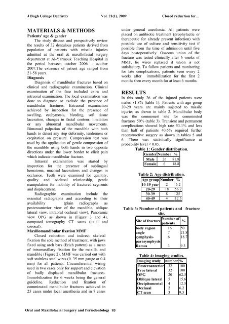
RESULTS<br />
In this study 26 of the injured patients were<br />
males 81.8% (table 1). Patients with age group<br />
20-29 years are mainly sujected to missile<br />
injuries as shown in table 2. Mandibular body<br />
was the commonest site for comminuted<br />
fractures 50% (table 3). Transient and permanent<br />
complications showed high rate 53.1% and less<br />
than half of patients 40.6% required further<br />
reconstructive surgery as shown in tables 5 and<br />
6. There was statistically significance at<br />
probability level < 0.05.<br />
Table 1: Gender distribution.<br />
Gender Number %<br />
Male 26 81.8<br />
Female 6 18.8<br />
Table 2: Age distribution.<br />
Age group Number %<br />
10-19 year 2 6.2<br />
20-29 18 56.2<br />
30-39 8 25<br />
40-49 4 12.5<br />
Table 3: Number of patients and fracture<br />
site.<br />
Number of<br />
Site of fracture %<br />
patients<br />
body region<br />
angle<br />
symphysisparasymphysis<br />
Ramus<br />
16<br />
7<br />
6<br />
3<br />
50<br />
<strong>21</strong>.8<br />
18.7<br />
9.3<br />
Table 4: imaging studies.<br />
Imaging study number %<br />
Posteroanterior<br />
True lateral<br />
OPG<br />
Oblique lateral<br />
Occipitomental<br />
Occlusal<br />
CT scan<br />
32<br />
32<br />
20<br />
5<br />
4<br />
2<br />
3<br />
100<br />
100<br />
62.5<br />
15.6<br />
12.5<br />
6.2<br />
9.3<br />
Oral and Maxillofacial Surgery and Periodontology 93