Vol 21 No. 1
Vol 21 No. 1
Vol 21 No. 1

You also want an ePaper? Increase the reach of your titles
YUMPU automatically turns print PDFs into web optimized ePapers that Google loves.
J Bagh College Dentistry <strong>Vol</strong>. <strong>21</strong>(1), 2009 Closed reduction for...<br />
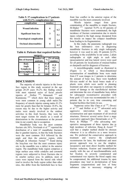
Table 5: 37 complications in 17 patients<br />
with 53.1% complication rate .<br />
complication number<br />
Wound infection<br />
Significant bone loss<br />
Neurological complication<br />
Occlusal abnormalities<br />
6<br />
3<br />
12<br />
6<br />
1<br />
6.2<br />
5.1<br />
2.4<br />
6.2<br />
Table 6: Patients that required further<br />
surgery .<br />
Number of % of total<br />
Site of fracture %<br />
patients number<br />
Body<br />
Angle<br />
Symphysisparasymphysis<br />
7<br />
2<br />
4<br />
53.8<br />
15.3<br />
30.7<br />
<strong>21</strong>.8<br />
6.2<br />
12.5<br />
Total 13 100 40.6<br />
DISCUSSION<br />
The majority of missile injuries to the lower<br />
face region in this study occurred in the age<br />
groups 20-29 years 56.2% this finding concur<br />
with most reported series of facial missile<br />
injuries of Hollier<br />
(20) (<strong>21</strong>)<br />
, Motamedi and<br />
Kummoona (22) which show that these injuries<br />
were most common in young adult males.The<br />
frequency of missile injuries among males 81.2%<br />
more far greatly than that for females 18.8%, the<br />
reason may be due to the higher activity and<br />
males were mostly involved in the outdoor<br />
activities in this Arab Society, they were the<br />
most target victims for attacks as a result of<br />
deterioration in the circumstances at the present<br />
time in our country due to occupation.<br />
Mandibular body was the most common<br />
fracture site 50%, Newlands et al (23) reported the<br />
distribution of a total 67 mandibular fractures<br />
due to gunshot injuries, in that the body fractures<br />
were the most common 38.8 followed by the<br />
angle and then the anterior region, while<br />
Akhlaghi & Aframian- Farnad (24) found during<br />
the Iraq-Iran war that the most common injury<br />
site to the mandible was a defect in the anterior<br />
region. The mandibular body region comprises a<br />
large surface area while condyles and coronoid<br />
regions were subjected to fewer fractures due to<br />
its smaller surface area. In addition, most of<br />
cases were injured from the lateral side which<br />
tend to involve the body region rather than other<br />
sites, this differ from the battlefield face to face<br />
front line conflict in the anterior region of the<br />
mandible was the most commonly involved.<br />
Missile injuries ranges from gross<br />
comminution of the mandible to simple fracture,<br />
M.Shaker (25) reported 76.9% in 60 patients of<br />
mandibular fractures were comminuted, the high<br />
incidence of fracture comminution due to missile<br />
injury related to the high energy dissipated from<br />
the missile on impact the compact mandibular<br />
bone leading to its fragmentation .<br />
In this study, the panoramic radiograph was<br />
the best informative view in diagnosing<br />
mandibular fractures in only single radiograph,<br />
however it was used in only 20 patients 62.5%<br />
according to the availability in our center, 2 plain<br />
radiographs at right angle to each other<br />
(poseroanterior and true lateral views) were used<br />
for all patients for localization of retained bullets<br />
or sharpnells and for diagnosis of fractures.<br />
A stereolithographic model as illustrated in<br />
(Figure 1) in which a three-dimentional<br />
reconstruction of mandibular bone were made<br />
from CT scan images in 2 patients to determine<br />
the amount of bone loss, these were handheld<br />
life-size model of the facial bones made of a<br />
plastic resin which were useful in planning<br />
treatment and allow our surgeons to estimate the<br />
extent of damage to the maxillofacial skeleton<br />
and determine the size of mandibular bony defect<br />
for subsequent reconstructive procedure with<br />
bone graft. CT scan was recommended for only 3<br />
patients due to its cost, time consuming and<br />
hospital facilities that limits its use.<br />
Numerous series like Chen et al (26) , Deveci<br />
et al (27) and Hollier et al (20) advocated early<br />
aggressive intervention for one stage<br />
reconstruction and open treatment of all involved<br />
structures. However several series favor a more<br />
coservative approach likes Demetriades et al (28) ,<br />
(29)<br />
Perry & Phillips who advocating nonoperative<br />
management of these injuries due to<br />
high incidence of infection. Haug & Assael (30)<br />
said that a simpler method should be choosen<br />
whenever it is as effective as a more invasive<br />
one. We preferred the conservative approach in<br />
treatment and reserve the complicated<br />
reconstructon for later, the traditional and most<br />
proven approach was to use the simplest,<br />
inexpensive and most direct feasible method for<br />
fracture reduction, fixation and immobilization<br />
usually with the use of maxillomandibular arch<br />
bar fixation. In severely comminuted and<br />
contaminated war injuries the blood supply may<br />
be compromised, the additional trauma by open<br />
procedures may lead to devitalization of bone<br />
fragments (sequestration) and loss of bony<br />
substance (due to necessary stripping of<br />
Oral and Maxillofacial Surgery and Periodontology<br />
94