programma & abstracts - Nederlandse Vereniging voor Radiologie
programma & abstracts - Nederlandse Vereniging voor Radiologie
programma & abstracts - Nederlandse Vereniging voor Radiologie

Create successful ePaper yourself
Turn your PDF publications into a flip-book with our unique Google optimized e-Paper software.
3<br />
<strong>programma</strong> <strong>abstracts</strong> & <strong>abstracts</strong><br />
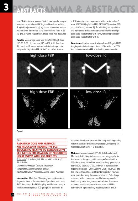
on a 64 detector-row scanner. Diastolic and systolic images<br />
were reconstructed with FBP (high and low-dose) and the<br />
IR algorithm (low-dose only). Hypo- and hyperdense artifact<br />
volumes were determined using two threshold filters (≤-50<br />
HU and ≥175 HU, respectively). Image noise was measured.<br />
Results: Mean image noise was 16.3±1.6 HU (high-dose<br />
FBP), 23.2±2.3 HU (low-dose FBP) and 16.5±1.7 (low-dose<br />
IR). Low-dose IR reconstructions had similar image noise<br />
compared to high-dose FBP (16.5±1.7 vs. 16.3±1.6, mean<br />
± SD). Mean hypo- and hyperdense artifact volumes (mm 3 )<br />
were 1235/5346 (high-dose FBP); 2405/6877 (low-dose FBP)<br />
and 1218/5333 (low-dose IR). For all PHV types, hypodense<br />
and hyperdense artifact volumes were similar for the highdose<br />
scans reconstructed with FBP when compared to lowdose<br />
scans reconstructed with IR.<br />
Conclusion: Iterative reconstruction allows ECG-gated PHV<br />
imaging with similar image noise and PHV artifacts at 50%<br />
less dose compared to FBP in an in vitro pulsatile model.<br />
Figure 1<br />
O3.5<br />
RADIATION DOSE AND ARTIFACTS<br />
ARE REDUCED BY PROSPECTIVE ECG-<br />
TRIGGERING RELATIVE TO RETROSPECTIVE<br />
ECG-GATING FOR IMAGING OF PROSTHETIC<br />
HEART VALVES WITH 256-SLICE CTS<br />
P. Symersky 1 , J. Habets 2 , B.A.J.M. de Mol 1 , M. Prokop 3 ,<br />
R.P.J. Budde 2<br />
1<br />
Academisch Medisch Centrum, Amsterdam<br />
2<br />
Universitair Medisch Centrum, Utrecht<br />
3<br />
Radboud University Nijmegen Medical Center, Nijmegen<br />
Introduction: Multislice CT imaging has complementary<br />
diagnostic value in the evaluation of prosthetic heart valve<br />
(PHV) dysfunction. For PHV imaging, modified coronary protocols<br />
with retrospective ECG-gating have been used at<br />
considerable radiation exposure. We compared image noise,<br />
radiation dose and artifacts with prospective triggering to<br />
retrospective gating for PHV evaluation.<br />
Methods: Two mechanical PHVs (St Jude bileaflet and<br />
Medtronic Hall tilting disc) were scanned using a pulsatile<br />
in vitro model. Image acquisition was performed with a<br />
256-slice scanner with either a retrospectively gated helical<br />
scan (120kV, 600mAs, CTDI vol<br />
39.8mGy) or a prospectively<br />
triggered axial scan (120kV, 200mAs, CTDI vol<br />
13.3mGy), rotation<br />
time 0.27sec. Hypo- and hyperdense artifact volumes<br />
were quantified using thresholds of -45 and 175HU. Image<br />
noise and artifacts were compared between protocols.<br />
Additionally, mean image noise and radiation dose were<br />
compared between 6 patients with mechanical PHVs<br />
scanned with a prospectively triggered protocol and 20<br />
30<br />
k i j k o o k o p w w w . c o n g r e s s c o m p a n y . c o m<br />
o f w w w . r a d i o l o g e n . n l