programma & abstracts - Nederlandse Vereniging voor Radiologie
programma & abstracts - Nederlandse Vereniging voor Radiologie
programma & abstracts - Nederlandse Vereniging voor Radiologie

You also want an ePaper? Increase the reach of your titles
YUMPU automatically turns print PDFs into web optimized ePapers that Google loves.
Kinderradiologie/Diversen 7<br />
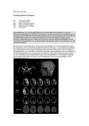
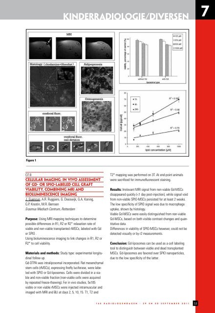
Figure 1<br />
O7.8<br />
CELLULAR IMAGING: IN VIVO ASSESSMENT<br />
OF GD- OR SPIO-LABELED CELL GRAFT<br />
VIABILITY, COMBINING MRI AND<br />
BIOLUMINESCENCE IMAGING<br />
J. Guenoun, A.R. Ruggiero, G. Doeswijk, G.A. Koning,<br />
G.P. Krestin, M.R. Bernsen<br />
Erasmus Medisch Centrum, Rotterdam<br />
Purpose: Using MRI mapping techniques to determine<br />
possible differences in R1, R2 or R2* relaxation rate of<br />
viable and non-viable transplanted rMSCs, labeled with Gd<br />
or SPIO .<br />
Using bioluminescence imaging to link changes in R1, R2 or<br />
R2* to cell viability.<br />
Materials and methods: Study type: experimental longitudinal<br />
follow up.<br />
Gd-DTPA was intraliposomal incorporated. Rat mesenchymal<br />
stem cells (rMSCs), expressing firefly luciferase, were labeled<br />
with SPIO or Gd-liposomes. Cells were divided in a viable<br />
and non-viable fraction (non-viable cells were acquired<br />
by repeated freeze-thawing). For in vivo studies, 5x10⁵<br />
viable or non viable rMSCs were injected intramuscular and<br />
imaged with MRI and BLI at days 2, 5, 10, 15. T1, T2 and<br />
T2* mapping was performed on 3T. At end-point animals<br />
were sacrificed for immunofluorescent staining.<br />
Results: Irrelevant MRI signal from non-viable Gd-MSCs<br />
disappeared quickly (