programma & abstracts - Nederlandse Vereniging voor Radiologie
programma & abstracts - Nederlandse Vereniging voor Radiologie
programma & abstracts - Nederlandse Vereniging voor Radiologie

Create successful ePaper yourself
Turn your PDF publications into a flip-book with our unique Google optimized e-Paper software.
9 <strong>programma</strong> <strong>abstracts</strong> & <strong>abstracts</strong><br />
Sessie 9<br />
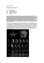
Neuro- en Hoofdhals radiologie<br />
Vrijdag 30 september, 10.45 - 12.15 uur<br />
O9.1<br />
ARTERIAL CALCIFICATION IN RELATION<br />
TO COGNITION AND STRUCTURAL BRAIN<br />
CHANGES<br />
D. Bos, M.W. Vernooij, S.E. Elias-Smale, G.P. Krestin,<br />
A. Hofman, W.J. Niessen, J.C.M. Witteman, A. van der Lugt,<br />
M.A. Ikram<br />
Erasmus Medisch Centrum, Rotterdam<br />
Purpose: Atherosclerosis plays an important role in the<br />
pathogenesis of cognitive decline and dementia. Calcified<br />
plaque measured with CT is a marker of atherosclerosis.<br />
This study investigates associations between CT-measured<br />
arterial calcifications measured at four locations, with cognition<br />
and macro- and microstructural brain changes.<br />
Method and materials: From the general population, 2437<br />
participants underwent CT of the coronary arteries, aortic<br />
arch, extracranial and intracranial carotid arteries to quantify<br />
calcification volume. Cognitive function was assessed in the<br />
following domains: memory, executive function, information<br />
processing speed and motor speed. In a random subgroup of<br />
844 participants brain MRI was performed to obtain measures<br />
of brain atrophy. Automated quantification of brain MRI<br />
scans yielded tissue-specific brain volumes. Furthermore,<br />
microstructural integrity of white matter was quantified<br />
using diffusion tensor imaging (DTI). Associations between<br />
arterial calcification and cognition, brain tissue volumes and<br />
DTI-measures were assessed with linear regression, adjusted<br />
for relevant confounders.<br />
Results: Larger calcification load was associated with<br />
worse cognitive scores in all domains. Calcification in all<br />
vessel beds was also associated with smaller total brain<br />
volume. Specifically, coronary calcification was associated<br />
with smaller grey matter volume, whilst both extra- and<br />
intracranial carotid artery calcification was associated with<br />
smaller white matter volume. Calcification in all vessel beds<br />
was associated with worse microstructural integrity of white<br />
matter.<br />
Conclusion: Arterial calcification load is associated with<br />
worse cognitive performance. Moreover, larger calcification<br />
load is associated with smaller brain tissue volumes and<br />
with worse white matter microstructural quality, elucidating<br />
possible mechanisms through which atherosclerosis leads to<br />
poorer cognition.<br />
O9.2<br />
TIA AND STROKE PATIENTS WITH CAROTID<br />
STENOSIS: PRESENCE OF COMPLICATED<br />
PLAQUE FEATURES AT MRI IS ASSOCIATED<br />
WITH RECURRENT EVENTS<br />
R.M. Kwee 1 , R.J. van Oostenbrugge 1 , W.H. Mess 1 ,<br />
M.H. Prins 1 , R.J. van der Geest 2 , J.W.M. ter Berg 3 ,<br />
C.L. Franke 4 , A.G.G.C. Korten 5 , B.J. Meems 6 ,<br />
J.M.A. van Engelshoven 1 , J.E. Wildberger 1 , M.E. Kooi 1<br />
1<br />
Maastricht Universitair Medisch Centrum, Maastricht<br />
2<br />
Leids Universitair Medisch Centrum, Leiden<br />
3<br />
Orbis Medisch Centrum, Sittard<br />
4<br />
Atrium Medisch Centrum Parkstad, Heerlen<br />
5<br />
Laurentius Ziekenhuis, Roermond<br />
6<br />
VieCuri Medisch Centrum, Venlo<br />
Purpose: There is a need for improved risk stratification of<br />
TIA and stroke patients with carotid atherosclerosis. The<br />
purpose of the present study was to prospectively investigate<br />
whether certain MRI-based carotid plaque characteristics<br />
are associated with recurrent ischemic events.<br />
Materials and Methods: One hundred TIA/stroke patients<br />
with ipsilateral 30-69% carotid stenosis underwent multisequence<br />
MRI, including contrast-enhanced images, of the<br />
carotid plaque within 32.7±19.9 days after the initial event.<br />
For each plaque, vessel wall volume and volumes of lipidrich<br />
necrotic core (LRNC), calcifications, and fibrous tissue<br />
were assessed. Maximum vessel wall thickness, minimum<br />
lumen area, fibrous cap status, and intraplaque hemorrhage<br />
(IPH) were also assessed. Patients were followed by structured<br />
interviews and chart review to determine the recurrence<br />
of ipsilateral TIA and/or ischemic stroke within one year.<br />
62<br />
k i j k o o k o p w w w . c o n g r e s s c o m p a n y . c o m<br />
o f w w w . r a d i o l o g e n . n l