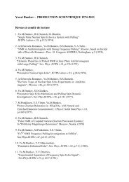
Activity Report 2010 - CNRS
Activity Report 2010 - CNRS
Activity Report 2010 - CNRS

Create successful ePaper yourself
Turn your PDF publications into a flip-book with our unique Google optimized e-Paper software.
7 – NANO APPROACHES<br />
TO LIFE SCIENCES<br />
Life sciences are one of the main<br />
application fields of nanosciences, as<br />
evidenced by the increasing number of<br />
proposals received along the successive<br />
call for proposals. Biological cells and<br />
molecules can be manipulated and<br />
analyzed with the greatest specificity in<br />
small volumes. Innovative devices will<br />
help building tomorrow’s medicine.<br />
MICRO- AND NANO<br />
FABRICATION FOR THE<br />
LIFE SCIENCES<br />
Contribution of 3D microenvironment<br />
to cell<br />
adhesion<br />
“New comers” Project 2008: Martial<br />
BALLAND (LIPhy)<br />
PhD student: Kalpana MANDAL<br />
Biological tissues are complex composite<br />
materials made of cells and intercellular<br />
matrix molecules secreted by the cells.<br />
Their formation and maintenance depend<br />
on both chemical and mechanical cues<br />
present in the microenvironment of each<br />
cell. Tissue biology involves therefore<br />
complex feedback loops. The aim of this<br />
project is to reproduce the organized<br />
geometry of biological tissues and to<br />
analyse quantitatively the mechanical<br />
forces developed at the cell/substrate<br />
and cell/cell interfaces.<br />
In order to measure mechanical forces,<br />
thin hydrogels are prepared that contain<br />
a homogeneous distribution of<br />
fluorescent 200nm beads (Fig. 1). The<br />
hydrogel surface is coated with<br />
fluorescent adhesion proteins, using an<br />
innovative deep-UV irradiation method<br />
developed in collaboration with Manuel<br />
Thery (IRTSV).<br />
This setup paves the way to a functional<br />
analysis of tumor cell invasiveness, which<br />
will be very useful to analyze biopsies<br />
and thus help cancer therapy.<br />
Fig. 1: Top: pictures of the adhesive protein<br />
micropattern (left) and the nanobeads<br />
distribution (right) used to calculate the cellsubstrate<br />
force distribution.<br />
Bottom: distribution of actin microfilaments<br />
(left) and traction forces (right) in a single<br />
micropatterned cell. Barscale: 20 µm<br />
Nanodroplet chip for<br />
controlled assembly of lipid<br />
bilayers and electrical<br />
detection of single-protein<br />
activity<br />
RTRA Project 2008: “Nanobiodrop"<br />
Benjamin CROSS (LEGI)<br />
Post-doctoral fellow: Anne MARTEL (IBS<br />
& LEGI)<br />
Transmembrane channels form a large<br />
class of molecules that play essential<br />
roles in cell physiology by allowing polar<br />
molecules, for instance ions, to cross<br />
biological lipid bilayer membranes. They<br />
are the target of numerous drugs and<br />
toxins. Nevertheless, conventional<br />
electrophysiological methods used to<br />
study ion channel activity are laborious<br />
and slow.<br />
FURTHER READING:<br />
SCIENTIFIC REPORT<br />
Front Biosci (Elite Ed). Jan 1;3:476-88 (2011)<br />
Multi-confocal fluorescence correlation<br />
spectroscopy<br />
The traction force distribution is<br />
computed from the bead displacement<br />
map by a Fast Fourier Traction Cytometry<br />
software. The forces developed by the<br />
micropatterned cells can thus be<br />
calculated and averaged, taking<br />
advantage of the similar geometry of the<br />
patterned cells. Significant differences<br />
between tumor cells have been<br />
evidenced, that are modulated by<br />
different tumorigenic signals.<br />
This innovative project called<br />
‘Nanobiodrop’ uses digital microfluidics to<br />
reconstitute lipid bilayers from two<br />
nanodroplets covered with phospholipids.<br />
Electrowetting is used to manipulate<br />
these droplets and put them into contact.<br />
Some of the nanodroplets are filled with<br />
membrane proteins, which are able to<br />
spontaneously insert in the membrane,<br />
once it is formed. The electrodes are then<br />
used to monitor the ionic current that<br />
flows from one droplet into the apposed<br />
one through the incorporated<br />
transmembrane channels.<br />
CONTACTS<br />
Franz BRUCKERT<br />
franz.bruckert@inpg.fr<br />
Tel: +33 4 56 52 93 21<br />
Julian GARCIA<br />
julian.garcia@ujf-grenoble.fr<br />
Tel: +33 4 56 52 08 31<br />
27