Activity Report 2010 - CNRS
Activity Report 2010 - CNRS
Activity Report 2010 - CNRS

Create successful ePaper yourself
Turn your PDF publications into a flip-book with our unique Google optimized e-Paper software.
CRG:<br />
The French Cooperative Research Group<br />
(CRG) at ESRF operates equipments for<br />
the structural and chemicals analysis of<br />
nanostructures by hard X-rays diffraction<br />
and diffusion.<br />
CMTC:<br />
Grenoble INP’s ‘Consortium de Moyens<br />
Technologiques Communs’ (CMTC) offers<br />
laboratory equipments for X-ray<br />
characterization and electron microscopy<br />
analysis of nanomaterials.<br />
The nanocharacterization facility is open<br />
to a large part of the Foundation<br />
members while there are no uniform<br />
access rules because some top level<br />
equipments can only be operated by<br />
trained experts or because the selection<br />
by Program Committees is required<br />
(ESRF).<br />
In 2008, the Foundation contributed to<br />
the acquisition of a new dual beam<br />
Focused Ion Beam (FIB) that is located at<br />
the PFNC. This NVision ZEISS FIB was<br />
installed during summer 2009. Since<br />
then, numerous “expert” users have been<br />
trained to use this equipment. Now,<br />
more than 10 users from the 3 partner’s<br />
laboratories (CMTC, PTA and PFNC) are<br />
working every day to perform advanced<br />
experiments:<br />
Nanomanipulation of nanoobjects<br />
as tripod ZnO, nanowires or tores<br />
using the 4 nanomanipulators<br />
Fig. 3: MEB-FIB nanomanipulators<br />
Electrical testing on nanodevices<br />
(carbon nanotubes, memories,<br />
transistors)<br />
Thin lamella preparation for TEM<br />
observations: classical lift out with<br />
optimised end milling at low voltage to<br />
reduced implantation /amorphisation;<br />
back end preparation to avoid ion beam<br />
damage on critical structures<br />
Ion milling for nano-object<br />
making (nanopillar for 3D X-ray<br />
tomography, hole in thin layers for<br />
Synchrotron experiments, others…)<br />
3D reconstruction using the “slice<br />
and view” method. Experiment of series<br />
of images is done overnight to perform<br />
3D reconstruction with nm resolution.<br />
Tests were done on a large range of<br />
materials: SC, porous materials,<br />
Biological sample (rat heart tissues),<br />
devices for microelectronic, fuel cell …<br />
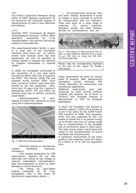
Fig. 4: TEM images of YBaCuO/LaZrO thin bilayer<br />
on NiW substrate obtained from a single<br />
12 µm TEM slice cut using the FIB (Coll.<br />
CMTC/Grenoble INP)<br />
Please read the corresponding Highlight<br />
at the end of this report for further<br />
information<br />
These experiments are done for various<br />
kinds of projects: ANR, Nanosciences<br />
Foundation, Carnot Institutes, PhD<br />
Thesis, Nano2012 for nanoscience and<br />
nanodevices applications.<br />
Additional experiments were also<br />
conducted by Earth Science Institute<br />
(‘Institut des Sciences de la Terre’ or<br />
‘ISTerre’) and ‘Grenoble Institute of<br />
Neuroscience’ or ‘GIN’ for biological<br />
applications.<br />
In <strong>2010</strong>, the Foundation has decided to<br />
partially support (~50%) the purchase of<br />
a new X-ray optics for the D2AM French<br />
CRG beamline at ESRF. This operation,<br />
which was also supported by Léti, will<br />
enable to extend the X ray photon energy<br />
up to 40 keV, and cover an energy range<br />
that is complementary to the available<br />
one at synchrotron Soleil in Saint-Aubin.<br />
This high energy range is particularly well<br />
suited to the characterization of buried<br />
interfaces and nanostructures.<br />
A significant improvement of the beam<br />
quality is also expected, which will reduce<br />
by a factor of 10 to 100 the acquisition<br />
time.<br />
SCIENTIFIC REPORT<br />
39