Activity Report 2010 - CNRS
Activity Report 2010 - CNRS
Activity Report 2010 - CNRS

You also want an ePaper? Increase the reach of your titles
YUMPU automatically turns print PDFs into web optimized ePapers that Google loves.
SCIENTIFIC REPORT<br />
The Nano-Chemistry and<br />
Biology Facility<br />
The objectives of these facilities are to<br />
provide to the community the tools for<br />
the synthesis and characterisation of<br />
molecules as well as the equipments<br />
required for analysis of organic/inorganic<br />
heterostructures and interfaces.<br />
NanoBio:<br />
The "Plateforme NanoBio-Chimie" located<br />
on the campus of Grenoble University<br />
offers equipments and tools for the<br />
synthesis of molecules, the grafting on<br />
surfaces and their characterisation (mass<br />
spectroscopy, AFM, IR spectroscopy...)<br />
and the imagery.<br />
IBS:<br />
The electron microscopy team of the<br />
‘Institut de Biologie Structurale’ (IBS) has<br />
developed the tools to study fragile<br />
materials such as living cells, proteins ,…<br />
grafted on inorganic nanostructures.<br />
The nano-chemistry and biology facility<br />
initiated in 2006 is dedicated to the<br />
conception and synthesis of bio molecules<br />
and to surface grafting. It is now well<br />
equipped for analytical characterization of<br />
final products, functionalized surfaces<br />
and molecular interactions, and is largely<br />
open to the community - the number of<br />
users increasing regularly.<br />
In 2008, the Foundation funded the<br />
purchase of an atomic force microscope<br />
AFM that works in biological media. Set<br />
up at the ‘Interdisciplinary Physics<br />
Laboratory’ (LIPhy), this new equipment<br />
belongs to the Nanobio facility (East<br />
Campus) and scientific activities have<br />
really started in September 2009.<br />
Coupled to an inverted Zeiss optical<br />
microscope, the JKP-Berlin AFM-Bio<br />
instrument allows various measurements<br />
such as cell-cell adhesion properties, the<br />
elasticity of gels or the topography of<br />
biological cells (Fig.5).<br />
The types of molecules analyzed by this<br />
technique are:<br />
biopolymers (oligonucleotides,<br />
peptides, carbohydrates)<br />
synthetic polymers<br />
various modified molecular<br />
objects functionalized by bio-organic<br />
molecules tag<br />
bio organic and inorganic<br />
complexes (non-covalent structure).<br />
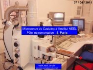
MALDI-TOF mass spectral analysis was<br />
used to characterize the relative<br />
molecular weight distribution of low<br />
molecular weight polymers - decanoate<br />
-CD ester (-CD-C 10 ) - presenting a<br />
low polydispersity of substitution. The<br />
correlation between the nanoparticle<br />
ultrastructure and the total degree of<br />
substitution has been demonstrated in<br />
the case of -CD-C 10 derivatives. (DPM-<br />
ICMG).<br />
It will also be possible to perform surface<br />
imaging (laser Smartbeam TM available<br />
with this new instrument). This feature<br />
has been used to discriminate between<br />
different in vitro bacteria culture<br />
(preliminary work, CERMAV-ICMG)<br />
Finally, using modified supports by<br />
adjunction of carbon nanotubes or others<br />
nanoparticles (gold), it will be possible to<br />
increase the capabilities of the<br />
instrument. The mass spectrometry<br />
technical staff is composed by 1 research<br />
engineer (<strong>CNRS</strong>), 1 design engineer<br />
(UJF) and 2 Technicians (<strong>CNRS</strong> and UJF).<br />
In 2009, the NanoBio facility acquired a<br />
new Maldi-ToF mass spectrometer<br />
(standing for ‘Matrix assisted laser<br />
desorption/ionisation – Time of Flight’).<br />
The objectives of this facility are to<br />
provide to the community the tools for<br />
the characterization by mass<br />
spectrometry of various kinds of<br />
molecules of high mass (superior limit<br />
500 000 Da).<br />
Fig. 5: Example of AFM topography on<br />
biological cell.<br />
40