Principles of cell signaling - UT Southwestern
Principles of cell signaling - UT Southwestern
Principles of cell signaling - UT Southwestern

Create successful ePaper yourself
Turn your PDF publications into a flip-book with our unique Google optimized e-Paper software.
39057_ch14_<strong>cell</strong>bio.qxd 8/28/06 5:11 PM Page 600<br />
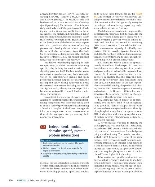
activated protein kinase (MAPK) cascade, including<br />
a MAP3K (Ste11p), a MAP2K (Ste7p)<br />
and a MAPK (Fus3p). (The MAPK cascade will<br />
be discussed in 14.32 MAPKs are central to many<br />
<strong>signaling</strong> pathways). The function <strong>of</strong> Ste5p is partially<br />
retained even if the positions <strong>of</strong> its binding<br />
sites for the kinases are shuffled in the linear<br />
sequence <strong>of</strong> the protein, indicating that a major<br />
role is to bring the enzymes into proximity, rather<br />
than to precisely orient them. Ste5p also binds<br />
to the subunits <strong>of</strong> the heterotrimeric G protein<br />
that mediates the actions <strong>of</strong> mating<br />
pheromones, linking the membrane signal to<br />
the intra<strong>cell</strong>ular transducers. Yeast that lack<br />
Ste5p cannot mate, demonstrating that Ste5p is<br />
required for this biological function (but not all<br />
functions) carried out by the pathway.<br />
In addition to facilitating <strong>signaling</strong> in their<br />
own pathways, scaffolds can enhance <strong>signaling</strong><br />
specificity by limiting interactions with other<br />
<strong>signaling</strong> proteins. Scaffolds thus insulate components<br />
<strong>of</strong> a <strong>signaling</strong> pathway both from activation<br />
by inappropriate signals and from<br />
producing incorrect outputs. For example, the<br />
mating and osmosensing pathways in yeast<br />
share several components, including the MAP3K<br />
Ste11p, but each pathway maintains specificity<br />
because it employs different scaffolds that restrict<br />
signal transmission.<br />
In contrast, the presence <strong>of</strong> excess scaffold<br />
can inhibit <strong>signaling</strong> because the individual <strong>signaling</strong><br />
components will more frequently bind<br />
to distinct scaffold proteins rather than forming<br />
a functional complex. Such dilution among scaffolds<br />
causes separation rather than concentration<br />
<strong>of</strong> the components, preventing their<br />
productive interaction.<br />
14.9<br />
Independent, modular<br />
domains specify proteinprotein<br />
interactions<br />
Key concepts<br />
• Protein interactions may be mediated by small,<br />
conserved domains.<br />
• Modular interaction domains are essential for<br />
signal transmission.<br />
• Adaptors consist exclusively <strong>of</strong> binding domains or<br />
motifs.<br />
Modular protein interaction domains or motifs<br />
occur in many <strong>signaling</strong> proteins and confer the<br />
ability to bind structural motifs in other molecules,<br />
including proteins, lipids, and nucleic<br />
acids. Some <strong>of</strong> these domains are listed in FIGURE<br />
14.9. In contrast to scaffolds, which bind specific<br />
proteins with considerable selectivity, modular<br />
interaction domains generally recognize<br />
not a single molecule but a group <strong>of</strong> targets that<br />
share related structural features.<br />
Modular interaction domains important for<br />
signal transduction were first discovered in the<br />
protein tyrosine kinase proto-oncogene Src,<br />
which contains a protein tyrosine kinase domain<br />
and two domains named Src homology<br />
(SH) 2 and 3 domains. The modular SH2 and<br />
SH3 domains were originally identified by comparison<br />
<strong>of</strong> Src to two other tyrosine kinases, Fps<br />
and Abl. One or both <strong>of</strong> these domains appear<br />
in numerous proteins and both are critically involved<br />
in protein-protein interactions.<br />
SH3 domains, which consist <strong>of</strong> approximately<br />
50 residues, bind to specific short proline-rich<br />
sequences. Many cytoskeletal proteins<br />
and proteins found in focal adhesion complexes<br />
contain SH3 domains and proline rich sequences,<br />
suggesting that this targeting motif<br />
may send proteins with these domains to these<br />
sites <strong>of</strong> action within <strong>cell</strong>s. In contrast to phosphotyrosine-SH2<br />
binding, the proline-rich binding<br />
sites for SH3 domains are present in resting<br />
and activated <strong>cell</strong>s. However, SH3-proline interactions<br />
may be negatively regulated by phosphorylation<br />
within the proline-rich motif.<br />
SH2 domains, which consist <strong>of</strong> approximately<br />
100 residues, bind to Tyr phosphorylated<br />
proteins, such as cytoplasmic tyrosine<br />
kinases and receptor tyrosine kinases. Thus, Tyr<br />
phosphorylation regulates the appearance <strong>of</strong><br />
SH2 binding sites and, thereby, regulates a set<br />
<strong>of</strong> protein-protein interactions in a stimulusdependent<br />
manner.<br />
A clever strategy was used to identify the<br />
binding specificity <strong>of</strong> SH2 domains. An isolated<br />
recombinant SH2 domain was incubated with<br />
<strong>cell</strong> lysates and then recovered from the lysates<br />
using a purification tag. The proteins associated<br />
with the SH2 domain were some <strong>of</strong> the same<br />
proteins that were recognized by antiphosphotyrosine<br />
antibodies. By this and other methods,<br />
it was discovered that SH2 domains recognize<br />
sequences surrounding Tyr phosphorylation<br />
sites and require phosphorylation <strong>of</strong> the included<br />
Tyr for high affinity binding.<br />
Information on specific amino acid sequences<br />
that recognize and bind to modular<br />
binding domains is being accumulated as these<br />
individual interactions are identified. In addition,<br />
screening programs using cDNA and/or<br />
peptide libraries to assess binding capabilities<br />
600 CHAPTER 14 <strong>Principles</strong> <strong>of</strong> <strong>cell</strong> <strong>signaling</strong>